
CBSE Class 9 Science Tissues Notes

Download CBSE Class 9 Science Useful Resources

Review targeted academic resources with the CBSE Class 9 Science Tissues Notes. Built according to official CBSE standards for the 2026-27 term, these downloadable Class 9 Science study materials support effective daily practice and deeper conceptual understanding for Chapter 06 Tissues.

Access CBSE Study Guides and Summaries

Access the complete useful resource PDF for Class 9 Science below. Regular practice with these targeted academic notes builds familiarity with complex topics and helps secure higher marks in final school evaluations.

CBSE Class 9 Science Tissues Notes. There are many more useful educational material which the students can download in pdf format and use them for studies. Study material like concept maps, important and sure shot question banks, quick to learn flash cards, flow charts, mind maps, teacher notes, important formulas, past examinations question bank, important concepts taught by teachers. Students can download these useful educational material free and use them to get better marks in examinations.  Also refer to other worksheets for the same chapter and other subjects too. Use them for better understanding of the subjects.

TISSUES

TISSUES

The body of plants and animals is made up of different types of cells. These cells originate from a single cell by repeated divisions and get differentiated during development. In unicellular organisms all the body functions are performed by a single cell, but in multicellular organisms different functions are performed by different groups of cells.

useful-resources-science-cbse-class-9-2

A group of cells having a common origin and performing similar functions is called a tissue. Several tissues are organised to form a tissue system, tissue systems form organs, and several organs form an organism. The study of tissues is called histology.

CellsTissuesTissue systemsOrgansOrganisms

Tissue and division of labour. In complex organisms, different tasks are carried out by different organs and organ systems. Tissues are the first step towards division of labour in complex organisms.

INTEXT QUESTIONS (PAGE 69)

What is a tissue?

Answer: A group of cells that are similar in structure and/or work together to achieve a particular function is called a tissue.

What is the utility of tissues in multicellular organisms?

Answer: In multicellular organisms the body is based on division of labour: cells performing a specific function are grouped together to form a particular tissue. The different tissues are organised in a way that gives the highest efficiency in the functioning of the body.

PLANT TISSUES

Plant tissues are of two main types: meristematic tissue and permanent tissue.

MERISTEMATIC TISSUE

The growth of plants occurs only in certain specific regions, because the dividing tissue, called meristematic tissue (meristos = divisible), is located only at these points. Meristematic tissues are made up of groups of similar, immature cells which can divide and form new cells. Meristematic cells divide continuously and thus help in increasing the length and thickness of the plant. Depending upon their position, meristematic tissues are of three types:

Apical MeristemIntercalary MeristemLateral Meristem
  1. Apical meristem: present at the growing tips of stems and roots; it increases the length of the plant body (primary growth).
  2. Intercalary meristem: occupies the base of leaves and the base of the internodal regions in plants such as grasses (mostly monocotyledonous plants); it helps in elongation of the internodes.
  3. Lateral meristem: the meristematic tissue occupying the lateral regions of stems and roots, which brings about an increase in the width of the plant body, e.g. cork cambium and vascular cambium.

i) Apical meristems: Apical meristem is present at the growing tips of stems and roots and increases the length of the plant body. They are responsible for growth in length, i.e. primary growth. ii) Intercalary meristems: These meristems occupy base of the leaves and the base of the internodal regions in plants such as grasses (mostly in monocotyledonous plants). These help in elongation of the internodes. iii) Lateral meristems: This includes the meristematic tissues occupying the lateral regions of the stems and roots which bring about increase in the width of the plant body. (e.g. Cork cambium and Vascular cambium). Characteristic features of Meristematic tissues The meristematic cells may be round, oval, polygonal or rectangular in shape. Their cell walls are thin, elastic and made up of cellulose. They are closely arranged without intercellular spaces. They have dense cytoplasm with large nucleus. PERMANENT TISSUE: Once the cells of meristematic tissue divide to a certain extent, they become specialized for a particular function. This process is called differentiation. Once differentiation is accomplished, the cells lose their capability to divide and the tissue becomes permanent tissue. Some cells produced by meristematic tissues stop dividing and form a permanent tissue. They have definite structure and function. They are differentiated into various types to perform different functions. The permanent tissues are classified as i) Simple tissues and ii) Complex tissues SIMPLE TISSUES A tissue with the cells of similar structure (one type of cells) and function is called simple tissue. It is of three types. 1. Parenchyma 2. Collenchyma 3. Sclerenchyma PARENCHYMA The cells of parenchyma have thin cell wall. They are loosely packed; with lot of intercellular spaces between them. They are living cells. They are generally present in all organs of a plant. They are oval or spherical or rectangular or cylindrical in shape. The cell wall is made of cellulose and pectic materials. Parenchyma makes the largest portion of a plant body. Parenchyma mainly works are packing material in plant parts. The main function of parenchyma is to provide support and to store food. In some plant parts, parenchyma has chlorophyll as well. In that case, parenchyma carries out photosynthesis and is then termed as chlorenchyma. In aquatic plants, large air cavities are present in parenchyma. This provides buoyancy to the plant, and then the parenchyma is known as aerenchyma. COLLENCHYMA The cells of collenchyma are polygonal in cross section and have unevenly thickened walls. These thickenings are due to the deposition of more cellulose, hemi-cellulose and pectin. The thickening is confined to the corners of the cells. They generally occur in the dicot stem in two or more layers below the epidermis. It is absent in the roots. It also occurs in petiole and pedicel. Like Parenchyma, Collenchyma is also a living tissue. The main function of Collenchyma is to provide strength and flexibility to the growing organs like young stem. SCLERENCHYMA It is a dead tissue. The cells are thick with lignified walls. They give mechanical support to the organs. This has two types of cells – Sclereids and Fibres. Sclereids Sclereids are stone cells which are commonly found in shells of the nut, pulp of certain fruits such as Pear and Sapota. Fibres The fibres are elongated strands with simple pits throughout its length. COMPLEX PERMANENT TISSUES XYLEM Xylem is mainly concerned with the transport of nutrients, water and minerals upwards in the plant body. It forms a continuous tube through the roots, stems, leaves, flowers and fruits by the fusion of elongated cells. It is composed of different kinds of cells namely, 1. Tracheids 2. Xylem vessels. 3. Xylem fibres 4. Xylem parenchyma. Tracheids Tracheids are elongated, tapering cells with blunt ends. They have lignified secondary wall. They are the chief water conducting elements in Pteridophytes and Gymnosperms. Xylem vessels Xylem vessels have perforations at the end and are placed one above the other like a long pipe line. They are seen in the xylem of angiosperms. They conduct water, mineral nutrients and also provide mechanical strength to the plant body. Xylem Fibres The fibres of Sclerenchyma associated with the xylem are known as xylem fibres. They give additional mechanical strength to the plant. They are also called wood fibres. Xylem Parenchyma The parenchyma cells associated with xylem are known as xylem parenchyma. It is the only living tissue amongst xylem cells. They store food reserves in the form of starch and fat. They also help in conduction of water. PHLOEM Phloem conducts food materials from leaves to the other parts of the plant. It is made up of four types of cells. 1. Sieve elements 2. Companion cells 3. Phloem fibres 4. Phloem parenchyma Sieve elements Sieve elements are the conducting elements of the phloem. Sieve elements are of two types –sieve cells and sieve tubes. Sieve cells are present in Pteridophytes and Gymnosperms where as sieve tubes are present in Angiosperms. Companion cells Companion cells are thin walled elongated specialized Parenchyma cells. They are associated with sieve elements. They have a prominent nucleus and cytoplasm. They help the sieve tube in conduction of food materials in angiosperms. Phloem fibres The fibres of sclerenchyma associated with phloem are called phloem fibres. They are also called bast-fibres. They give mechanical support to the plant. Among the four types of phloem cells, phloem fibres are the only dead tissues. Phloem parenchyma The parenchyma cells associated with phloem are called phloem parenchyma. They store starch and fats. ANIMAL TISSUES Animal tissues are of four types, viz. epithelial tissue, connective tissue, muscular tissue and nervous tissue. EPITHELIAL TISSUE: The epithelial tissue forms the covering or lining of most of the organs. The cells of epithelial tissue are tightly packed and form a continuous sheet. There is small amount of cementing materials between the cells and no intercellular space is present. Permeability of the epithelial tissue plays a great role in exchange of materials among various organs it also plays an important role in osmoregulation. All epithelial tissues are separated by the underlying tissue by an extracellular fibrous basement membrane. Epithelial tissues are of following types: 1. Simple Epithelium 2. Cuboidal Epithelium 3. Columnar Epithelium 4. Stratified Epithelium

Characteristic features of meristematic tissues

  • The cells may be round, oval, polygonal or rectangular in shape.
  • Their cell walls are thin, elastic and made of cellulose.
  • The cells are closely arranged, without intercellular spaces.
  • They have dense cytoplasm with a large nucleus.

PERMANENT TISSUE

Once the cells of meristematic tissue divide to a certain extent, they become specialised for a particular function. This process is called differentiation. Once differentiation is accomplished, the cells lose their capability to divide and the tissue becomes permanent tissue. Permanent tissue has a definite structure and function, and is differentiated into various types to perform different functions. Permanent tissues are classified as (i) simple tissues and (ii) complex tissues.

i) Apical meristems: Apical meristem is present at the growing tips of stems and roots and increases the length of the plant body. They are responsible for growth in length, i.e. primary growth. ii) Intercalary meristems: These meristems occupy base of the leaves and the base of the internodal regions in plants such as grasses (mostly in monocotyledonous plants). These help in elongation of the internodes. iii) Lateral meristems: This includes the meristematic tissues occupying the lateral regions of the stems and roots which bring about increase in the width of the plant body. (e.g. Cork cambium and Vascular cambium). Characteristic features of Meristematic tissues The meristematic cells may be round, oval, polygonal or rectangular in shape. Their cell walls are thin, elastic and made up of cellulose. They are closely arranged without intercellular spaces. They have dense cytoplasm with large nucleus. PERMANENT TISSUE: Once the cells of meristematic tissue divide to a certain extent, they become specialized for a particular function. This process is called differentiation. Once differentiation is accomplished, the cells lose their capability to divide and the tissue becomes permanent tissue. Some cells produced by meristematic tissues stop dividing and form a permanent tissue. They have definite structure and function. They are differentiated into various types to perform different functions. The permanent tissues are classified as i) Simple tissues and ii) Complex tissues SIMPLE TISSUES A tissue with the cells of similar structure (one type of cells) and function is called simple tissue. It is of three types. 1. Parenchyma 2. Collenchyma 3. Sclerenchyma PARENCHYMA The cells of parenchyma have thin cell wall. They are loosely packed; with lot of intercellular spaces between them. They are living cells. They are generally present in all organs of a plant. They are oval or spherical or rectangular or cylindrical in shape. The cell wall is made of cellulose and pectic materials. Parenchyma makes the largest portion of a plant body. Parenchyma mainly works are packing material in plant parts. The main function of parenchyma is to provide support and to store food. In some plant parts, parenchyma has chlorophyll as well. In that case, parenchyma carries out photosynthesis and is then termed as chlorenchyma. In aquatic plants, large air cavities are present in parenchyma. This provides buoyancy to the plant, and then the parenchyma is known as aerenchyma. COLLENCHYMA The cells of collenchyma are polygonal in cross section and have unevenly thickened walls. These thickenings are due to the deposition of more cellulose, hemi-cellulose and pectin. The thickening is confined to the corners of the cells. They generally occur in the dicot stem in two or more layers below the epidermis. It is absent in the roots. It also occurs in petiole and pedicel. Like Parenchyma, Collenchyma is also a living tissue. The main function of Collenchyma is to provide strength and flexibility to the growing organs like young stem. SCLERENCHYMA It is a dead tissue. The cells are thick with lignified walls. They give mechanical support to the organs. This has two types of cells – Sclereids and Fibres. Sclereids Sclereids are stone cells which are commonly found in shells of the nut, pulp of certain fruits such as Pear and Sapota. Fibres The fibres are elongated strands with simple pits throughout its length. COMPLEX PERMANENT TISSUES XYLEM Xylem is mainly concerned with the transport of nutrients, water and minerals upwards in the plant body. It forms a continuous tube through the roots, stems, leaves, flowers and fruits by the fusion of elongated cells. It is composed of different kinds of cells namely, 1. Tracheids 2. Xylem vessels. 3. Xylem fibres 4. Xylem parenchyma. Tracheids Tracheids are elongated, tapering cells with blunt ends. They have lignified secondary wall. They are the chief water conducting elements in Pteridophytes and Gymnosperms. Xylem vessels Xylem vessels have perforations at the end and are placed one above the other like a long pipe line. They are seen in the xylem of angiosperms. They conduct water, mineral nutrients and also provide mechanical strength to the plant body. Xylem Fibres The fibres of Sclerenchyma associated with the xylem are known as xylem fibres. They give additional mechanical strength to the plant. They are also called wood fibres. Xylem Parenchyma The parenchyma cells associated with xylem are known as xylem parenchyma. It is the only living tissue amongst xylem cells. They store food reserves in the form of starch and fat. They also help in conduction of water. PHLOEM Phloem conducts food materials from leaves to the other parts of the plant. It is made up of four types of cells. 1. Sieve elements 2. Companion cells 3. Phloem fibres 4. Phloem parenchyma Sieve elements Sieve elements are the conducting elements of the phloem. Sieve elements are of two types –sieve cells and sieve tubes. Sieve cells are present in Pteridophytes and Gymnosperms where as sieve tubes are present in Angiosperms. Companion cells Companion cells are thin walled elongated specialized Parenchyma cells. They are associated with sieve elements. They have a prominent nucleus and cytoplasm. They help the sieve tube in conduction of food materials in angiosperms. Phloem fibres The fibres of sclerenchyma associated with phloem are called phloem fibres. They are also called bast-fibres. They give mechanical support to the plant. Among the four types of phloem cells, phloem fibres are the only dead tissues. Phloem parenchyma The parenchyma cells associated with phloem are called phloem parenchyma. They store starch and fats. ANIMAL TISSUES Animal tissues are of four types, viz. epithelial tissue, connective tissue, muscular tissue and nervous tissue. EPITHELIAL TISSUE: The epithelial tissue forms the covering or lining of most of the organs. The cells of epithelial tissue are tightly packed and form a continuous sheet. There is small amount of cementing materials between the cells and no intercellular space is present. Permeability of the epithelial tissue plays a great role in exchange of materials among various organs it also plays an important role in osmoregulation. All epithelial tissues are separated by the underlying tissue by an extracellular fibrous basement membrane. Epithelial tissues are of following types: 1. Simple Epithelium 2. Cuboidal Epithelium 3. Columnar Epithelium 4. Stratified Epithelium

Permanent TissueSimple TissueComplex TissueParenchymaCollenchymaSclerenchymaXylem

Simple tissues: parenchyma, collenchyma, sclerenchyma. Complex tissues: xylem, phloem.

SIMPLE TISSUES

A tissue whose cells are of a similar structure (one type of cell) and function is called a simple tissue. It is of three types: parenchyma, collenchyma and sclerenchyma.

i) Apical meristems: Apical meristem is present at the growing tips of stems and roots and increases the length of the plant body. They are responsible for growth in length, i.e. primary growth. ii) Intercalary meristems: These meristems occupy base of the leaves and the base of the internodal regions in plants such as grasses (mostly in monocotyledonous plants). These help in elongation of the internodes. iii) Lateral meristems: This includes the meristematic tissues occupying the lateral regions of the stems and roots which bring about increase in the width of the plant body. (e.g. Cork cambium and Vascular cambium). Characteristic features of Meristematic tissues The meristematic cells may be round, oval, polygonal or rectangular in shape. Their cell walls are thin, elastic and made up of cellulose. They are closely arranged without intercellular spaces. They have dense cytoplasm with large nucleus. PERMANENT TISSUE: Once the cells of meristematic tissue divide to a certain extent, they become specialized for a particular function. This process is called differentiation. Once differentiation is accomplished, the cells lose their capability to divide and the tissue becomes permanent tissue. Some cells produced by meristematic tissues stop dividing and form a permanent tissue. They have definite structure and function. They are differentiated into various types to perform different functions. The permanent tissues are classified as i) Simple tissues and ii) Complex tissues SIMPLE TISSUES A tissue with the cells of similar structure (one type of cells) and function is called simple tissue. It is of three types. 1. Parenchyma 2. Collenchyma 3. Sclerenchyma PARENCHYMA The cells of parenchyma have thin cell wall. They are loosely packed; with lot of intercellular spaces between them. They are living cells. They are generally present in all organs of a plant. They are oval or spherical or rectangular or cylindrical in shape. The cell wall is made of cellulose and pectic materials. Parenchyma makes the largest portion of a plant body. Parenchyma mainly works are packing material in plant parts. The main function of parenchyma is to provide support and to store food. In some plant parts, parenchyma has chlorophyll as well. In that case, parenchyma carries out photosynthesis and is then termed as chlorenchyma. In aquatic plants, large air cavities are present in parenchyma. This provides buoyancy to the plant, and then the parenchyma is known as aerenchyma. COLLENCHYMA The cells of collenchyma are polygonal in cross section and have unevenly thickened walls. These thickenings are due to the deposition of more cellulose, hemi-cellulose and pectin. The thickening is confined to the corners of the cells. They generally occur in the dicot stem in two or more layers below the epidermis. It is absent in the roots. It also occurs in petiole and pedicel. Like Parenchyma, Collenchyma is also a living tissue. The main function of Collenchyma is to provide strength and flexibility to the growing organs like young stem. SCLERENCHYMA It is a dead tissue. The cells are thick with lignified walls. They give mechanical support to the organs. This has two types of cells – Sclereids and Fibres. Sclereids Sclereids are stone cells which are commonly found in shells of the nut, pulp of certain fruits such as Pear and Sapota. Fibres The fibres are elongated strands with simple pits throughout its length. COMPLEX PERMANENT TISSUES XYLEM Xylem is mainly concerned with the transport of nutrients, water and minerals upwards in the plant body. It forms a continuous tube through the roots, stems, leaves, flowers and fruits by the fusion of elongated cells. It is composed of different kinds of cells namely, 1. Tracheids 2. Xylem vessels. 3. Xylem fibres 4. Xylem parenchyma. Tracheids Tracheids are elongated, tapering cells with blunt ends. They have lignified secondary wall. They are the chief water conducting elements in Pteridophytes and Gymnosperms. Xylem vessels Xylem vessels have perforations at the end and are placed one above the other like a long pipe line. They are seen in the xylem of angiosperms. They conduct water, mineral nutrients and also provide mechanical strength to the plant body. Xylem Fibres The fibres of Sclerenchyma associated with the xylem are known as xylem fibres. They give additional mechanical strength to the plant. They are also called wood fibres. Xylem Parenchyma The parenchyma cells associated with xylem are known as xylem parenchyma. It is the only living tissue amongst xylem cells. They store food reserves in the form of starch and fat. They also help in conduction of water. PHLOEM Phloem conducts food materials from leaves to the other parts of the plant. It is made up of four types of cells. 1. Sieve elements 2. Companion cells 3. Phloem fibres 4. Phloem parenchyma Sieve elements Sieve elements are the conducting elements of the phloem. Sieve elements are of two types –sieve cells and sieve tubes. Sieve cells are present in Pteridophytes and Gymnosperms where as sieve tubes are present in Angiosperms. Companion cells Companion cells are thin walled elongated specialized Parenchyma cells. They are associated with sieve elements. They have a prominent nucleus and cytoplasm. They help the sieve tube in conduction of food materials in angiosperms. Phloem fibres The fibres of sclerenchyma associated with phloem are called phloem fibres. They are also called bast-fibres. They give mechanical support to the plant. Among the four types of phloem cells, phloem fibres are the only dead tissues. Phloem parenchyma The parenchyma cells associated with phloem are called phloem parenchyma. They store starch and fats. ANIMAL TISSUES Animal tissues are of four types, viz. epithelial tissue, connective tissue, muscular tissue and nervous tissue. EPITHELIAL TISSUE: The epithelial tissue forms the covering or lining of most of the organs. The cells of epithelial tissue are tightly packed and form a continuous sheet. There is small amount of cementing materials between the cells and no intercellular space is present. Permeability of the epithelial tissue plays a great role in exchange of materials among various organs it also plays an important role in osmoregulation. All epithelial tissues are separated by the underlying tissue by an extracellular fibrous basement membrane. Epithelial tissues are of following types: 1. Simple Epithelium 2. Cuboidal Epithelium 3. Columnar Epithelium 4. Stratified Epithelium

Parenchyma. The cells of parenchyma have thin cell walls. They are loosely packed, with a lot of intercellular spaces between them. They are living cells and are generally present in all organs of a plant. They are oval, spherical, rectangular or cylindrical in shape. The cell wall is made of cellulose and pectic materials. Parenchyma makes up the largest portion of the plant body. It mainly works as packing material in plant parts, and its main functions are to provide support and to store food. In some plant parts, parenchyma has chlorophyll as well; it then carries out photosynthesis and is called chlorenchyma. In aquatic plants, large air cavities are present in the parenchyma; these provide buoyancy to the plant, and the parenchyma is then known as aerenchyma.

Collenchyma. The cells of collenchyma are polygonal in cross section and have unevenly thickened walls. The thickening is due to the deposition of more cellulose, hemicellulose and pectin, and is confined to the corners of the cells. Collenchyma generally occurs in the dicot stem, in two or more layers below the epidermis. It is absent in roots. It also occurs in the petiole and pedicel. Like parenchyma, collenchyma is a living tissue. Its main function is to provide strength and flexibility to growing organs such as the young stem.

i) Apical meristems: Apical meristem is present at the growing tips of stems and roots and increases the length of the plant body. They are responsible for growth in length, i.e. primary growth. ii) Intercalary meristems: These meristems occupy base of the leaves and the base of the internodal regions in plants such as grasses (mostly in monocotyledonous plants). These help in elongation of the internodes. iii) Lateral meristems: This includes the meristematic tissues occupying the lateral regions of the stems and roots which bring about increase in the width of the plant body. (e.g. Cork cambium and Vascular cambium). Characteristic features of Meristematic tissues The meristematic cells may be round, oval, polygonal or rectangular in shape. Their cell walls are thin, elastic and made up of cellulose. They are closely arranged without intercellular spaces. They have dense cytoplasm with large nucleus. PERMANENT TISSUE: Once the cells of meristematic tissue divide to a certain extent, they become specialized for a particular function. This process is called differentiation. Once differentiation is accomplished, the cells lose their capability to divide and the tissue becomes permanent tissue. Some cells produced by meristematic tissues stop dividing and form a permanent tissue. They have definite structure and function. They are differentiated into various types to perform different functions. The permanent tissues are classified as i) Simple tissues and ii) Complex tissues SIMPLE TISSUES A tissue with the cells of similar structure (one type of cells) and function is called simple tissue. It is of three types. 1. Parenchyma 2. Collenchyma 3. Sclerenchyma PARENCHYMA The cells of parenchyma have thin cell wall. They are loosely packed; with lot of intercellular spaces between them. They are living cells. They are generally present in all organs of a plant. They are oval or spherical or rectangular or cylindrical in shape. The cell wall is made of cellulose and pectic materials. Parenchyma makes the largest portion of a plant body. Parenchyma mainly works are packing material in plant parts. The main function of parenchyma is to provide support and to store food. In some plant parts, parenchyma has chlorophyll as well. In that case, parenchyma carries out photosynthesis and is then termed as chlorenchyma. In aquatic plants, large air cavities are present in parenchyma. This provides buoyancy to the plant, and then the parenchyma is known as aerenchyma. COLLENCHYMA The cells of collenchyma are polygonal in cross section and have unevenly thickened walls. These thickenings are due to the deposition of more cellulose, hemi-cellulose and pectin. The thickening is confined to the corners of the cells. They generally occur in the dicot stem in two or more layers below the epidermis. It is absent in the roots. It also occurs in petiole and pedicel. Like Parenchyma, Collenchyma is also a living tissue. The main function of Collenchyma is to provide strength and flexibility to the growing organs like young stem. SCLERENCHYMA It is a dead tissue. The cells are thick with lignified walls. They give mechanical support to the organs. This has two types of cells – Sclereids and Fibres. Sclereids Sclereids are stone cells which are commonly found in shells of the nut, pulp of certain fruits such as Pear and Sapota. Fibres The fibres are elongated strands with simple pits throughout its length. COMPLEX PERMANENT TISSUES XYLEM Xylem is mainly concerned with the transport of nutrients, water and minerals upwards in the plant body. It forms a continuous tube through the roots, stems, leaves, flowers and fruits by the fusion of elongated cells. It is composed of different kinds of cells namely, 1. Tracheids 2. Xylem vessels. 3. Xylem fibres 4. Xylem parenchyma. Tracheids Tracheids are elongated, tapering cells with blunt ends. They have lignified secondary wall. They are the chief water conducting elements in Pteridophytes and Gymnosperms. Xylem vessels Xylem vessels have perforations at the end and are placed one above the other like a long pipe line. They are seen in the xylem of angiosperms. They conduct water, mineral nutrients and also provide mechanical strength to the plant body. Xylem Fibres The fibres of Sclerenchyma associated with the xylem are known as xylem fibres. They give additional mechanical strength to the plant. They are also called wood fibres. Xylem Parenchyma The parenchyma cells associated with xylem are known as xylem parenchyma. It is the only living tissue amongst xylem cells. They store food reserves in the form of starch and fat. They also help in conduction of water. PHLOEM Phloem conducts food materials from leaves to the other parts of the plant. It is made up of four types of cells. 1. Sieve elements 2. Companion cells 3. Phloem fibres 4. Phloem parenchyma Sieve elements Sieve elements are the conducting elements of the phloem. Sieve elements are of two types –sieve cells and sieve tubes. Sieve cells are present in Pteridophytes and Gymnosperms where as sieve tubes are present in Angiosperms. Companion cells Companion cells are thin walled elongated specialized Parenchyma cells. They are associated with sieve elements. They have a prominent nucleus and cytoplasm. They help the sieve tube in conduction of food materials in angiosperms. Phloem fibres The fibres of sclerenchyma associated with phloem are called phloem fibres. They are also called bast-fibres. They give mechanical support to the plant. Among the four types of phloem cells, phloem fibres are the only dead tissues. Phloem parenchyma The parenchyma cells associated with phloem are called phloem parenchyma. They store starch and fats. ANIMAL TISSUES Animal tissues are of four types, viz. epithelial tissue, connective tissue, muscular tissue and nervous tissue. EPITHELIAL TISSUE: The epithelial tissue forms the covering or lining of most of the organs. The cells of epithelial tissue are tightly packed and form a continuous sheet. There is small amount of cementing materials between the cells and no intercellular space is present. Permeability of the epithelial tissue plays a great role in exchange of materials among various organs it also plays an important role in osmoregulation. All epithelial tissues are separated by the underlying tissue by an extracellular fibrous basement membrane. Epithelial tissues are of following types: 1. Simple Epithelium 2. Cuboidal Epithelium 3. Columnar Epithelium 4. Stratified Epithelium

Sclerenchyma. It is a dead tissue. Its cells are thick, with lignified walls, and give mechanical support to the organs. It has two types of cells: sclereids and fibres. Sclereids are stone cells, commonly found in the shells of nuts and in the pulp of certain fruits such as pear and sapota. Fibres are elongated strands with simple pits throughout their length.

i) Apical meristems: Apical meristem is present at the growing tips of stems and roots and increases the length of the plant body. They are responsible for growth in length, i.e. primary growth. ii) Intercalary meristems: These meristems occupy base of the leaves and the base of the internodal regions in plants such as grasses (mostly in monocotyledonous plants). These help in elongation of the internodes. iii) Lateral meristems: This includes the meristematic tissues occupying the lateral regions of the stems and roots which bring about increase in the width of the plant body. (e.g. Cork cambium and Vascular cambium). Characteristic features of Meristematic tissues The meristematic cells may be round, oval, polygonal or rectangular in shape. Their cell walls are thin, elastic and made up of cellulose. They are closely arranged without intercellular spaces. They have dense cytoplasm with large nucleus. PERMANENT TISSUE: Once the cells of meristematic tissue divide to a certain extent, they become specialized for a particular function. This process is called differentiation. Once differentiation is accomplished, the cells lose their capability to divide and the tissue becomes permanent tissue. Some cells produced by meristematic tissues stop dividing and form a permanent tissue. They have definite structure and function. They are differentiated into various types to perform different functions. The permanent tissues are classified as i) Simple tissues and ii) Complex tissues SIMPLE TISSUES A tissue with the cells of similar structure (one type of cells) and function is called simple tissue. It is of three types. 1. Parenchyma 2. Collenchyma 3. Sclerenchyma PARENCHYMA The cells of parenchyma have thin cell wall. They are loosely packed; with lot of intercellular spaces between them. They are living cells. They are generally present in all organs of a plant. They are oval or spherical or rectangular or cylindrical in shape. The cell wall is made of cellulose and pectic materials. Parenchyma makes the largest portion of a plant body. Parenchyma mainly works are packing material in plant parts. The main function of parenchyma is to provide support and to store food. In some plant parts, parenchyma has chlorophyll as well. In that case, parenchyma carries out photosynthesis and is then termed as chlorenchyma. In aquatic plants, large air cavities are present in parenchyma. This provides buoyancy to the plant, and then the parenchyma is known as aerenchyma. COLLENCHYMA The cells of collenchyma are polygonal in cross section and have unevenly thickened walls. These thickenings are due to the deposition of more cellulose, hemi-cellulose and pectin. The thickening is confined to the corners of the cells. They generally occur in the dicot stem in two or more layers below the epidermis. It is absent in the roots. It also occurs in petiole and pedicel. Like Parenchyma, Collenchyma is also a living tissue. The main function of Collenchyma is to provide strength and flexibility to the growing organs like young stem. SCLERENCHYMA It is a dead tissue. The cells are thick with lignified walls. They give mechanical support to the organs. This has two types of cells – Sclereids and Fibres. Sclereids Sclereids are stone cells which are commonly found in shells of the nut, pulp of certain fruits such as Pear and Sapota. Fibres The fibres are elongated strands with simple pits throughout its length. COMPLEX PERMANENT TISSUES XYLEM Xylem is mainly concerned with the transport of nutrients, water and minerals upwards in the plant body. It forms a continuous tube through the roots, stems, leaves, flowers and fruits by the fusion of elongated cells. It is composed of different kinds of cells namely, 1. Tracheids 2. Xylem vessels. 3. Xylem fibres 4. Xylem parenchyma. Tracheids Tracheids are elongated, tapering cells with blunt ends. They have lignified secondary wall. They are the chief water conducting elements in Pteridophytes and Gymnosperms. Xylem vessels Xylem vessels have perforations at the end and are placed one above the other like a long pipe line. They are seen in the xylem of angiosperms. They conduct water, mineral nutrients and also provide mechanical strength to the plant body. Xylem Fibres The fibres of Sclerenchyma associated with the xylem are known as xylem fibres. They give additional mechanical strength to the plant. They are also called wood fibres. Xylem Parenchyma The parenchyma cells associated with xylem are known as xylem parenchyma. It is the only living tissue amongst xylem cells. They store food reserves in the form of starch and fat. They also help in conduction of water. PHLOEM Phloem conducts food materials from leaves to the other parts of the plant. It is made up of four types of cells. 1. Sieve elements 2. Companion cells 3. Phloem fibres 4. Phloem parenchyma Sieve elements Sieve elements are the conducting elements of the phloem. Sieve elements are of two types –sieve cells and sieve tubes. Sieve cells are present in Pteridophytes and Gymnosperms where as sieve tubes are present in Angiosperms. Companion cells Companion cells are thin walled elongated specialized Parenchyma cells. They are associated with sieve elements. They have a prominent nucleus and cytoplasm. They help the sieve tube in conduction of food materials in angiosperms. Phloem fibres The fibres of sclerenchyma associated with phloem are called phloem fibres. They are also called bast-fibres. They give mechanical support to the plant. Among the four types of phloem cells, phloem fibres are the only dead tissues. Phloem parenchyma The parenchyma cells associated with phloem are called phloem parenchyma. They store starch and fats. ANIMAL TISSUES Animal tissues are of four types, viz. epithelial tissue, connective tissue, muscular tissue and nervous tissue. EPITHELIAL TISSUE: The epithelial tissue forms the covering or lining of most of the organs. The cells of epithelial tissue are tightly packed and form a continuous sheet. There is small amount of cementing materials between the cells and no intercellular space is present. Permeability of the epithelial tissue plays a great role in exchange of materials among various organs it also plays an important role in osmoregulation. All epithelial tissues are separated by the underlying tissue by an extracellular fibrous basement membrane. Epithelial tissues are of following types: 1. Simple Epithelium 2. Cuboidal Epithelium 3. Columnar Epithelium 4. Stratified Epithelium

COMPLEX PERMANENT TISSUES

Xylem. Xylem is mainly concerned with the transport of nutrients, water and minerals upwards in the plant body. It forms a continuous tube through the roots, stems, leaves, flowers and fruits by the fusion of elongated cells. It is composed of four kinds of cells: tracheids, xylem vessels, xylem fibres and xylem parenchyma.

  • Tracheids are elongated, tapering cells with blunt ends. They have a lignified secondary wall. They are the chief water-conducting elements in pteridophytes and gymnosperms.
  • Xylem vessels have perforations at their ends and are placed one above the other like a long pipeline. They are seen in the xylem of angiosperms. They conduct water and mineral nutrients and also provide mechanical strength to the plant body.
  • Xylem fibres are the sclerenchyma fibres associated with the xylem. They give additional mechanical strength to the plant and are also called wood fibres.
  • Xylem parenchyma consists of the parenchyma cells associated with the xylem. It is the only living tissue among the xylem cells. It stores food reserves in the form of starch and fat and also helps in the conduction of water.

i) Apical meristems: Apical meristem is present at the growing tips of stems and roots and increases the length of the plant body. They are responsible for growth in length, i.e. primary growth. ii) Intercalary meristems: These meristems occupy base of the leaves and the base of the internodal regions in plants such as grasses (mostly in monocotyledonous plants). These help in elongation of the internodes. iii) Lateral meristems: This includes the meristematic tissues occupying the lateral regions of the stems and roots which bring about increase in the width of the plant body. (e.g. Cork cambium and Vascular cambium). Characteristic features of Meristematic tissues The meristematic cells may be round, oval, polygonal or rectangular in shape. Their cell walls are thin, elastic and made up of cellulose. They are closely arranged without intercellular spaces. They have dense cytoplasm with large nucleus. PERMANENT TISSUE: Once the cells of meristematic tissue divide to a certain extent, they become specialized for a particular function. This process is called differentiation. Once differentiation is accomplished, the cells lose their capability to divide and the tissue becomes permanent tissue. Some cells produced by meristematic tissues stop dividing and form a permanent tissue. They have definite structure and function. They are differentiated into various types to perform different functions. The permanent tissues are classified as i) Simple tissues and ii) Complex tissues SIMPLE TISSUES A tissue with the cells of similar structure (one type of cells) and function is called simple tissue. It is of three types. 1. Parenchyma 2. Collenchyma 3. Sclerenchyma PARENCHYMA The cells of parenchyma have thin cell wall. They are loosely packed; with lot of intercellular spaces between them. They are living cells. They are generally present in all organs of a plant. They are oval or spherical or rectangular or cylindrical in shape. The cell wall is made of cellulose and pectic materials. Parenchyma makes the largest portion of a plant body. Parenchyma mainly works are packing material in plant parts. The main function of parenchyma is to provide support and to store food. In some plant parts, parenchyma has chlorophyll as well. In that case, parenchyma carries out photosynthesis and is then termed as chlorenchyma. In aquatic plants, large air cavities are present in parenchyma. This provides buoyancy to the plant, and then the parenchyma is known as aerenchyma. COLLENCHYMA The cells of collenchyma are polygonal in cross section and have unevenly thickened walls. These thickenings are due to the deposition of more cellulose, hemi-cellulose and pectin. The thickening is confined to the corners of the cells. They generally occur in the dicot stem in two or more layers below the epidermis. It is absent in the roots. It also occurs in petiole and pedicel. Like Parenchyma, Collenchyma is also a living tissue. The main function of Collenchyma is to provide strength and flexibility to the growing organs like young stem. SCLERENCHYMA It is a dead tissue. The cells are thick with lignified walls. They give mechanical support to the organs. This has two types of cells – Sclereids and Fibres. Sclereids Sclereids are stone cells which are commonly found in shells of the nut, pulp of certain fruits such as Pear and Sapota. Fibres The fibres are elongated strands with simple pits throughout its length. COMPLEX PERMANENT TISSUES XYLEM Xylem is mainly concerned with the transport of nutrients, water and minerals upwards in the plant body. It forms a continuous tube through the roots, stems, leaves, flowers and fruits by the fusion of elongated cells. It is composed of different kinds of cells namely, 1. Tracheids 2. Xylem vessels. 3. Xylem fibres 4. Xylem parenchyma. Tracheids Tracheids are elongated, tapering cells with blunt ends. They have lignified secondary wall. They are the chief water conducting elements in Pteridophytes and Gymnosperms. Xylem vessels Xylem vessels have perforations at the end and are placed one above the other like a long pipe line. They are seen in the xylem of angiosperms. They conduct water, mineral nutrients and also provide mechanical strength to the plant body. Xylem Fibres The fibres of Sclerenchyma associated with the xylem are known as xylem fibres. They give additional mechanical strength to the plant. They are also called wood fibres. Xylem Parenchyma The parenchyma cells associated with xylem are known as xylem parenchyma. It is the only living tissue amongst xylem cells. They store food reserves in the form of starch and fat. They also help in conduction of water. PHLOEM Phloem conducts food materials from leaves to the other parts of the plant. It is made up of four types of cells. 1. Sieve elements 2. Companion cells 3. Phloem fibres 4. Phloem parenchyma Sieve elements Sieve elements are the conducting elements of the phloem. Sieve elements are of two types –sieve cells and sieve tubes. Sieve cells are present in Pteridophytes and Gymnosperms where as sieve tubes are present in Angiosperms. Companion cells Companion cells are thin walled elongated specialized Parenchyma cells. They are associated with sieve elements. They have a prominent nucleus and cytoplasm. They help the sieve tube in conduction of food materials in angiosperms. Phloem fibres The fibres of sclerenchyma associated with phloem are called phloem fibres. They are also called bast-fibres. They give mechanical support to the plant. Among the four types of phloem cells, phloem fibres are the only dead tissues. Phloem parenchyma The parenchyma cells associated with phloem are called phloem parenchyma. They store starch and fats. ANIMAL TISSUES Animal tissues are of four types, viz. epithelial tissue, connective tissue, muscular tissue and nervous tissue. EPITHELIAL TISSUE: The epithelial tissue forms the covering or lining of most of the organs. The cells of epithelial tissue are tightly packed and form a continuous sheet. There is small amount of cementing materials between the cells and no intercellular space is present. Permeability of the epithelial tissue plays a great role in exchange of materials among various organs it also plays an important role in osmoregulation. All epithelial tissues are separated by the underlying tissue by an extracellular fibrous basement membrane. Epithelial tissues are of following types: 1. Simple Epithelium 2. Cuboidal Epithelium 3. Columnar Epithelium 4. Stratified Epithelium

Phloem. Phloem conducts food materials from the leaves to the other parts of the plant. It is made up of four types of cells: sieve elements, companion cells, phloem fibres and phloem parenchyma.

  • Sieve elements are the conducting elements of the phloem, of two types: sieve cells and sieve tubes. Sieve cells are present in pteridophytes and gymnosperms, whereas sieve tubes are present in angiosperms.
  • Companion cells are thin-walled, elongated, specialised parenchyma cells associated with the sieve elements. They have a prominent nucleus and cytoplasm. They help the sieve tubes in the conduction of food materials in angiosperms.
  • Phloem fibres are the sclerenchyma fibres associated with the phloem, also called bast fibres. They give mechanical support to the plant. Among the four types of phloem cells, phloem fibres are the only dead cells.
  • Phloem parenchyma consists of the parenchyma cells associated with the phloem. They store starch and fats.

ANIMAL TISSUES

Animal tissues are of four types: epithelial tissue, connective tissue, muscular tissue and nervous tissue.

i) Apical meristems: Apical meristem is present at the growing tips of stems and roots and increases the length of the plant body. They are responsible for growth in length, i.e. primary growth. ii) Intercalary meristems: These meristems occupy base of the leaves and the base of the internodal regions in plants such as grasses (mostly in monocotyledonous plants). These help in elongation of the internodes. iii) Lateral meristems: This includes the meristematic tissues occupying the lateral regions of the stems and roots which bring about increase in the width of the plant body. (e.g. Cork cambium and Vascular cambium). Characteristic features of Meristematic tissues The meristematic cells may be round, oval, polygonal or rectangular in shape. Their cell walls are thin, elastic and made up of cellulose. They are closely arranged without intercellular spaces. They have dense cytoplasm with large nucleus. PERMANENT TISSUE: Once the cells of meristematic tissue divide to a certain extent, they become specialized for a particular function. This process is called differentiation. Once differentiation is accomplished, the cells lose their capability to divide and the tissue becomes permanent tissue. Some cells produced by meristematic tissues stop dividing and form a permanent tissue. They have definite structure and function. They are differentiated into various types to perform different functions. The permanent tissues are classified as i) Simple tissues and ii) Complex tissues SIMPLE TISSUES A tissue with the cells of similar structure (one type of cells) and function is called simple tissue. It is of three types. 1. Parenchyma 2. Collenchyma 3. Sclerenchyma PARENCHYMA The cells of parenchyma have thin cell wall. They are loosely packed; with lot of intercellular spaces between them. They are living cells. They are generally present in all organs of a plant. They are oval or spherical or rectangular or cylindrical in shape. The cell wall is made of cellulose and pectic materials. Parenchyma makes the largest portion of a plant body. Parenchyma mainly works are packing material in plant parts. The main function of parenchyma is to provide support and to store food. In some plant parts, parenchyma has chlorophyll as well. In that case, parenchyma carries out photosynthesis and is then termed as chlorenchyma. In aquatic plants, large air cavities are present in parenchyma. This provides buoyancy to the plant, and then the parenchyma is known as aerenchyma. COLLENCHYMA The cells of collenchyma are polygonal in cross section and have unevenly thickened walls. These thickenings are due to the deposition of more cellulose, hemi-cellulose and pectin. The thickening is confined to the corners of the cells. They generally occur in the dicot stem in two or more layers below the epidermis. It is absent in the roots. It also occurs in petiole and pedicel. Like Parenchyma, Collenchyma is also a living tissue. The main function of Collenchyma is to provide strength and flexibility to the growing organs like young stem. SCLERENCHYMA It is a dead tissue. The cells are thick with lignified walls. They give mechanical support to the organs. This has two types of cells – Sclereids and Fibres. Sclereids Sclereids are stone cells which are commonly found in shells of the nut, pulp of certain fruits such as Pear and Sapota. Fibres The fibres are elongated strands with simple pits throughout its length. COMPLEX PERMANENT TISSUES XYLEM Xylem is mainly concerned with the transport of nutrients, water and minerals upwards in the plant body. It forms a continuous tube through the roots, stems, leaves, flowers and fruits by the fusion of elongated cells. It is composed of different kinds of cells namely, 1. Tracheids 2. Xylem vessels. 3. Xylem fibres 4. Xylem parenchyma. Tracheids Tracheids are elongated, tapering cells with blunt ends. They have lignified secondary wall. They are the chief water conducting elements in Pteridophytes and Gymnosperms. Xylem vessels Xylem vessels have perforations at the end and are placed one above the other like a long pipe line. They are seen in the xylem of angiosperms. They conduct water, mineral nutrients and also provide mechanical strength to the plant body. Xylem Fibres The fibres of Sclerenchyma associated with the xylem are known as xylem fibres. They give additional mechanical strength to the plant. They are also called wood fibres. Xylem Parenchyma The parenchyma cells associated with xylem are known as xylem parenchyma. It is the only living tissue amongst xylem cells. They store food reserves in the form of starch and fat. They also help in conduction of water. PHLOEM Phloem conducts food materials from leaves to the other parts of the plant. It is made up of four types of cells. 1. Sieve elements 2. Companion cells 3. Phloem fibres 4. Phloem parenchyma Sieve elements Sieve elements are the conducting elements of the phloem. Sieve elements are of two types –sieve cells and sieve tubes. Sieve cells are present in Pteridophytes and Gymnosperms where as sieve tubes are present in Angiosperms. Companion cells Companion cells are thin walled elongated specialized Parenchyma cells. They are associated with sieve elements. They have a prominent nucleus and cytoplasm. They help the sieve tube in conduction of food materials in angiosperms. Phloem fibres The fibres of sclerenchyma associated with phloem are called phloem fibres. They are also called bast-fibres. They give mechanical support to the plant. Among the four types of phloem cells, phloem fibres are the only dead tissues. Phloem parenchyma The parenchyma cells associated with phloem are called phloem parenchyma. They store starch and fats. ANIMAL TISSUES Animal tissues are of four types, viz. epithelial tissue, connective tissue, muscular tissue and nervous tissue. EPITHELIAL TISSUE: The epithelial tissue forms the covering or lining of most of the organs. The cells of epithelial tissue are tightly packed and form a continuous sheet. There is small amount of cementing materials between the cells and no intercellular space is present. Permeability of the epithelial tissue plays a great role in exchange of materials among various organs it also plays an important role in osmoregulation. All epithelial tissues are separated by the underlying tissue by an extracellular fibrous basement membrane. Epithelial tissues are of following types: 1. Simple Epithelium 2. Cuboidal Epithelium 3. Columnar Epithelium 4. Stratified Epithelium

EPITHELIAL TISSUE

Epithelial tissue forms the covering or lining of most of the organs. Its cells are tightly packed and form a continuous sheet. There is only a small amount of cementing material between the cells and no intercellular space. The permeability of epithelial tissue plays a great role in the exchange of materials among various organs, and it also plays an important role in osmoregulation. All epithelial tissues are separated from the underlying tissue by an extracellular fibrous basement membrane. Epithelial tissues are of the following types:

SimpleApical surfaceBasal surfaceApical surfaceBasal surfaceStratified(a) Classification based on number of cell layersNote that basal cells regenerate; as apical cells slough off, they are replaced by basal cellsSquamousCuboidalColumnar(b) Classification based on cell shape
TYPES OF EPITHELIAL TISSUE – different epithelia show different structures as they perform different functions
Type of epitheliumStructureLocation in the bodyFunction
Squamous epitheliumCells are thin, flat, irregular cells which fit like floor tiles to form a delicate lining called pavement epithelium. Nuclei in centre.Oesophagus, lining of mouth, alveoli of the lungs, blood vesselsProtects the underlying tissue from injury, germs. Exchange of gases in lungs and materials between cells and blood.
Cuboidal epitheliumCells are cuboidal with round nucleus in centre. Nuclei in centre.Kidney tubules, duct of salivary glandsGives mechanical support. At times the epithelial tissue folds, forms a gland that secretes substances. Such epithelium is called glandular epithelium.
Columnar epitheliumCells are more tall and less wide (pillar like), placed side by side. Nucleus is situated near the base. Nuclei near base.Inner lining of intestine. In respiratory tract, cells have cilia (hair like) that move and push the mucous to clear it. Such epithelium is called ciliated columnar epithelium.Helps in absorption, excretion and secretion.
Striated squamous epitheliumSquamous flat cells arranged in many layers to prevent wear and tear of parts.Skin (to prevent wear and tear), tongue, oesophagus, lining of mouth.Protection, prevent wear and tear.
  • Simple squamous epithelium: a single layer of thin, flat cells. It forms the lining of blood vessels and alveoli. The thin layer of cells facilitates the exchange of substances.
  • Cuboidal epithelium: the cells are cube-shaped. The linings of kidney tubules and the ducts of salivary glands are made of cuboidal epithelium. It provides mechanical support. Epithelial cells that secrete substances are called glandular epithelium.
  • Columnar epithelium: the cells are column-shaped. It facilitates secretion and absorption, e.g. the lining of the intestine. In some organs the columnar epithelium has cilia on its outer surface, which help to move certain substances; the ciliated epithelium in the respiratory tract pushes the mucus forward.
  • Stratified epithelium: the cells are arranged in many layers. Skin is an example of stratified epithelium. Stratification prevents wear and tear.

CONNECTIVE TISSUE

The cells of a connective tissue are loosely scattered in a matrix. The matrix can be fluid, jelly-like, dense or rigid; its nature depends on the function the connective tissue serves. The various connective tissues are:

  • Areolar (loose) connective tissue: found between the skin and muscles, around blood vessels and nerves, and in the bone marrow. It fills the gaps between tissues, provides support, and helps in the repair of tissues.
  • Dense (fibrous) connective tissue: has thicker, denser fibres and fewer cells. The matrix is made up mostly of collagen fibres, with fibroblasts arranged in rows. It forms tendons and ligaments, which attach muscle to bone and bone to bone respectively.
  • Adipose tissue: composed of fat globules. It is found below the skin and beneath the organs, and provides insulation and works as a cushion.
  • Bone: a rigid connective tissue made of bone cells (osteocytes, formed from osteoblasts) in a hard matrix. Bone makes up the skeletal system, which gives a structural framework to the body, protects important organs and facilitates movement.
  • Cartilage: a connective tissue made of cartilage cells (chondrocytes, formed from chondroblasts) in a firm, flexible matrix. It is present at the ends of articulating bones, and in the external ear, bronchi, etc.
  • Blood: composed of blood cells, platelets and plasma. Blood plays an important role in the transportation of various substances in the body, and also helps in osmoregulation and temperature control.

MUSCULAR TISSUE

Muscular tissue is composed of muscle cells, which are specialised cells with the capability to contract and relax. Because of contraction and relaxation, muscles facilitate various kinds of movements in the body. Muscular tissues are of three types:

  • Striated muscles: the cells are long, unbranched fibres and are multinucleate. Light and dark bands (striations) are present on the muscle fibres, which gives them the name striated muscles. They are found in organs where voluntary movement is possible, e.g. hands, legs, back and neck.
  • Smooth muscles: the cells are spindle-shaped and each has one nucleus. Smooth muscle is found in organs where involuntary movement takes place, e.g. the alimentary canal.
  • Cardiac muscles: the cells are branched fibres. Striations are present and the cells are uninucleate. They are found in the heart and are capable of continuous contraction and relaxation throughout life.

Muscle comparison figure (labels):

  • Smooth muscle: has spindle-shaped, nonstriated, uninucleated fibers; occurs in walls of internal organs; is involuntary.
  • Cardiac muscle: has striated, branched, uninucleated fibers; occurs in walls of heart; is involuntary.
  • Skeletal muscle: has striated, tubular, multinucleated fibers; is usually attached to skeleton; is voluntary.

Muscle cell figure labels: Cardiac muscle cell, Skeletal muscle cell, Smooth muscle cell.

NERVOUS TISSUE

All cells possess the ability to respond to stimuli. However, the cells of nervous tissue are highly specialised for being stimulated and then transmitting the stimulus very rapidly from one place to another within the body. The brain, spinal cord and nerves are all composed of nervous tissue. The cells of this tissue are called nerve cells or neurons.

A neuron consists of a cell body with a nucleus and cytoplasm, from which long, thin, hair-like parts arise. Usually each neuron has a single long part, called the axon, and many short, branched parts called dendrites. An individual nerve cell may be up to a metre long. Many nerve fibres bound together by connective tissue make up a nerve.

NucleusDendriteAxonNerve endingCell body

CLASSIFICATION OF PLANT TISSUE (Summary)

Plant TissueMeristematic Tissue(immature cells, capable of division)Permanent Tissue(mature cells, incapable of division)Apical MeristemLateral MeristemIntercalary MeristemSimple(Protecting and supporting)Complex(Conducting)XylemPhloemParenchymaCollenchymaSclerenchyma

CLASSIFICATION OF ANIMAL TISSUE (Summary mind map)
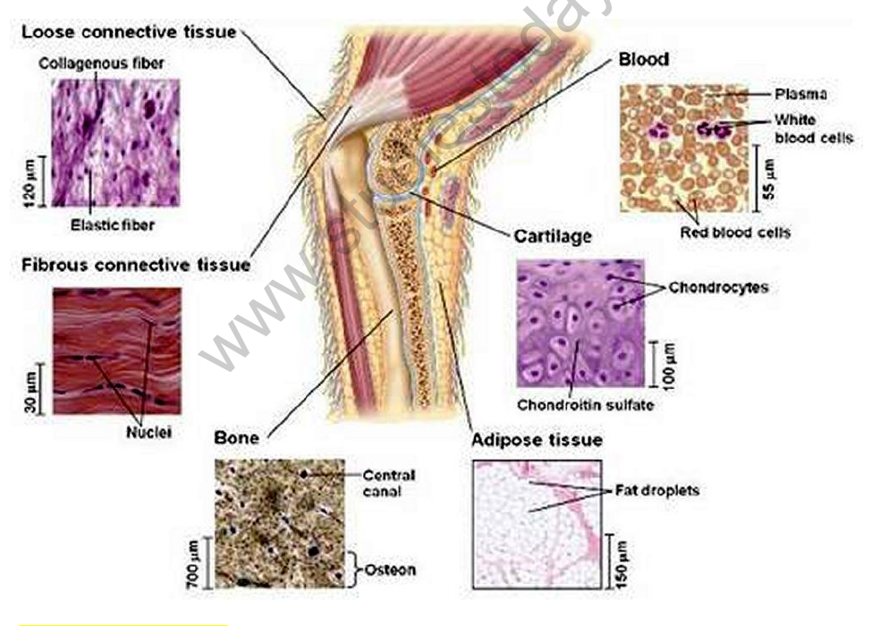
the connective tissue composite figure (loose connective tissue, fibrous connective tissue, bone, cartilage, b

  • Tissues – a group of cells similar in structure and function; a group of cells similar in structure together perform a particular function. Animal tissues:
    • Epithelial tissue (protective tissues)
      • Uni layer:
        • Squamous (mouth lining)
        • Cuboidal (kidney tubules)
        • Columnar: ciliated (respiratory tract); non-ciliated (intestines)
        • Glandular (specialized tissue) – salivary glands
      • Multi layer: stratified squamous – example: skin
    • Connective tissue
      • Ligament
      • Areolar – function: supports internal organs and repairs tissues; bone marrow
      • Bone
      • Cartilage – ear
      • Adipose (fat storing) – below the skin, contour of the body
      • Blood – fluid connective tissue
      • Tendon
    • Muscular tissue (responsible for movement)
      • Striated: voluntary muscles, e.g. limbs; cylindrical, multinucleated, unbranched
      • Unstriated: involuntary muscles; spindle shaped, uninucleated, unbranched, e.g. muscles of intestines
      • Cardiac: involuntary; uninucleated, branched, e.g. heart
    • Nervous tissue – made up of neurons that receive and conduct impulses; transmits stimulus very rapidly

INTEXT QUESTIONS (PAGE 74)

Name the types of simple tissues.

Answer: The three types of simple tissues are (i) parenchyma, (ii) collenchyma and (iii) sclerenchyma.

Where is apical meristem found?

Answer: Apical meristem is present at the growing tips of stems and roots. It helps in increasing the length of the stem and the root.

Which tissue makes up the husk of a coconut?

Answer: The husk of a coconut is made up of sclerenchymatous tissue.
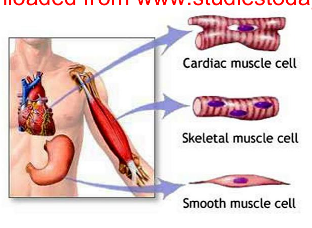
muscle cell images (cardiac, skeletal, smooth) and the muscle comparison drawing

What are the constituents of phloem?

Answer: The constituents of phloem are:

  1. Sieve tubes (tubular living cells with perforated end walls).
  2. Companion cells (living cells).
  3. Phloem parenchyma (living cells).
  4. Phloem fibres (non-living sclerenchyma cells).

INTEXT QUESTIONS (PAGE 78)

Name the tissue responsible for movement in our body.

Answer: Muscular tissue. It consists of elongated cells (muscle fibres) that can contract and relax.

What does a neuron look like?

Answer: A neuron consists of a cell body with a nucleus and cytoplasm. It has two kinds of extensions, the axon and the dendrites. The axon is a long, thread-like extension that transmits impulses away from the cell body. The dendrites are short, branched, thread-like extensions of the cell body that receive nerve impulses. This coordinated function helps in transmitting impulses very quickly.

NucleusDendriteAxonNerve endingCell body

Give three features of cardiac muscles.

Answer: Three features of cardiac muscles:

  1. Cardiac muscles are involuntary muscles that contract rapidly and rhythmically and do not get fatigued.
  2. The cells of cardiac muscles are cylindrical, branched and uninucleate.
  3. They control the contraction and relaxation of the heart.

What are the functions of areolar tissue?

Answer: Functions of areolar tissue:
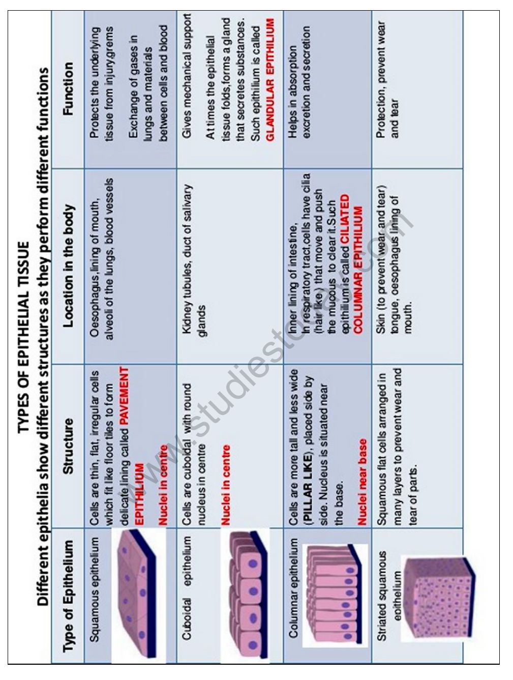
the epithelium table images

  1. It fills the space between organs and supports internal organs.
  2. It helps in repairing tissues (for example of the skin and muscles).

EXERCISE QUESTIONS

Define the term “tissue”.

Answer: A group of cells that are similar in structure and/or work together to achieve a particular function is called a tissue.

How many types of elements together make up the xylem tissue? Name them.

Answer: Four types of elements make up xylem tissue:

  1. Tracheids (tubular, unicellular).
  2. Xylem vessels (multicellular).
  3. Xylem parenchyma (stores food and helps in sideways conduction of water).
  4. Xylem fibres (provide mechanical support).

How are simple tissues different from complex tissues in plants?

Simple tissueComplex tissue
These tissues consist of only one type of cells.These tissues are made up of more than one type of cells.
The cells are more or less similar in structure and perform similar functions.Different types of cells perform different functions. For example, in the xylem tissue, tracheids help in water transport, whereas parenchyma stores food.
Three types of simple tissues in plants are parenchyma, collenchyma, and sclerenchyma.Two types of complex permanent tissues in plants are xylem and phloem.

Answer: The differences are given in the table.

Simpletissue
Complextissue

Differentiate between parenchyma, collenchyma and sclerenchyma on the basis of their cell wall.

ParenchymaCollenchymaSclerenchyma
Cell walls are relatively thin, and the cells in parenchyma tissues are loosely packed.The cell wall is irregularly thickened at the corners, and there is very little space between the cells.The cell walls are uniformly thickened, and there are no intercellular spaces.
The cell wall in this tissue is made up of cellulose.Pectin and hemicellulose are the major constituents of the cell wall.An additional layer of the cell wall composed mainly of lignin is found.

Answer: The differences are given in the table.

Parenchyma
Collenchyma
Sclerenchyma

What are the functions of the stomata?

Answer: Functions of the stomata:

  1. They allow the exchange of gases (CO2 and O2) with the atmosphere.
  2. Water evaporates from the leaf surface through the stomata, so the stomata help in transpiration.

Diagrammatically show the difference between the three types of muscle fibres.

Answer: The three types of muscle fibres are striated (skeletal) muscle, smooth (unstriated) muscle and cardiac muscle. Striated muscle fibres are long, cylindrical, unbranched and multinucleate, with alternate light and dark bands and blunt ends. Smooth muscle fibres are long and spindle-shaped, unbranched, uninucleate, with no bands and tapering ends. Cardiac muscle fibres are cylindrical and branched, uninucleate, with faint bands and flat, wavy ends (intercalated discs).

Smooth muscle• has spindle-shaped,nonstriateduninucleated fibers.• occurs in walls ofinternal organs.• is involuntary.Cardiac muscle• has striated, branched,uninucleated fibers.• occurs in walls ofheart.• is involuntary.Skeletal muscle• has striated,tubular,multinucleatedfibers.• is usuallyattached toskeleton.• is voluntary.

What is the specific function of the cardiac muscle?

Answer: The specific function of cardiac muscle is to control the contraction and relaxation of the heart, i.e. the pumping of blood.

Differentiate between striated, unstriated and cardiac muscles on the basis of their structure and site/location in the body.

Striated muscleUnstriated muscleCardiac muscle
On the basis of structure:
Cells are cylindricalCells are longCells are cylindrical
Cells are not branchedCells are not branchedCells are branched
Cells are multinucleateCells are uninucleateCells are uninucleate
Alternate light and dark bands are presentThere are no bands presentFaint bands are present
Its ends are bluntIts ends are taperingIts ends are flat and wavy
On the basis of location:
These muscles are present in body parts such as hands, legs, tongue, etc.These muscles control the movement of food in the alimentary canal, the contraction and relaxation of blood vessels, etc.These muscles control the contraction and relaxation of the heart

Answer: The differences are given in the table.

Basis
Striatedmuscle
Unstriated(smooth)muscle
Cardiacmuscle

Draw a labelled diagram of a neuron.

Answer: A neuron has: (1) a cell body (cyton) containing the nucleus and cytoplasm; (2) dendrites, short branched extensions of the cell body that receive impulses; (3) an axon, a single long extension that carries impulses away from the cell body; (4) nerve endings at the end of the axon.

NucleusDendriteAxonNerve endingCell body

Name the following. (a) The tissue that forms the inner lining of our mouth. (b) The tissue that connects muscle to bone in humans. (c) The tissue that transports food in plants. (d) The tissue that stores fat in our body. (e) The connective tissue with a fluid matrix. (f) The tissue present in the brain.

Answer: (a) Epithelial tissue (stratified squamous epithelium); (b) dense regular connective tissue (tendons); (c) phloem; (d) adipose tissue; (e) blood; (f) nervous tissue.

Identify the type of tissue in the following: skin, bark of tree, bone, lining of kidney tubule, vascular bundle.

Answer: Skin: stratified squamous epithelial tissue. Bark of a tree: protective tissue (cork), a simple permanent tissue made of dead cells. Bone: connective tissue. Lining of kidney tubule: cuboidal epithelial tissue. Vascular bundle: complex permanent tissue.

Name the regions in which parenchyma tissue is present.

Answer: Parenchyma is present in leaves, fruits, flowers, the cortex and pith of stems, and roots, i.e. in almost all the soft parts of a plant. (The source named only leaves, fruits and flowers.)

What is the role of epidermis in plants?

Answer: The epidermis is present on the outer surface of the entire plant body. Its cells form a continuous layer without intercellular spaces. It performs the following functions:

  1. It is a protective tissue of the plant body.
  2. It protects the plant against mechanical injury.
  3. It allows the exchange of gases through the stomata.

How does the cork act as a protective tissue?

Answer: The outer protective layer, or bark, of a tree is known as cork. It is made up of dead cells, with a thick deposit of a waxy substance (suberin) on their walls, so it protects the plant against mechanical injury and temperature extremes. It also prevents the loss of water by evaporation.

Complete the table.

Permanent tissueSimpleComplexCollenchyma

Answer: The completed table:

Permanent tissueSimpleComplexParenchymaCollenchymaSclerenchymaPhloem

Permanent tissue → Simple: Parenchyma, Collenchyma, Sclerenchyma; Complex: Xylem, Phloem.

Tissue
Location
Function

Epithelial tissues are of following types:
1. Simple Epithelium
2. Cuboidal Epithelium
3. Columnar Epithelium
4. Stratified Epithelium

Please click the link below to download CBSE Class 9 Science Tissues Notes.

CBSE Class 9 Science Study Material: Chapter 06 Tissues

Quick Revision Material for Chapter 06 Tissues

Access comprehensive study material for Chapter 06 Tissues, including revision notes, concept maps, and high-probability questions. These resources are designed in alignment with the latest 2026 CBSE syllabus for Class 9 Science to support effective exam preparation.

Expert Notes and Solved Exam Questions

Designed around the official curriculum, these study guides guarantee standard compliance. Reviewing step-by-step solutions clarifies complex sub-topics within Chapter 06 Tissues and demystifies standard marking schemes for Science evaluations.

Additional Study Resources for Class 9

For peak performance in upcoming evaluations, integrate official Science sample papers directly into your study schedule. Follow up your revision by attempting online MCQ tests for Chapter 06 Tissues to refine calculation speed and precision.

FAQs

What is included in the advanced study material for Class 9 Science Chapter 06 Tissues?

Our advanced study package for Chapter 06 Tissues includes detailed concepts, diagrams, Mind Maps, and explanation of complex topics to ensure Class 9 students learn as per syllabus for 2026 exams.

How do Mind Maps for Science Chapter 06 Tissues help in revision?

The Mind Maps provided for Chapter 06 Tissues act as visual anchors which will help faster recall during high-pressure exams.

Are these Science resources suitable for both classroom teaching and self-study?

Yes, teachers use our Class 9 Science resources for lesson planning as they are in simple language and have lot of solved examples.

Is this advanced study material for Chapter 06 Tissues free to download in PDF?

Yes, You can download the complete, mobile-friendly PDF of the Science Chapter 06 Tissues advanced resources for free.

Does this material cover rationalized content for the 2026-27 CBSE session?

Yes, our subject matter experts have updated the Chapter 06 Tissues material to align with the rationalized NCERT textbooks and have removed deleted topics and added new competency-based questions.