

Official NEET Study Materials for NEET Biology
Explore structured advanced study materials through the NEET Biology Biological Classification Notes. Tailored for NEET learners, utilizing these Biology resources ensures thorough preparation and strengthens foundational knowledge before final NEET evaluations.
Advanced Resources for Biology
Access the complete useful resource PDF for NEET Biology below. Regular practice with these targeted academic notes builds familiarity with complex topics and helps secure higher marks in final school evaluations.
Biological classification is the grouping of organisms into kingdoms based on cell structure, body organisation, mode of nutrition and evolutionary relationships. This NEET Biology notes page for Class 11 covers the history of classification from Aristotle to Whittaker's five kingdoms, the features of Monera, Protista, Fungi, Plantae and Animalia, and the main groups of bacteria, cyanobacteria, mycoplasma, diatoms, dinoflagellates, euglenoids and slime moulds, with their diseases, uses and examples.
At a Glance (chapter contents)
- Monera
- Archaebacteria
- Bacteria
- Cyanobacteria
- Mycoplasma
- Actinomycetes
- Protista
- Chrysophytes (Diatoms)
- Dinoflagellates
- Euglenoids
- Slime moulds
- Protozoa
- Fungi
- Classification of fungi
- Plantae
- Animalia
- Viruses
- Viroids
- Lichens
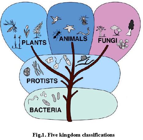
Chapter opening figure (tree of life, five kingdom groupings): Plants: eukaryotic, multicellular, photosynthesize, nonmotile, sexual. Animals: eukaryotic, multicellular, ingest, motile, sexual. Fungi: eukaryotic, multicellular, absorb, nonmotile, sexual. Protists: eukaryotic, unicellular or multicellular, absorb, ingest or photosynthesize, sexual and asexual. Monera: prokaryotic, unicellular, absorb or photosynthesize, motile or nonmotile, asexual.
Terms and Definitions to Know
1. Phylogenetic relationship
2. Methanogens
3. Thermoacidophiles
4. Halophiles
5. Colicinogenic factor
6. Trichomes
7. Heterocyst
8. Diatomite
9. Capsomeres
Classification of Organisms
Scientists have classified organisms into different kingdoms at different times. Biological classification was first proposed by Aristotle, who used simple morphological characters to classify plants and animals.
Two Kingdom Classification
Linnaeus initially separated plants and animals into two kingdoms, Kingdom Plantae and Kingdom Animalia. For a long time this system was universally accepted, but later it was found to be insufficient for the following reasons:
(i) Prokaryotes and eukaryotes were grouped together. For example, bacteria and cyanobacteria were placed under the plant kingdom.
(ii) Heterotrophs and autotrophs were placed together. For example, fungi, which are heterotrophs, were placed with plants, and Euglena, which has chlorophyll, was placed under protozoa, which are heterotrophs.
(iii) It did not differentiate between unicellular and multicellular organisms. For example, protozoans are unicellular but were placed with multicellular animals.
(iv) Simple organisms were placed with highly complex organisms. For example, protozoans were placed with human beings in the animal kingdom, and bacteria (prokaryotes) were placed with highly evolved angiosperms in the plant kingdom.
(v) Unicellular aquatic organisms are difficult to distinguish as plants or animals.
Three Kingdom Classification
Haeckel gave the three kingdom classification by adding Kingdom Protista to Kingdom Plantae and Kingdom Animalia. He placed all unicellular eukaryotes in Protista, whether they were plants or animals. This system was not accepted because it grouped prokaryotic and eukaryotic, chlorophyllous and non-chlorophyllous organisms together.
Four Kingdom Classification
Copeland classified organisms into four kingdoms: Monera (all prokaryotes), Protista (unicellular eukaryotes), Metaphyta (multicellular plants) and Metazoa (multicellular eukaryotes).
Five Kingdom Classification
The most accepted system of classification is the five kingdom classification given by R.H. Whittaker in 1969. In this system a separate kingdom was created for Fungi. The five kingdoms are Monera, Protista, Fungi, Plantae and Animalia.
The five kingdom classification is based on the following criteria:
1. Complexity of cell structure: prokaryotes or eukaryotes
2. Complexity of organism body: unicellular or multicellular
3. Mode of obtaining nutrition: autotrophs or heterotrophs
4. Phylogenetic relationships
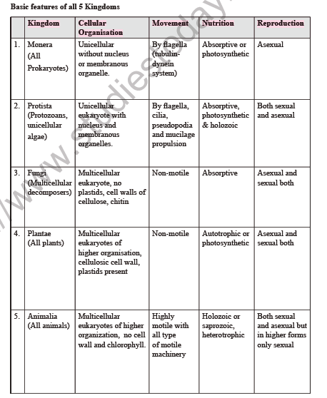
Basic Features of the Five Kingdoms
| Kingdom | Cellular Organisation | Movement | Nutrition | Reproduction |
|---|---|---|---|---|
| 1. Monera (all prokaryotes) | Unicellular without nucleus or membranous organelles | By flagella (tubulin-dynein system) | Absorptive or photosynthetic | Asexual |
| 2. Protista (protozoans, unicellular algae) | Unicellular eukaryote with nucleus and membranous organelles | By flagella, cilia, pseudopodia and mucilage propulsion | Absorptive, photosynthetic and holozoic | Both sexual and asexual |
| 3. Fungi (multicellular decomposers) | Multicellular eukaryote, no plastids, cell walls of cellulose and chitin | Non-motile | Absorptive | Asexual and sexual both |
| 4. Plantae (all plants) | Multicellular eukaryotes of higher organisation, cellulosic cell wall, plastids present | Non-motile | Autotrophic or photosynthetic | Asexual and sexual both |
| 5. Animalia (all animals) | Multicellular eukaryotes of higher organisation, no cell wall and no chlorophyll | Highly motile with all type of motile machinery | Holozoic or saprozoic, heterotrophic | Both sexual and asexual but in higher forms only sexual |
Whittaker's Five Kingdoms and Their Phylogenetic Relationships
[Figure: Fig. 2.1 Whittaker's five kingdoms of life with major groups and their phylogenetic relationships. Kingdom Plantae includes Tracheophyta (vascular plants), Bryophyta (mosses and liverworts), Phaeophyta (brown algae), Chlorophyta (green algae) and Rhodophyta (red algae). Kingdom Fungi includes Basidiomycota (club fungi), Ascomycota (sac fungi), Zygomycota (conjugation fungi) and Oomycota (water moulds). Kingdom Animalia includes Arthropoda, Annelida (segmented worms), Mollusca, Nematelminthes (roundworms), Platyhelminthes (flatworms), Cnidaria (coelenterates), Porifera (sponges), Echinodermata (echinoderms) and Chordata (chordates). Kingdom Protista includes Euglenoids, Dinoflagellates, Chrysophytes, Slime moulds, Sarcodina (Amoeba), Ciliophora (ciliates), Sporozoa and Zoomastigina. Monera includes Cyanobacteria, Eubacteria and Archaebacteria. All kingdoms arise from the Progenote]
Labels of Fig. 2.1 (phylogenetic tree):
- Progenote gives rise to Archaebacteria (ancient bacteria), Eubacteria (bacteria) and Cyanobacteria (blue-green algae), which form Kingdom Monera.
- Primitive Eucaryotic Flagellate (Kingdom Protista) gives rise to:
- Pyrrophyta (Dinoflagellates)
- Euglenophyta (Euglenoids)
- Chrysophyta (Diatoms and related algae)
- Myxomycota (Slime moulds)
- Sporozoa (Sporozoans)
- Zoomastigina (Animal flagellates)
- Sarcodina (Amoebae)
- Ciliophora (Ciliates)
- Kingdom Plantae: Chlorophyta (Green algae), Rhodophyta (Red algae), Phaeophyta (Brown algae), Bryophyta (Mosses and liverworts), Tracheophyta (Vascular plants)
- Kingdom Fungi: Oomycota (water moulds), Ascomycota (Sac fungi), Basidiomycota (Club fungi), Zygomycota (Conjugation fungi)
- Kingdom Animalia: Platyhelminthes (Flatworms), Nematelminthes (Roundworms), Cnidaria (Coelenterates), Porifera (Sponges), Mollusca (Molluscs), Annelida (Segmented worms), Arthropoda (Arthropods), Echinodermata (Echinoderms), Chordata (Chordates)
Connecting Concepts: Phylogenetic Relationships The earliest living forms produced prokaryotic organisms or monerans. They developed chemoheterotrophy, photoautotrophy, anaerobic respiration and aerobic respiration at different times. Monera gave rise to protista, most probably through association of several types of primitive and advanced monerans. There are several types of protista. Fungi, plants and animals have developed from different types of early protistans.
Kingdom Monera (Kingdom of Prokaryotes)
The kingdom includes all prokaryotes: mycoplasma, bacteria, actinomycetes and cyanobacteria or blue green algae.
Characteristics of Monera
1. All monerans evolved from an ancient life form called progenote. They are the most primitive and important decomposers and mineralisers in the biosphere.
2. They are unicellular, colonial or multicellular with prokaryotic cellular organisation. They are devoid of plastids, mitochondria and advanced (9 + 2) flagella.
3. They have no nuclear membrane, nucleoplasm or nucleolus.
4. They reproduce asexually by transverse binary fission or budding.
5. Protosexual phenomenon is found.
6. The cell wall contains peptidoglycan. The exceptions are archaebacteria and mycoplasma.
7. These organisms are non-motile or move by beating of simple flagella or by gliding.
8. They show both autotrophic and heterotrophic modes of nutrition.
Archaebacteria
1. Archaebacteria are one of the most primitive groups of bacteria and are called the oldest living fossils.
2. They are found in harsh habitats such as extremely salty areas (halophiles), hot springs (thermoacidophiles) and marshy areas (methanogens).
3. Their cell wall is adapted to extreme conditions. Peptidoglycan is absent; the wall contains protein and non-cellulosic polysaccharides. Pseudomurein is found in some methanogens.
4. Their cell membranes contain branched chain lipids, which make them highly resistant to heat and low pH.
The three main groups of archaebacteria are:
(i) Methanogens: These are anaerobic bacteria that mainly occur in muddy areas and in the stomach of ruminant animals such as cattle. They produce methane gas (CH4) from CO2 or formic acid (HCOOH), and are used in bio-gas plants. They are autotrophic but obtain energy and carbon from the end products of decomposition, and typical carbon assimilation is absent.
(ii) Halophiles: These are anaerobic bacteria that live in extreme saline or salty conditions, up to 35% salt (NaCl) in culture medium. Their intracellular salt concentration is high, and their enzymes and ribosomes work only at high intracellular salt concentration. A purple pigmented membrane containing bacteriorhodopsin is developed in sunlight, which uses light energy for metabolic activities, different from photosynthesis.
(iii) Thermoacidophiles: These are aerobic bacteria found in hot sulphur springs (up to 80°C). They oxidise sulphur to H2SO4 at high temperature and high acidity (pH 2.0), so they are named thermoacidophiles, meaning temperature and acid loving. Some of them can reduce sulphur to H2S under anaerobic conditions.
Connecting Concepts
1. Archaebacteria live under extremely low pH, high temperature, high salinity and high pressure. These properties are used in modern biotechnology in a number of fields: (i) generation of biogas, (ii) thermophilic enzymes, (iii) restriction enzymes, (iv) bioleaching of poor mines, (v) biosensors.
2. Bacteria were abundant for over 2 billion years before the appearance of eukaryotes in the world. They were responsible for creating the properties of the present atmosphere and soil. These bacteria also make life on earth possible as they perform an integral function as decomposers of organic materials.
3. Microorganisms are small-sized microscopic organisms with a size of 1 mm or less. They cannot be observed with the naked eye. Microorganisms belong to several taxonomic groups: viruses, bacteria, algae, fungi, protistans and metazoa.
Bacteria
1. Anton van Leeuwenhoek (1683), a Dutch scientist, observed small or tiny animalcules in sewage water, saliva and tooth scum under his crude microscope. These were later known as bacteria. He is called the father of bacteriology.
2. Louis Pasteur gave the germ theory of diseases, reporting that certain diseases in plants and animals are caused by bacteria.
3. Robert Koch was the first to culture bacteria.
4. John Lister started antiseptic surgery using carbolic acid as a disinfectant.
5. The branch of biology that deals with the study of bacteria is called bacteriology.
6. The term bacterium (plural: bacteria) was coined by the German microbiologist C.G. Ehrenberg in 1828.
7. Bacteria are the simplest and most primitive unicellular organisms. They are found everywhere, in air, water, soil, food and inside our body.
Characteristics of Bacteria
1. They are prokaryotes. They lack a nucleus and other complex cell organelles. They have 70S prokaryotic ribosomes.
2. Like plants, they have a cell wall. It is made of sugar derivatives which are not present anywhere else, namely N-acetyl glucosamine (NAG) and N-acetyl muramic acid (NAM).
3. They are mostly unicellular but may form colonies or filaments of independent cells.
4. Each bacterial cell contains a single chromosome (nuclear material) that is not enclosed in a nuclear membrane. This primitive nucleus is called nucleoid, genophore or incipient nucleus.
5. Besides nuclear DNA, some bacteria have extranuclear or extrachromosomal DNA called plasmids. Plasmids are small, circular, double-stranded DNA molecules that exist separately from the genomic chromosome and replicate independently.
Three types of plasmids are:
(1) F-factor or fertility factor: It is responsible for the transfer of genetic material.
(2) R-factor or resistance factor: It provides resistance against drugs.
(3) Colicinogenic factor: It produces colicins, which kill other bacteria.
Shapes of Bacteria
According to shape, bacteria are grouped into four types:
1. Coccus: spherical shaped
2. Bacillus: rod-shaped
3. Spirillum: spiral or curved shaped
4. Vibrio: comma-shaped
[Figure: Fig. 2.2 Different forms of bacteria, showing coccus, bacillus, spirillum and vibrio forms]
Labels in the figure: Coccus, Bacillus, Spirillum (with Flagellum), Vibrio.
E. coli as a Research Organism
E. coli is an important research material because of its short life cycle, haploid nature and genetic recombination. The mechanism of gene action, protein synthesis and the operon model of regulation were worked out in E. coli, and it is an ideal host in genetic engineering experiments. In a donor cell, the F-factor may join the main genome or nuclear DNA; such a donor cell is called a Hfr donor cell (high frequency donor cell), and transfer of DNA from it is rapid.
Motility of Bacteria
Most bacteria are immotile, meaning they cannot move. Some bacteria such as bacilli and spirilla can move on their own. These bacteria may have a single polar flagellum at one end of the cell, or clusters or many flagella at one or both ends.
Nutrition in Bacteria
Bacteria show both autotrophic and heterotrophic modes of nutrition.
1. Autotrophic nutrition means synthesis of organic material from inorganic raw materials with energy obtained from outside sources. It is of two types: chemosynthesis and photosynthesis. Bacteria performing these are called chemoautotrophs and photoautotrophs respectively.
2. Heterotrophic nutrition involves obtaining ready-made organic nutrients from outside sources.
Photoautotrophic Bacteria
These bacteria possess two types of photosynthetic pigments, bacteriochlorophyll and bacteriophaeophytin. The pigments are present in the membranes of chromatophores dispersed in the cytoplasm, because chloroplasts are absent. No oxygen is evolved in bacterial photosynthesis, so it is called anoxygenic photosynthesis.
Chemoautotrophic Bacteria
These bacteria manufacture their organic food from inorganic raw materials, using energy from exergonic chemical reactions in which an inorganic substance in the external medium is oxidised. The chemical energy from the oxidation is trapped in ATP molecules.
Respiration in bacteria can be aerobic, in the presence of atmospheric oxygen, or anaerobic, in its absence.
Endospore
The bacterial spore or endospore is perhaps the most resistant living structure in the biosphere. It is highly resistant to very high and very low temperatures, strong chemicals and acids, because of calcium, dipicolinic acid (DPA) and peptidoglycan in the cell wall. DPA helps in stabilising its proteins, and DPA with calcium ions provides resistance to heat.
Reproduction in Bacteria
Bacteria reproduce both asexually and by genetic recombination. Asexual reproduction is by binary fission, which takes place in favourable conditions, or by endospore formation. True sexual reproduction is absent, but genetic recombination occurs, which brings together the genetic material of two bacterial cells. There are three main methods of genetic recombination:
(1) Transformation
(2) Transduction
(3) Conjugation
(1) Transformation: The genetic material of one bacterial cell moves into another cell by an unknown mechanism and converts one type of bacterium into another, for example from non-capsulated to capsulated form. It was first studied by Griffith (1928) in Diplococcus pneumoniae, and so it is called the Griffith effect. It was later studied in detail by Avery, McCarty and McLeod.
[Figure: Fig. 2.3 A typical bacterial cell]
Labels in the figure. Structures sometimes present: flagellum (one or many, usually longer than cell); mucilaginous capsule (if diffuse, called slime layer); photosynthetic membranes (either tubular or sac-like; possess pigments, present throughout the cell if photosynthetic); pili (or fimbriae); mesosome (folded invagination of plasma membrane). Structures always present: cell wall (contains murein); plasma membrane; ribosomes (70S); food reserve (lipid, glycogen, volutin granules); cytoplasm (contains enzymes, etc.); DNA (circular molecule, double stranded).
(2) Transduction: The genetic material of one bacterial cell is carried to another bacterial cell by bacteriophages, which are viruses that infect bacteria. Transduction was first reported in Salmonella typhimurium by Zinder and Lederberg (1952).
(3) Conjugation: Two bacterial cells come into physical contact and the DNA of one cell moves into the other through a conjugation tube formed by sex pili. It was first reported by Lederberg and Tatum (1946) in E. coli. The donor bacterium has sex pili and the F-factor, while the recipient bacterium lacks both.
Useful and Harmful Effects of Bacteria
Bacteria have both useful and harmful effects. They play an important role in nitrogen fixation, decay and putrefaction, and are used in industries, in the curdling of milk, setting of fibres, tanning of leather, and in the production of vitamins, antibiotics and medicines. Bacteria cause harmful diseases in plants, animals and humans. Denitrifying bacteria reduce the fertility of soil by converting nitrates into free nitrogen.
Some Common Bacterial Diseases
| Human Diseases | ||
|---|---|---|
| S. No. | Disease | Pathogen |
| 1. | Cholera | Vibrio cholerae |
| 2. | Typhoid | Salmonella typhosa |
| 3. | Dysentery | Shigella dysenteriae |
| 4. | Peptic Ulcer | Helicobacter pylori |
| 5. | Bubonic Plague | Yersinia (= Pasteurella) pestis |
| 6. | Bacterial Influenza | Haemophilus influenzae |
| 7. | Whooping Cough | Bordetella pertussis |
| 8. | Syphilis | Treponema pallidum |
| 9. | Gonorrhoea | Neisseria gonorrhoeae |
| 10. | Bacterial Meningitis | Neisseria meningitidis |
| 11. | Vaginitis | Gardnerella vaginalis |
| 12. | Tuberculosis (TB) | Mycobacterium tuberculosis |
| 13. | Leprosy | Mycobacterium leprae |
| 14. | Diphtheria | Corynebacterium diphtheriae |
| 15. | Pneumonia | Pneumococcus or Streptococcus pneumoniae |
| 16. | Tetanus | Clostridium tetani |
| 17. | Boils | Staphylococcus aureus |
| Animal Diseases | ||
|---|---|---|
| S. No. | Disease | Pathogen |
| 1. | Anthrax (Cattle, Sheep) | Bacillus anthracis |
| 2. | Brucellosis (Cattle, Pig, Goat) | Brucella melitensis, B. suis |
| Plant Diseases | ||
|---|---|---|
| S. No. | Disease | Pathogen |
| 1. | Soft Rot of Turnip | Erwinia carotovora |
| 2. | Fire Blight of Apple, Pear | Erwinia amylovora |
| 3. | Bacterial Blight of Rice | Xanthomonas oryzae |
| 4. | Citrus Canker | Xanthomonas citri |
| 5. | Crown Gall | Agrobacterium tumefaciens |
| 6. | Angular Leaf Spot of Cotton | Xanthomonas malvacearum |
Connecting Concepts on Bacteria
1. Pasteurization is a process named after Louis Pasteur which uses heat to destroy human pathogens in food. Milk is heated at 62.8°C for 30 minutes, or 71.7°C for 15 seconds, and then cooled immediately. This kills all non-spore forming bacteria, so unboiled milk turns sour earlier than boiled milk.
2. Lactic acid bacteria unite the casein protein of milk into small droplets and form curd and butter.
3. Poly β-hydroxybutyrate (PHB) is found in several bacteria as reserve food and can be used to make biodegradable plastics.
4. Waksman was the first person to give the term antibiotics to chemical substances produced by one micro-organism that kill other micro-organisms. The first antibiotic produced from bacteria by Waksman was thiothricin, from Bacillus brevis.
5. The first antibiotic, penicillin (the wonder drug), was obtained from the fungus Penicillium notatum by Sir Alexander Fleming (1923). It checks the growth of Gram-positive bacteria by inhibiting cell wall or peptidoglycan synthesis.
6. A genetically engineered strain of Pseudomonas putida, the superbug, was developed by Dr. Ananda Mohan Chakraborty (USA) to biodegrade spilled oil.
Kingdom Monera: Cyanobacteria
1. The new name cyanobacteria is given to blue green algae, myxophyceae or cyanophyceae, because of their simple prokaryotic organisation like bacteria.
2. Cyanobacteria are the largest and most diverse group of photosynthetic bacteria. There are about 2,000 or more species. They have chlorophyll-a like green plants.
3. They may be unicellular, colonial or filamentous. Each filament consists of a sheath of mucilage and one or more cellular strands called trichomes.
4. Cells are larger and more elaborate than bacteria, but the cell structure is prokaryotic: one envelope with a peptidoglycan wall, naked DNA, 70S ribosomes, and no membrane-bound structures such as endoplasmic reticulum, mitochondria, Golgi bodies, plastids, lysosomes or vacuoles.
5. They reproduce by binary fission, budding, fragmentation and multiple fission. Fragments of filamentous forms can grow into small motile filaments called hormogonia. Some species develop akinetes.
6. Heterocyst is a large, pale coloured, thick-walled cell found in the terminal, intercalary or lateral position of filamentous forms such as Nostoc. It contains the enzyme nitrogenase and is specialised for nitrogen fixation. About 5 to 10% of cells in a trichome become heterocysts. They take nutrients from adjacent vegetative cells and supply fixed nitrogen as the amino acid glutamine.
7. Cyanobacteria form symbiotic associations with lichens, protozoa, liverworts, mosses, gymnosperms and angiosperms, as photosynthetic partners.
Mycoplasma
1. Mycoplasma are the smallest known aerobic prokaryotes and they have no cell wall. The smallest cell so far discovered is Mycoplasma gallisepticum, with a diameter of 0.1 µ.
2. Since they lack a cell wall, they have no definite shape and are pleomorphic. They are therefore called the Jokers of microbiology. They may be filamentous or coccus (round) in form.
3. They have a typical colonial appearance and pass through 450 nm bacterial filters.
4. The outer side has a tripartite (three layered) cell membrane made of lipoproteins.
5. In human beings, Mycoplasma hominis causes pleuropneumonia, infertility in men and inflammation of the genitals. Mycoplasma pneumoniae causes Primary Atypical Pneumonia (PAP). Arthritis, respiratory infections and rheumatism are also caused by mycoplasma.
6. Mycoplasma are insensitive to penicillin and can be killed by chloramphenicol, streptomycin and erythromycin.
7. Important plant diseases caused by mycoplasma: little leaf of brinjal, bunchy top of papaya, mulberry dwarf, potato witches' broom, clover dwarf, maize stunt and aster yellows.
Actinomycetes
1. Actinomycetes are filamentous bacteria, like moulds or fungi.
2. They are generally present as decomposers in soil and reproduce asexually by conidia produced at the tips of filaments.
3. Most of them secrete antimicrobial chemicals called antibiotics. For example, Streptomyces species secrete streptomycin, neomycin, chloromycetin, terramycin and erythromycin.
Kingdom Protista (Kingdom of Unicellular Eukaryotes)
1. Haeckel (1886) created the kingdom Protista to include all unicellular eukaryotic microorganisms. Phylogenetically, protists link the prokaryotic kingdom Monera with the multicellular kingdoms of Fungi, Plantae and Animalia.
2. Protists have a typical eukaryotic structure with membrane-bound organelles and a nucleus.
3. They are classified into photosynthetic protists (such as diatoms and dinoflagellates) and protozoan protists (such as amoeba, paramecium and sporozoans), and also slime moulds.
4. Protists may be photosynthetic, heterotrophic or mixotrophic, as in Euglena.
5. Mesokaryotic condition of the nucleus is found in dinoflagellates.
6. Locomotion is by flagella (Euglena), cilia (Paramecium) and pseudopodia (Amoeba and slime moulds). Diatoms move by secreting mucilage. Sporozoans and non-flagellate euglenoids move by wriggling.
7. Flagella have a (9 + 2) microtubular arrangement.
Chrysophytes (Diatoms)
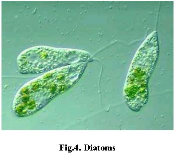
1. Chrysophytes are golden algae, which include diatoms such as Triceratium, Pleurosigma, Navicula, Cymbella and Amphipleura. They are golden brown photosynthetic microscopic protists.
2. They are basically unicellular but may form pseudofilaments and colonies.
3. Diatoms live in aquatic and moist terrestrial habitats and are very good pollution indicators.
4. The body is covered by a transparent siliceous shell (silica deposited in the cell wall) called the frustule. The frustule has two valves, the epitheca and the hypotheca.
5. Diatoms are microscopic unicells of various shapes, such as circular, semicircular, rectangular, triangular, spindle-shaped and boat-shaped. Cells may form zigzag (Diatoma), stellate or fan-shaped colonies. Incipient filaments occur in Melosira.
6. Based on symmetry, diatoms are of two types: pennate (bilateral symmetry, e.g., Pinnularia, Navicula) and centric (radial symmetry, e.g., Melosira).
7. They have chlorophyll-a, chlorophyll-c, α-carotenoids and fucoxanthin, which gives them a brown colour.
8. Each cell has a large central vacuole. Food reserves are oils and leucosin (a polysaccharide). Volutin globules, which are proteinaceous, also occur.
9. Flagella do not occur in somatic cells. Some diatoms glide by secreting mucilage. Mucilage and oil globules help planktonic forms float on the surface of water.
10. The common mode of multiplication is binary fission. Sexual reproduction varies from isogamy to oogamy. The zygote grows and forms a rejuvenescent cell called an auxospore.
11. The siliceous cells do not decay easily. They pile up at the bottom of water bodies and form deposits called diatomite, diatomaceous earth or kieselguhr.
Economic Importance of Diatoms
1. Diatoms are an important food source for aquatic animals.
2. The oils extracted from some fishes and whales are actually produced by diatoms.
3. Diatomite deposits are often found with petroleum fields.
4. Diatomite is porous and chemically inert, so it is used in the filtration of sugar, alcohols and antibiotics.
5. It is used as a cleaning agent in toothpastes and metal polishes.
6. It is added to paints to enhance night visibility.
7. It is used as an insulation material in refrigerators, boilers and furnaces, and to make sound proof rooms.
Dinoflagellates
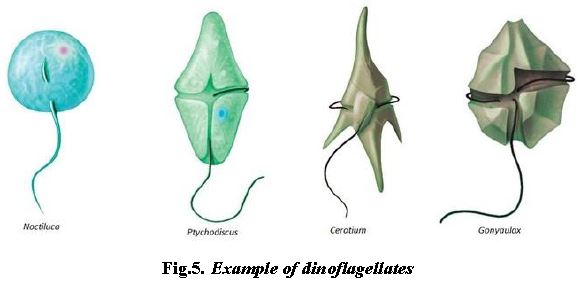
1. Dinoflagellates are biflagellate, unicellular, golden brown photosynthetic protists, mainly marine. They appear yellow, green, brown, blue or red depending on the pigments in their cells.
2. The cell wall has stiff cellulose plates on the outer surface.
3. The nucleus is distinctive, with an unusual combination of prokaryotic and eukaryotic characteristics. It is called a mesokaryon and has permanently condensed chromosomes. Some species have non-condensed chromosomes at some stages of their life cycle. Chromosomes are generally membrane attached and the nuclear membrane remains intact during mitosis.
4. Most have two flagella, one lying longitudinally and the other transversely in a furrow between the wall plates.
5. Many species have light-sensitive organelles called eye spots. Some are mere collections of carotenoid-containing lipid globules, while others also have a membrane; such an eye spot has a pigmented region and a lens-like refractive portion.
6. Red dinoflagellates such as Gonyaulax multiply so rapidly that they make the sea appear red.
7. Dinoflagellates show bioluminescence, the emission of light by living beings. About 18 genera emit light, for example Gonyaulax, Noctiluca, Ceratium and Peridinium.
8. Their toxins may kill marine animals such as fishes. Toxins accumulated in shellfish cause paralytic shell-fish poisoning (P.S.P.), which affects neuromuscular transmission and can be fatal to humans.
Check Point
Write the causative agent of the following diseases:
(i) Bunchy top of papaya: Mycoplasma (mycoplasma-like organism).
(ii) Diphtheria: Corynebacterium diphtheriae.
(iii) Citrus canker: Xanthomonas citri.
(iv) Syphilis: Treponema pallidum.
(v) Bubonic plague: Yersinia pestis.
Euglenoids
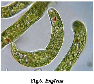
1. Euglenoids are flagellates with plant-like characteristics (chlorophyll) and some animal characteristics. They ingest food particles and carry out photosynthesis.
2. They are mostly unicellular and free living, found in fresh water ponds, ditches or damp soil.
3. Instead of a cell wall, they have a protein-rich layer called the pellicle, which makes the body flexible.
4. In sunlight they are photosynthetic. When deprived of sunlight, they behave like heterotrophs and feed on smaller organisms. Example: Euglena.
5. They have two tinsel flagella, and one is usually reduced. A single long flagellum seems to branch at the base, with each branch having its own basal granule.
6. They swim actively with the long flagellum, and can creep by expansion and contraction of the body, called metaboly or euglenoid movement.
7. A swelling at the base of the flagellum is the paraflagellar body. A stigma (eye spot) is attached at the level of the paraflagellar body, and both act as photoreceptors.
8. An osmoregulatory contractile vacuole is present.
9. Nutrition is holophytic (photoautotrophic), saprobic (e.g., Rhabdomonas) or holozoic (e.g., Peranema). Forms that take in organic compounds as well as photosynthesising are called mixotrophic.
10. Photoautotrophic forms have chloroplasts, with or without pyrenoids, and store carbohydrate as paramylum bodies.
11. Under unfavourable conditions some Euglenae come together, secrete a gelatinous covering and stay embedded in it. This is the palmella stage. When conditions improve, they regenerate flagella and resume their normal life.
[Figure: Fig. 2.4 Euglena, labelled with photoreceptor, contractile vacuole, flagellum and nucleus]
Slime Moulds
1. Slime moulds are a group of consumer-decomposer organisms that live on decaying organic matter in both terrestrial and aquatic habitats.
2. They are unicellular, saprophytic organisms that lack a cell wall and have only a plasma membrane as the outer covering.
3. They move by pseudopodia.
4. The cellular forms are called plasmodium and the acellular forms are called pseudoplasmodium. They have numerous diploid nuclei.
5. Their spores have a cell wall and are produced by reduction division. The spores act as gametes and are known as myxamoebae or swarm cells. The zygote undergoes free nuclear division to produce the multinucleate slime mould.
6. Slime moulds share characters of both animals and fungi, so they are commonly called fungus animals.
Acellular Slime Moulds
1. The free-living diploid somatic phase is one large mass of protoplasm with thousands of nuclei and no cell walls. This is called plasmodium.
2. It often appears as a slimy, fan-shaped network of living matter.
3. The mass flows along in an amoeboid manner on the forest soil, dead leaves or a rotting log, engulfing food particles and digesting them in food vacuoles.
4. Examples: Physarum, Physarella, Fuligo, Dictydium, Lycogala and Tubifera.
Cellular Slime Moulds
1. They lack flagellate cells, form a pseudoplasmodium, have no sporangial cover, have a cellulose wall around the spores, and reproduce sexually by anisogamy.
2. Numerous individual amoeboid cells aggregate and move together like a mass of protoplasm. This is called a pseudoplasmodium, because the individuals are not fused.
Protozoa
1. Classification of protozoa is mainly based on locomotory organelles. Protistan protozoa are divided into four major groups: Zooflagellata, Sarcodina, Sporozoa and Ciliata.
Zooflagellates
1. They are protozoan protists that possess one to several flagella for locomotion.
2. They are generally uninucleate, occasionally multinucleate. The body is covered by a firm pellicle. Cyst formation is also present.
3. Examples: Trichonympha, Lophomonas, Giardia, Trypanosoma, Leishmania and Trichomonas. Trichonympha and Lophomonas are cellulose-digesting symbionts in the body of termites.
Diseases Caused by Zooflagellates
1. Central African sleeping sickness, or Gambian sleeping sickness, is caused by Trypanosoma gambiense. Its vector is the tsetse fly, Glossina palpalis.
2. East African sleeping sickness, or Rhodesian sleeping sickness, is caused by Trypanosoma rhodesiense. It is transmitted by Glossina morsitans. Both are also called trypanosomiasis.
3. Trypanosoma cruzi causes Chagas disease, or American trypanosomiasis.
4. Giardia lamblia (Giardia intestinalis) causes giardiasis, also called black packer's disease. It is popularly known as the grand old man of the intestine.
5. Kala-azar, also called dumdum fever or visceral leishmaniasis, is caused by Leishmania donovani. Its vector is the blood-sucking sandfly, Phlebotomus.
6. Leishmania tropica causes dermal or skin leishmaniasis, also called oriental sore or Delhi sore (Delhi boil). Leishmania brasiliensis produces mucocutaneous leishmaniasis, or espundia.
7. Polymorphism in Trypanosoma: Trypanosoma has four forms. Three are developmental stages, namely crithidia, leptomonas and leishmania. The infective or metacyclic stage is trypanosoma. In human blood, the adult or trypanosoma form occurs.
Sarcodines
1. Sarcodines develop pseudopodia for locomotion and for ingestion of food. Pseudopodia are temporary protoplasmic outgrowths.
2. They are generally uninucleate, occasionally multinucleate. The body is without a periplast (only plasmalemma covers it) or has a shell.
Examples of Sarcodines
1. Entamoeba histolytica is a human parasite that lives in the upper part of the large intestine. It causes amoebic dysentery, or amoebiasis.
2. Entamoeba coli inhabits the human colon. It is a commensal parasite and does not produce any disease.
3. Entamoeba gingivalis, or mouth amoeba, is found in tartar of teeth and in pus pockets of bleeding gums. The condition is called pyorrhoea.
Sporozoans
1. They have a parasitic mode of life, usually as endoparasites.
2. Locomotory organs (cilia, flagella, pseudopodia) are absent.
3. The body is covered by an elastic pellicle or cuticle and has a single nucleus. Contractile vacuoles are absent.
4. Sporozoans occasionally form amoeba-like stages.
Examples of Sporozoans
1. Plasmodium vivax causes benign tertian fever.
2. Plasmodium falciparum causes malignant tertian fever, cerebral malaria, pernicious malaria or black-water fever.
3. Plasmodium ovale causes benign tertian fever.
4. Plasmodium is digenetic, meaning it has two hosts. Human beings are the secondary host and the female Anopheles mosquito is the primary host, because sexual reproduction occurs in it. Monkeys act as reservoir hosts.
Ciliates
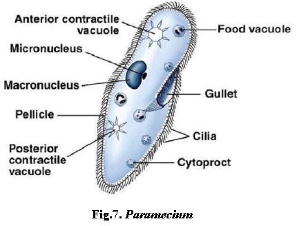
1. Ciliates are protistan protozoans that possess cilia as organelles of locomotion and food capture.
2. Most are free-living freshwater protists. A few are marine and some are parasites.
3. Free-living forms are generally motile. A few, such as Vorticella, are sedentary.
4. Though unicellular, ciliates have the highest degree of structural and physiological complexity, with specialised organelles for skin, muscles, sense organs, kidney, ingestion and egestion.
5. The food collecting apparatus consists of an oral groove, vestibule, buccal cavity, cytostome and cytopharynx. A cytopyge or cytoproct is formed for egestion.
6. Two types of nuclei are found: a larger, polyploid macronucleus that controls metabolism, and a smaller, diploid micronucleus that controls heredity.
7. Asexual reproduction occurs by binary fission, budding and cyst formation. Sexual reproduction is by conjugation. Autogamy also occurs.
8. Examples: Paramoecium, Tetrahymena, Vorticella and Balantidium. Tetrahymena is used in biological research. Balantidium coli causes balantidial or ciliary dysentery in humans, and infection comes from faecal contamination by pigs.
Kingdom Fungi (Kingdom of Multicellular Decomposers)
1. Fungi are achlorophyllous, heterotrophic, gametophytic, haploid, multicellular, eukaryotic, nucleated, spore-producing thallophytes. Their cell wall is made of chitin (fungus cellulose).
2. R.H. Whittaker (1969) gave fungi the status of a kingdom in his five kingdom system.
3. The study of fungi is called mycology or mycetology.
4. Pier Antonio Micheli, an Italian botanist, is regarded as the father of mycology. He wrote Nova Genera Plantarum.
5. E.J. Butler is regarded as the father of Indian mycology and phytopathology.
[Figure: Fig. 2.5 Some fungi, showing Saccharomyces, Agaricus, Alternaria, Penicillium, Aspergillus, Mucor, Rhizopus, Peziza and Polyporus]
General Characters of Fungi
1. Fungi are non-vascular, non-seeded, non-flowering, multicellular decomposers and mineralisers of organic wastes. They help in recycling of matter in the biosphere.
2. In true fungi the plant body is a thallus. It may be non-mycelial or mycelial.
3. Non-mycelial forms are unicellular, but they may form a pseudomycelium by budding, for example yeast.
4. Mycelial forms have a plant body made of thread-like structures called hyphae. Hyphae are usually branched tubes bounded by a cell wall of chitin. Hyphae may be septate (higher fungi) or aseptate (lower fungi).
5. Septate hyphae may be uninucleate (monokaryotic), dikaryotic with binucleate cells, or multinucleate. Some fungi are aseptate and called coenocytic fungi, with hundreds of nuclei in a continuous cytoplasmic mass.
6. The cell shows eukaryotic organisation but lacks chloroplasts and Golgi bodies. The genetic material is DNA and mitosis is intracellular (karyochorisis).
7. Fungi lack chlorophyll, so they do not prepare food by photosynthesis. They are heterotrophs that absorb their nutrients and store food as glycogen.
8. Primitive fungi have oogamous sexual reproduction, whereas the most advanced ones have no sexual reproduction.
9. Parasites obtain food from a living host, and some of their hyphae are modified into haustoria. Saprophytes derive food from dead and decaying organic matter.
10. Some fungi live symbiotically with algae in lichens. Some fungi live in the roots of higher plants, for example in Pinus. This relationship is called mycorrhiza.
11. Some fungi grow on cow dung and are called coprophilous fungi.
Types of Parasites and Saprophytes in Fungi
1. Obligate parasites thrive on a living host throughout their life, and die when the host dies. Example: Puccinia.
2. Facultative parasites live independently of a host but may occasionally be parasitic under certain conditions. Example: Fusarium.
3. Ectoparasites (ectophytic parasites) grow on the surface of host cells and absorb food through haustoria.
4. Endoparasites (endophytic parasites) enter the tissues of the host.
5. Obligate saprophytes cannot live without dead organic matter.
6. Facultative saprophytes are usually parasitic but, after the death of the host plant, absorb food from the decaying body of the plant.
7. Predaceous fungi trap and digest small animals. Example: Dactylella, which feeds on protozoans, eelworms and rotifers.
Reproduction in Fungi
Fungi have three types of reproduction: vegetative, asexual and sexual.
Vegetative Reproduction
1. Fragmentation: multiplication by breakage of the mycelium.
2. Fission: simple splitting of vegetative cells into two daughter cells.
3. Budding: small outgrowths called buds form on the vegetative body and are cut off to form new individuals.
4. Oidium formation: the hyphae break into small fragments called oidia, which give rise to new hyphae.
5. Chlamydospores: thick-walled resting spores formed under unfavourable conditions, which germinate when conditions improve. Chlamydospores are organs of perennation.
[Figure: Fig. 2.6 Mycelium of fungus with septate hyphae and mycelium of fungus with aseptate hyphae]
Labels in the figure: Hypha, Mycelium, Septa, Septate hypha (A); Sporangiophore, Sporangium, Spores, Stolon, Mycelium, Aseptate hypha.
[Figure: Fig. 2.7 Vegetative reproduction in fungi]
Labels in the figure: Budding (mother cell, buds); Fission (dividing cell, divided cell); Oidia (oidia, hypha); Chlamydospores (intercalary, terminal); Fragmentation.
Asexual Reproduction
1. Zoospore formation: zoospores are thin-walled, uninucleate, motile structures formed in zoosporangia. They may be uniflagellate (Synchytrium) or biflagellate (Saprolegnia).
2. Sporangiospores: non-flagellate spores developed inside sporangia, for example in Mucor and Rhizopus. They are dispersed by air currents and are produced in large numbers.
3. Conidia: non-motile, thin-walled exogenous spores produced on a conidiophore. They are arranged in chains in Aspergillus and Penicillium, and may be produced singly in Pythium.
Sexual Reproduction
Sexual reproduction is reduced in fungi and takes place by two fusing gametes. It includes three stages:
(a) Plasmogamy, or fusion of two protoplasts
(b) Karyogamy, or fusion of two nuclei
(c) Meiosis, or reduction division
In higher fungi, karyogamy is delayed and occurs just before meiosis. The stage between plasmogamy and karyogamy, when cells contain two nuclei (n + n), is called the dikaryophase, and such cells are called dikaryotic cells.
Types of sexual reproduction in fungi:
(i) Planogametic copulation: fusion of two motile gametes of opposite sex or strains. It is of two types, isogamy and heterogamy.
(ii) Gametangial contact: two gametangia come into contact but do not fuse, and keep their identity.
(iii) Gametangial copulation: the entire contents of two gametangia fuse.
(iv) Spermatisation: the uninucleate, non-motile male gamete called spermatium is transferred to the trichogyne of the ascogonium (female gametangium), and the dikaryotic condition is established.
(v) Somatogamy: formation of gametes is absent, and two hyphae of opposite strains fuse to bring about dikaryotisation.
[Figure: Fig. 2.8 Different types of sexual reproduction in fungi]
Labels in the figure: Planogametic copulation (isogamy, anisogamy, oogamy with sperm and egg); Gametangial contact (oogonium, antheridium, male gametangium, female gametangium, egg); Gametangial copulation (A, B, host cell); Spermatization (spermatia, receptive hypha); Somatogamy (hyphae of opposite mating types).
Heterothallism
Heterothallism (Greek: heteros, different; thallos, undifferentiated body) is the condition in which the fusing gametes belong to two genetically distinct strains of the same species, though there may be no morphological difference between the gametes or the structures bearing them. It was first discovered by Blakeslee in 1904. The strains are called (+) and (-), A and B, or a and α. Heterothallism introduces variation that helps fungi adapt to diverse habitats, unfavourable environments and toxic chemicals.
Connecting Concepts on Fungi
1. Roman emperor Caesar was poisoned by his wife by Amanita caesarea, a mushroom.
2. In karyochorisis, nuclear division involves formation of an internal spindle. The spindle may be connected with centrioles or spindle pole bodies (SPB). The nuclear envelope persists during nuclear division.
Classification of Fungi
1. Criteria used for classifying fungi include morphology of mycelium and reproductive structures, types of spores and their dispersal, and life cycle. Reproductive structures are the most important, because they show more variation.
2. Kingdom Fungi is divided into two divisions:
Division 1: Myxomycotina, for example slime moulds. They have no cell wall.
Division 2: Eumycotina, the true fungi, which have a cell wall. This division has four classes: Phycomycetes, Ascomycetes, Basidiomycetes and Deuteromycetes.
Phycomycetes (Algal Fungi)
1. They are aseptate, coenocytic fungi.
2. Asexual reproduction is by zoospores (motile) or aplanospores (non-motile), endogenously produced in a sporangium.
3. Sexual reproduction is by gametangial copulation (conjugation) between two similar, multinucleated coenogametangia containing multinucleated coenogametes, to produce a zygospore.
4. Examples: Mucor, Rhizopus (bread mould) and Albugo (parasitic fungus).
Ascomycetes (Sac Fungi)
1. The mycelium is branched and septate, except in yeast, which is unicellular. Each cell is uninucleate, binucleate or multinucleate. The cell wall is made of chitin.
2. Flagella are totally absent.
3. Asexual reproduction is by conidia, produced exogenously on a branched or unbranched conidiophore. Conidia are brown, green, blue, yellow or pink and give colour to the mycelium.
4. Sexual spores are ascospores, produced endogenously in sac-like asci (singular: ascus). The asci are arranged in fruiting bodies called ascocarps.
5. Examples: unicellular yeast (Saccharomyces), blue mould (Aspergillus), green mould (Penicillium), pink mould (Neurospora), Erysiphe which causes powdery mildews, and Claviceps purpurea which causes ergot of rye.
6. Aspergillus flavus is the "guinea pig" of the plant kingdom. Neurospora is the "Drosophila" of the plant kingdom and is used extensively in biochemical and genetic work.
Basidiomycetes (Club Fungi)
1. They are called club fungi because the basidia are club-shaped.
2. They are the most evolved, most complex and most advanced fungi, and the best decomposers of wood.
3. Their fruiting bodies are large and easily visible, for example mushrooms and puff balls. Rusts and smuts also belong here, but predaceous fungi do not produce fruiting bodies.
4. The mycelium is branched and septate. Asexual reproduction is generally absent, but vegetative reproduction by fragmentation is common.
5. There are no sex organs. Plasmogamy is brought about by fusion of two somatic cells of different strains, which gives a dikaryotic structure that gives rise to the basidium. Karyogamy and meiosis take place in the basidium, producing four basidiospores exogenously.
6. The basidia are arranged in fruiting bodies called basidiocarps.
7. Examples: Agaricus (mushroom), Ustilago (smut) and Puccinia (rust fungus).
8. Phanerochaete chrysosporium, a basidiomycete, can bleach paper pulp better than bleaching powder (biobleaching).
Deuteromycetes (Fungi Imperfecti)
1. The perfect (sexual) stage is either absent or not reported, so they are called fungi imperfecti.
2. Some are saprophytes or parasites, while many are decomposers of litter and help in mineral cycling.
3. The mycelium is septate and branched. Asexual reproduction is by conidia produced on conidiophores. Conidiophores may occur singly or aggregate to form complex structures such as acervulus, synnema, sporodochium and pycnidium.
4. An acervulus is an aggregation of hyphae developed beneath the surface of the host plant. Short conidiophores are packed closely to form a flat bed-like mass on the surface, and the conidia are not covered by fungal tissue.
5. Conidiophores that are free from each other and bear conidia, as in Colletotrichum, are an example of this type.
6. Examples: Alternaria, Colletotrichum and Trichoderma.
Check Point
Give the scientific term for:
(a) A cell with two haploid nuclei of different mating types: dikaryotic cell (dikaryon).
(b) The symbiotic association of a fungus with the root of a higher plant: mycorrhiza.
(c) Fruiting body of ascomycetes: ascocarp.
(d) Uninucleate non-motile gamete transferred to the female gametangium: spermatium.
(e) Aggregation of hyphae developed beneath the surface of the host plant that produces conidia without fungal covering: acervulus.
Kingdom Plantae (Kingdom of Plants)
1. Members are multicellular, eukaryotic, photosynthetic organisms with a cellulosic cell wall.
2. They are non-motile, except some aquatic forms.
3. They have chloroplasts and perform photosynthesis (oxygenic photosynthesis). Examples include green, brown and red algae, bryophytes, pteridophytes, gymnosperms and angiosperms.
4. Growth is usually indefinite.
5. Some plants are heterotrophic and mostly parasitic, and a few are saprobes.
6. A small group of autotrophic plants catches small animals and insects to obtain nitrogen. They are called carnivorous or insectivorous plants.
7. Reproduction is both asexual and sexual. Accessory spores are present in lower plants.
8. An embryo stage is absent in the algal group but present in the other groups.
Kingdom Animalia (Kingdom of Animals)
1. Members are multicellular eukaryotic organisms.
2. Their cells have no cell wall, only a cell membrane.
3. They do not perform photosynthesis and have heterotrophic nutrition.
4. They have the power of locomotion.
5. They show increased sensitivity through the nervous system.
Viruses
1. The study of viruses is called virology. The term virus comes from Latin and means poison, venom or viscous fluid.
2. A virus is defined as an infectious nucleoprotein.
3. Viruses are submicroscopic organisms, generally less than 200 mµ in size.
4. They are obligate parasites and can live only inside a living host.
5. Ivanowsky (1892) recognised certain microbes as the causal organisms of the mosaic disease of tobacco. They were smaller than bacteria because they passed through bacteria-proof filters.
6. M.W. Beijerinck (1898) showed that the extract of infected tobacco plants could infect healthy plants, and called the fluid Contagium vivum fluidum (infectious living fluid).
7. W.M. Stanley (1935) showed that viruses could be crystallised. Outside their specific host cell they are inert.
8. They have either RNA or DNA as genetic material.
Living and Non-living Characters of Viruses
Non-living characters:
1. No protoplasm
2. No enzyme system
3. No respiration
4. They can be crystallised
5. They do not grow in culture medium
Living characters:
1. They contain nucleic acid, so they can direct the synthesis of proteins.
2. They multiply inside a living host cell.
3. They cause disease in humans, plants and animals.
Once a virus enters a host cell, it takes control of the whole biochemical machinery of the host and makes the host synthesise viral components.
Bacteriophages
1. Bacteriophages, or bacterial viruses, are viruses that infect bacteria. They are usually double-stranded DNA viruses.
2. They are of two types:
(i) Prophages, or non-virulent (non-infective) phages, which do not cause lysis of the bacterium soon after their formation. A bacterial cell carrying a prophage is called a lysogenic bacterium.
(ii) Virulent phages, or infective phages, which cause lysis of the bacterial cell at once.
3. The most studied series of bacteriophages is the T-series, for example T2, T4 and T6.
Structure of Bacteriophage
1. A bacteriophage is tadpole-like, with a head and a tail. Nucleic acid, generally DNA, is inside the head.
2. The head is hexagonal, about 950 Å long and 650 Å broad. The tail is joined to the head by a neck and collar.
3. The tail has a hollow core surrounded by a tail sheath. At the end of the tail is an end plate, to which tail fibres are attached.
[Figure: Fig. 2.9 Structure of bacteriophage, showing head (DNA covered with protein coat), collar, tail, tail fibres and end plate]
Multiplication of Bacteriophage
Lytic Cycle
1. Adsorption: the bacteriophage attaches to the bacterial cell surface with the help of its tail fibres.
2. Penetration: only the nucleic acid of the phage enters the bacterial cell, after lysis of the cell wall by lysozyme secreted at the tail. The protein coat is left outside as a ghost or doughnut.
3. Biosynthesis: the viral nucleic acid takes over the metabolic activities of the bacterial cell and forms new phage components. These unite to form 100 to 300 new phages in 15 to 20 minutes.
4. Lysis: the bacterial cell bursts to release the new phages.
[Figure: Fig. 2.10 Lytic cycle of T2 bacteriophage]
Labels in the figure (top to bottom): bacteriophage attached to a bacterial cell (phage DNA, phage protein coat, bacterial DNA, bacterial cell); empty head with protein coat only, phage DNA inside bacterial cell; new protein coat formed; DNA of phage replicates; new phage particles with DNA and protein coat; lysis of bacterial cell and release of new phage particles.
Lysogenic Cycle
1. Entry of viral genome into the bacterial cell: the virus attaches to the host surface, and its genetic material passes into the host with the help of lysozyme.
2. Formation of prophage: the viral genome produces a repressor and an integrase enzyme. The repressor stops the viral genome from taking over the host's machinery. Integrase joins the viral genome to the host DNA at a specific site. The integrated viral genome is called a prophage or provirus. In this state the virus is non-virulent, and it is also called the temperate stage.
3. Lysogeny: the condition of a host cell carrying a prophage is called lysogeny. The prophage multiplies along with the host without harming it.
4. Conversion into lytic phage: occasionally, repressor formation is inhibited by physical factors such as UV radiation, or chemicals such as nitrogen gas and mustard gas. The viral genome then separates from the host genome and becomes virulent. It takes over the host machinery, replicates, forms viral proteins and assembles new virus particles. The host cell lyses to release them.
The inert particle of a virus outside its host is called a virion.
Importance of Bacteriophages
1. They are used in studying the mechanism of viral infection.
2. They are used to control certain bacterial diseases, because phages are very specific to their host.
3. The purity of the holy Ganges is attributed to the presence of bacteriophages.
Tobacco Mosaic Virus (TMV)
1. TMV is a rod-shaped virus. The rod has a core containing helically coiled single-stranded RNA.
2. A protective protein covering called the capsid surrounds the infective part. The capsid is made of 2130 subunits called capsomeres and has antigenic properties.
3. TMV causes mosaic disease in tobacco leaves and some other plants. Young leaves lose chlorophyll, and later non-green and green patches appear on mature leaves. The green areas grow thicker and give a blistered appearance. The infected leaves also curl and become distorted.
HIV (Human Immunodeficiency Virus, AIDS Virus)
1. HIV is a round retrovirus of about 100 nm in diameter.
2. Its surface is covered by an envelope derived from the host. The envelope bears spikes made of protein components called peplomers, which fit the CD4 (T4) antigen receptor on helper T-cells, monocytes and macrophages.
3. Inside the envelope is a viral wall made of two layers.
4. The core contains two single strands of genomic RNA, protein and the enzyme reverse transcriptase.
5. HIV causes AIDS, for which no remedy has been found.
Nucleic Acids in Viruses
1. In plant viruses, RNA is generally present, but in cauliflower mosaic virus DNA is present.
2. In animal viruses, DNA is generally present, but the following animal viruses have RNA: influenza virus (single-stranded RNA), Rous sarcoma virus (single-stranded RNA), AIDS virus (single-stranded RNA), poliomyelitis virus (single-stranded RNA) and reovirus (double-stranded RNA).
3. Single-stranded RNA viruses such as the AIDS virus carry the enzyme reverse transcriptase, which copies RNA into DNA (reverse transcription). These are called retroviruses.
4. In bacteriophages, DNA is generally present, but in MS2, F2 and r-17 bacteriophages RNA is present.
5. DNA is generally double-stranded, but in φX174 and S13 E. coli phages it is single-stranded.
6. RNA is generally single-stranded, but in reoviruses, wound tumour viruses and rice dwarf virus it is double-stranded.
7. A large virus, such as vaccinia (cowpox virus), has about a hundred genes, while the smallest virus has three or four.
8. In some mammalian viruses, outside the capsid there is another membrane called the limiting membrane or mantle.
Other Viruses
1. Coliphage is the virus that infects E. coli, discovered by F.H. d'Herelle.
2. Cyanophages infect blue-green algae (cyanobacteria). The first cyanophage was isolated from the blue-green algal genera Lyngbya, Phormidium and Plectonema, and was called LPP-1. Safferman and Morris (1963) discovered cyanophages, which have double-stranded DNA.
3. Mycophages infect fungi. They were first discovered by Sinden (1957) in Agaricus bisporus. They have double-stranded RNA and are spherical or polygonal in shape.
4. During the latent (lysogenic) mode of replication, the viral genome is not affected because it contains 5-hydroxymethyl cytosine (5-HMC) instead of cytosine. The viral genome modifies the bacterial nucleoid, which produces modified enzymes, modified mRNAs and ribosomes that take part in the replication and transcription of the viral genome.
Economic Importance of Viruses
Viruses cause diseases such as mumps, smallpox, herpes, rabies, chickenpox, poliomyelitis, hepatitis, bird flu (H5N1), swine flu (H1N1) and influenza. AIDS in humans is also caused by a virus.
Viroids
1. T.O. Diener (1971) discovered some infectious agents smaller than viruses. These sub-viral agents are called viroids.
2. Viroids contain only very low molecular weight RNA, with no protein coat.
3. The viroid genome is a rod-shaped circular RNA. It appears double-stranded due to complementary base pairing, and it does not code for any protein because it lacks an initiation codon.
4. A viroid can multiply by two methods, DNA-dependent and RNA-dependent.
5. Viroids cause potato spindle tuber disease (PSTV), chrysanthemum stunt, citrus exocortis and cucumber pale fruit.
6. In plants, the symptoms can be mosaic formation, leaf rolling and curling, yellowing, vein clearing, dwarfing and stunted growth.
7. Viroid infections are persistent, so the plant never recovers.
Prions
1. Prions are non-nucleic infectious particles formed by modification of a normal neuronal protein.
2. A prion can multiply by converting the normal neuronal protein into the prion form.
3. On accumulation, prions cause degeneration of neuronal tissue. Examples: scrapie in sheep, mad cow disease, kuru and Creutzfeldt-Jakob disease (CJD).
4. Prions are resistant to proteases, nucleases, the high temperature of an autoclave, UV rays, formaldehyde and most disinfectants, except diethyl pyrocarbonate. The diseases they cause are therefore incurable.
5. Eating meat from animals suffering from a prion disease can cause a similar disease in humans.
Fill in the Blanks
(1) The phages which do not cause lysis of bacteria soon after their formation inside the bacterial cell are known as prophages.
(2) Viruses which attack bacteria are called bacteriophages.
(3) Capsid is the protein covering of TMV.
(4) The AIDS virus contains single-stranded RNA.
(5) The integrated viral genome is called prophage (provirus).
Lichens
1. Lichens are a small and distinct group of plants with about 400 genera.
2. A lichen has two components: the algal partner, called the phycobiont, and the fungal partner, called the mycobiont.
3. The major part of the lichen body is formed by the fungus. The photosynthetic algal partner forms hardly 5% of the body.
4. The internal structure has an upper or outer cortex, an algal zone, a medulla, a lower cortex and rhizines. Rhizines are attaching devices made of fungal hyphae.
5. Only the algal zone contains the photosynthetic partner. The rest of the zones are made of fungus only.
6. In homoiomerous lichens the algal partner is distributed almost throughout the body, for example Collema.
7. In heteromerous lichens the alga is restricted to the algal zone, for example Parmelia and Physcia.
8. About 98% of lichens have mycobionts that belong to ascomycetes. The rest have basidiomycetes or deuteromycetes. Accordingly, lichens are placed in three groups: ascolichens, basidiolichens and deuterolichens (lichenes imperfecti).
9. Phycobionts belong either to cyanobacteria (Gloeocapsa, Scytonema, Nostoc, Calothrix) or to green algae (Coccomyxa, Trebouxia, Trentepohlia).
10. Lichens grow very slowly and show symbiotic relationships. They can grow in places where other plants cannot, such as bare rock and the arctic region.
11. Lichens reproduce vegetatively, sexually and asexually. Soredia and isidia are reproductive propagules.
12. Lichens are economically important as food, fodder, dyes and sources of medicines. Cladonia rangiferina (reindeer moss) is food for reindeer and wild animals of the tundra. Cetraria islandica (Iceland moss), Lecanora esculenta and Umbilicaria esculenta are eaten in different parts of the world. Evernia and Ramalina extracts are used in soaps and perfumes, and sweet-scented lichens give the fragrance of incense in dhup and havan samagri.
13. Orcein, a cytological stain used for chromosomes, is purified from orchil. Litmus was once obtained from Roccella montagnei.
14. Lichens are sensitive to sulphur dioxide pollution, so they are used as pollution indicators.
Biological classification
Biological classification is defined as the process of grouping the organisms according to certain similarities
Linnaeus proposed the two kingdoms of classification. He classified organism into animal kingdom as Animalia and plant kingdom as Plantae. There were certain limitations associated with two kingdom classification such as- it does not differentiate between eukaryotes and prokaryotes, unicellular and multicellular organisms, and photosynthetic and non-photosynthetic organisms.
Five kingdom classifications
R.H. Whittaker proposed the five kingdoms of classification. The five kingdoms classification is as follows- Monera, Protista, Fungi, Plantae, and Animalia. The classification was based on thallus organization, cell structure, mode of nutrition, phylogenetic relationship, and reproduction.
Kingdom Monera
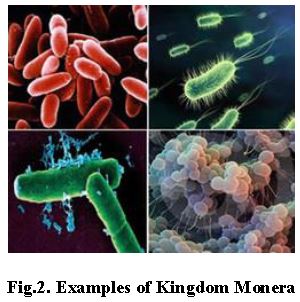
The most important members of Monera are Bacteria. All organisms present in this kingdom are prokaryotes. It includes bacteria as well as blue-green algae. They can be present in extreme habitats such as hot springs, deserts, snow as well as deep oceans. Certain organisms act as parasites also. All monerans are prokaryotes. They do not have the true nucleus, that is, nucleus not surrounded by nuclear membrane. They can be autotrophs, heterotrophs as well as parasites. They do have cell wall. There respiration can be aerobic as well as anaerobic. The movement occurs through flagella. Exchange of nutrients or gases occurs by diffusion.
Archaebacteria can be thermoacidophiles (hot springs), halophiles (survive in high salt concentration), and methanogens (marshy areas). There structure of cell wall is different from other monerans. Methanogens are found in the gut of some ruminants such as buffaloes and cows. This helps in production of biogas from the dung of the animal.
Eubacteria
They are referred as “True bacteria”. The presence of motile flagellum and a rigid cell wall characterize them. It includes cyanobacteria (blue-green algae). They are like plants in having chlorophyll a. So, known as photosynthetic autotrophs.
Cyanobacteria are unicellular, filamentous algae. They form colonies which are surrounded by gelatinous sheath. Some have specialized structures known as heterocyst which helps in fixing nitrogen. For example, Nostoc.
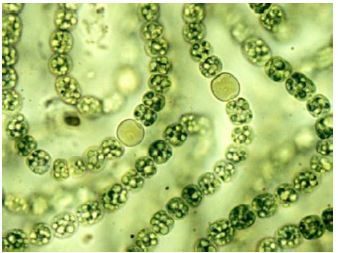
Fig.3. Nostoc
There are certain bacteria that can oxidize certain inorganic substances such as nitrites, nitrates, they are known as chemosynthetic autotrophs. The energy released during oxidation is used in the production of the ATP.
Bacteria can reproduce asexually by fission. They can also reproduce through spores under unfavorable conditions. Monera also includes Mycoplasma. They do not have cell wall and survive in anaerobic conditions. Mycoplasma are the smallest living organisms known.
Kingdom Protista
Single celled eukaryotes are kept under Protista. They are mostly aquatic in nature. For example, Algae serves as primary producers in aquatic ecosystem. They are unicellular, eukaryotic organisms. They have true nucleus, bounded by nuclear membrane. They can move by cilia as well as via flagella. Respiration can be both aerobic as well as anaerobic. Similarly, reproduction can be sexual as well as asexual.
They are divided into Chrysophytes, Dinoflagellates, Euglenoids, Slime moulds and Protozoans.
1. Chrysophytes
It consists of diatoms and golden algae. They can live in freshwater water as well as marine water. Mostly they are photosynthetic. Their cell wall is impregnated with silica. They are the major producers in the oceans. Mostly they are unicellular flagellates. But some are amoeboid.
2. Dinoflagellates
Dinoflagellates are marine and photosynthetic organisms. They can be of different colors such as yellow, brown, red, or blue in color. The color is due to the presence of different colored pigments present in the cells of the dinoflagellates. The cell wall is made up of cellulose. They are biflagellate with one flagella placed longitudinally and other flagella placed transversely. For example, Gonyaulax catenella, Noctiluca scintillans.
3. Euglenoids
Euglenoids are freshwater organisms that are found in stagnant water. They are surrounded by a protein rich layer known as pellicle. They have one short flagella and one long flagella. They can behave as autotrophic as well as heterotrophic organisms. For example, Euglena.
Most of the euglenoids have chloroplast so they can synthesize their own food. The reserve food material found is euglenoids are Paramylon (carbohydrate).
4. Slime moulds
They are saprophytic in nature. Under favorable conditions, they form aggregates which are known as Plasmodium. During unfavorable condition, they form fruiting bodies with spores. They possess true cell walls. They are extremely resistant to adverse conditions.
5. Protozoan like protists
They are heterotrophs. They can live as predators as well as parasites. There are four major types of protozoans found-
• Amoeboid protozoans are found in fresh water, moist soil, or sea water. They possess pseudopodia for engulfing the food particles, such as Amoeba.
• Flagellated protozoans can be free-living or parasitic. This group of organisms are responsible for different parasitic diseases. For example, Trypanosoma causes sleeping sickness.
• Ciliated protozoans possess thousands of cilia. The movement of cilia helps in propelling forward or backward. It also helps in obtaining food from the outside. For example, Paramecium.
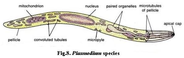
• Sporozoans form reproductive cells which are known as spores. They are parasitic and pathogenic in nature. For example, Plasmodium species causes Malaria. Reproduction can be asexual or sexual.
Kingdom Fungi
Fungi are heterotrophic in nature. Some acts on dead organic matter and so known as saprophytes. Some can feed on living organisms so known as parasites. Some fungi can live in mutual relationship with other organisms such as algae, they are known as lichens. Such fungi are known as symbionts. Lichens are symbiotic association between the fungi and the algae. Mycorrhiza are the symbiotic association between the fungi and the roots of the higher plants.
Reproduction in fungi can occur by- budding, fragmentation, and fission. Asexual reproduction occurs by means of spores known as conidia, zoospores or sporangiospores.
Sexual reproduction occurs by oospores, ascospores and basidiospores. There are three steps of sexual cycle in fungi-the first step is the fusion of the protoplasm which is known as plasmogamy. Then, the fusion of nuclei occurs which is known as karyogamy. This results in the formation of the zygote. Then finally, the meiosis of zygote results in the formation of the spores.
The vegetative part of the fungus is known as mycelium. The network of hyphae is known as mycelium. During sexual cycle of fungi, two haploid hyphae fuses to form diploid cells. Kingdom fungi is divided into different classes-
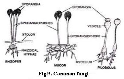
• Phycomycetes (lower fungi)- Asexual reproduction occurs by motile spores known as zoospores and non-motile spores are known as aplanospores. Spores are produced inside the sporangium. Fusion of gametes give rise to zygospore. Mycelium is coenocytic (multiple nucleus) and multicellular. For example, such as Mucor, Rhizopus, Albugo.
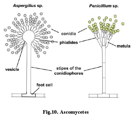
• Ascomycetes (sac fungi)- They can be saprophytic as well as parasitic. Asexual reproduction occurs by conidia. Sexual reproduction occurs through ascospores. Branched and septate mycelium is found in Ascomycetes. For example, Penicillium, Aspergillus, Claviceps etc.
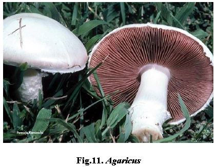
• Basidiomycetes (club fungi)- Vegetative reproduction occurs through fragmentation. Absence of sex organs. Plasmogamy between two vegetative or somatic cells results in the formation of basidium. Basidium undergo karyogamy and meiosis to form four basidiospores. For example, Agaricus (mushrooms), Ustilago (smut) etc.
• Deuteromycetes are commonly known as imperfect fungi as they do not have sexual reproduction at any stage of the life. The asexual reproduction occurs by conidia. Mycelium is septate and branched. For example, Alternaria, Trichoderma etc.
• Zygomycetes (conjugation fungi) are primitive group of fungi. Asexual reproduction occurs by non-motile sporangiospores. For example, Rhizopus.
Kingdom Plantae
It includes all photosynthetic, multicellular, eukaryotic plants. Characteristics of members of Plantae are as follows-
• Mostly they are eukaryotic in nature.
• The main pigment present is chlorophyll.
• Their cell wall is made up of cellulose.
• Photosynthesis helps in the synthesis of the food.
• Reproduction can be sexual as well as asexual.
• They exhibit the phenomenon of alternation of generation, that is, diploid sporophyte and haploid gametophyte.
Kingdom Animalia
They are heterotrophic, eukaryotic organisms. Some of the characteristics of members of Animalia are as follows-
• They are multicellular organisms with variable size.
• They have well developed organ system such as skeleton system, circulatory system, respiratory system etc
• They are bilaterally symmetrical.
• They have well developed locomotory organs.
• Respiration occurs by gills, book lungs, book gills, skin, lungs etc.
• Membrane bound cell organelles with nucleus bounded by a nuclear membrane.
• Circulation occurs via blood, blood vessels, and heart.
• Reproduction occurs by the formation of the haploid gametes. The fusion of gametes give rise to a new diploid organism.
• Kidneys are the main respiratory organs.
Viruses:
They are acellular structures and therefore do not find a place in Whittaker’s five kingdom classification. They consist of a nucleic acid (either DNA or RNA) that is surrounded by a protein coat. They can grow and multiply only inside a host cell. Outside the host cell the viruses exist as crystals. They cause diseases and damage to the host. Examples of common viruses are the viruses causing common cod, influenza, polio, AIDS, etc.
Viroids:
They are the smallest structures known that are capable of infection. They consist only the nucleic acid without the protein coat.
Lichens:
They are symbiotic associations of algae and fungi. The algal partner is autotrophic and synthesizes and provides food. The fungal partner offers protection and shelter.
Biology NEET Exam Resources: Biological Classification
Core Study Kit: NEET Biology Biological Classification
Explore essential learning tools for NEET Biology Biological Classification. This curated collection features in-depth notes and targeted practice questions built around the active 2026 curriculum to streamline your daily revision.
NCERT-Aligned Solutions for Biological Classification
Each resource draws directly from authorized textbooks to maintain academic accuracy. Evaluating solved examples allows NEET students to master the formal presentation and answer-writing standards expected in upcoming school exams.
Next Steps in Your Exam Preparation
Maximize your grade potential by utilizing NEET Biology practice papers in tandem with these structured notes. All printable assignments and interactive mock tests on our platform are updated for the 2026 session and available free of charge.
FAQs
Our advanced study package for Biological Classification includes detailed concepts, diagrams, Mind Maps, and explanation of complex topics to ensure NEET students learn as per syllabus for 2026 exams.
The Mind Maps provided for Biological Classification act as visual anchors which will help faster recall during high-pressure exams.
Yes, teachers use our NEET Biology resources for lesson planning as they are in simple language and have lot of solved examples.
Yes, You can download the complete, mobile-friendly PDF of the Biology Biological Classification advanced resources for free.
Yes, our subject matter experts have updated the Biological Classification material to align with the rationalized NCERT textbooks and have removed deleted topics and added new competency-based questions.