

Official NEET Study Materials for NEET Biology
Review targeted academic resources with the NEET Biology Photosynthesis Notes. Built according to official NEET standards for the 2026-27 term, these downloadable NEET Biology study materials support effective daily practice and deeper conceptual understanding for Photosynthesis.
Advanced Resources for Biology
View or download the dedicated NEET Biology Photosynthesis Notes resource below. Engaging with these advanced study guides under focused conditions ensures continuous academic progress and mastery of the 2026-27 curriculum for Photosynthesis.
NEET Biology notes on Chapter 13 Photosynthesis, covering the early experiments and history of photosynthesis, the site of photosynthesis and the structure of the chloroplast, photosynthetic pigments (chlorophyll a and b, carotenoids), absorption and action spectra, the light reaction (photosystems I and II, electron transport, photolysis of water, cyclic and non-cyclic photophosphorylation, chemiosmotic hypothesis), the dark reaction (Calvin cycle, C4 cycle, CAM pathway), photorespiration, the factors affecting photosynthesis, and bacterial photosynthesis and chemosynthesis.
AT A GLANCE
Terms and definitions to learn:
1. Photosynthesis
2. Quantasome
3. Action and absorption spectrum
4. Quantum yield and quantum requirement
5. Light reaction and dark reaction
6. Photosystem I and photosystem II
7. Cyclic and non-cyclic photophosphorylation
8. Photolysis of water
9. Chemiosmotic hypothesis
10. Calvin cycle, C4 cycle, CAM and photorespiration
Differences to learn:
1. Chlorophyll a and chlorophyll b
2. Light reaction and dark reaction
3. Photosystem I and photosystem II
4. Cyclic and non-cyclic photophosphorylation
5. C3 and C4 plants
Flow chart to learn: Mechanism of photosynthesis.
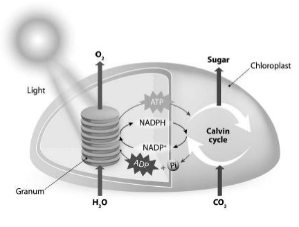
At a Glance (chapter contents):
- Early experiments
- Site of photosynthesis
- Photosynthetic pigments: chlorophyll, carotenoids, absorption and action spectra
- Mechanism of photosynthesis
- Light reaction: transfer of energy; Emerson's effect and red drop; two pigment system; electron transport system; photolysis of water; photophosphorylation; chemiosmotic hypothesis
- Dark reaction: Calvin cycle; C4 cycle; CAM
- Photorespiration
- Factors affecting photosynthesis
INTRODUCTION
Photosynthesis is the process by which plants and other organisms, such as algae and some bacteria, synthesize their own food in the presence of light. So, they are called autotrophs.
It is the primary source of all food on earth. It is also responsible for the release of oxygen into the atmosphere by green plants.
The process of photosynthesis can also be defined as the transformation of photonic energy (i.e., light or radiant energy) into chemical energy (locked in the high energy bonds of carbohydrate molecules) by the green parts of plants.
The simplest equation for photosynthesis in higher plants and algae is:
6CO2 + 12H2O → C6H12O6 + 6O2 ↑ + 6H2O

Photosynthesis is an anabolic or constructive process.
Green plants capture energy from the sun and convert it into chemical energy (food) with the help of CO2 taken from the atmosphere and water from the soil, and release oxygen. This food is consumed by other animals, which are therefore called heterotrophs. Thus, photosynthesis is the single most important biological process required for human existence. During photosynthesis, photoautotrophs utilize only 0.2% of the solar energy incident on the earth's surface.
EXPERIMENTAL PROOF OF REQUIREMENTS OF PHOTOSYNTHESIS
Some experiments prove that certain factors are essential for photosynthesis, such as chlorophyll, light and CO2.
Chlorophyll: Take a fresh variegated leaf of Coleus or Croton plants growing in bright sunlight. Trace the outline of the leaf and its non-green parts on a tracing paper. Boil the leaf in 70% alcohol till it is decolourized. Take out the leaf and wash it with water. Immerse the leaf in iodine solution. The green chlorophyll-containing parts of the leaf turn blue, while the non-green parts remain pale. This shows that chlorophyll is essential for photosynthesis.
[Figure: Fig. 13.1 Experiment to demonstrate that chlorophyll is essential for photosynthesis]
Fig. 13.1 labels: (A) green or chlorophyllous portion; non-green or colourless portion. (B) bluish portion (starch present); colourless portion.
Light: Take two leaves: a variegated leaf, or a leaf that was partially covered with black paper, and one that was exposed to light. On testing these leaves for starch, it was clear that photosynthesis occurred only in the green parts of the leaves in the presence of light.
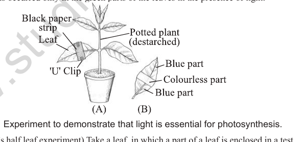
[Figure: Fig. 13.2 Experiment to demonstrate that light is essential for photosynthesis]
Fig. 13.2 labels: (A) black paper strip, leaf, 'U' clip, potted plant (destarched). (B) blue part, colourless part, blue part.
CO2: (Moll's half leaf experiment) Take a leaf in which a part of the leaf is enclosed in a test tube containing some KOH solution (which absorbs CO2), while the other half is exposed to air. The set-up is placed in light for some time. On testing for starch later in the two halves of the leaf, the exposed part of the leaf showed a positive result for starch, while the portion that was in the tube tested negative. This concluded that CO2 was required for photosynthesis.
[Figure: Fig. 13.3 Moll's half leaf experiment. A potted plant (destarched) with a split cork and wide mouth bottle containing KOH solution enclosing part of the leaf; the bluish part and colourless parts are shown in (A) and (B)]
Fig. 13.3 labels: split cork, wide mouth bottle, KOH solution, potted plant (destarched); in the leaf, the bluish part is c, and the colourless parts are b and a.
Check Point
Fill in the blanks.
1. Moll's half leaf experiment shows that ______ is essential for photosynthesis.
2. ________ plotted the action spectrum of photosynthesis.
3. John Priestley termed the impure air as ________ and the pure air as ________.
4. Purple and green sulphur bacteria use ________ as the hydrogen donor.
5. __________ showed that the process of photosynthesis takes place in chloroplast and results in the synthesis of starch.
EARLY EXPERIMENTS AND HISTORY OF PHOTOSYNTHESIS
It is important to know about the simple experiments that led to the gradual development of our understanding of the process of photosynthesis.
Joseph Priestley (1733-1804)
He performed a series of experiments (1770) that revealed the essential role of air in the growth of plants.
He concluded that a burning candle, or an animal that breathes the air, somehow damages the air. He observed that a candle burning in a closed space gets extinguished soon, and a mouse would suffocate soon in a closed space.
But when he placed a mint plant in the same bell jar, the mouse stayed alive and the candle continued to burn.
So, Priestley hypothesized that plants restore to the air whatever breathing animals and burning candles remove.
He termed the impure air as 'phlogiston' and the pure air as 'dephlogiston'.
Jan Ingenhousz (1730-1799)
He conducted a similar experiment with an aquatic plant, one kept in bright sunlight and the other in the dark.
He showed that in the plant kept in light, small bubbles were formed around the green parts, while no bubbles appeared in the plant kept in the dark.
These bubbles were later identified as oxygen bubbles.
He concluded that plants purify air only in the presence of light.
Julius von Sachs
He showed that the green substance (chlorophyll) is located in special bodies (chloroplasts) within the plant cell.
He provided evidence (1854) for the production of glucose when plants grow, and that glucose is stored in the form of starch.
T. W. Engelmann (1843-1900)
He determined the action spectrum of chlorophyll by using the green alga Cladophora.
He split light using a prism into its component parts and illuminated a green alga, Cladophora, placed in a suspension of aerobic bacteria. The bacteria were used to detect the sites of oxygen evolution.
He observed that the bacteria accumulated mainly in the regions of blue and red light of the split spectrum. A first action spectrum of photosynthesis was thus described.
Cornelius van Niel (1897-1985)
He was a microbiologist. Based on his experiments with purple and green bacteria, he made a contribution, a milestone, in the understanding of photosynthesis.
He demonstrated that photosynthesis is essentially a light-dependent reaction in which hydrogen from a hydrogen donor reduces carbon dioxide to carbohydrates:
Light: 2H2A + CO2 → 2A + CH2O + H2O
In green plants, water (H2O) is the hydrogen donor and is oxidized to oxygen. Purple and green sulphur bacteria use H2S as the hydrogen donor.
He inferred that the oxygen evolved by green plants during photosynthesis comes from water (H2O) and not from carbon dioxide (CO2). This was later proved by using radioactive isotopes of oxygen (H218O).
Light: 6CO2 + 12H2O → C6H12O6 + 6H2O + 6O2
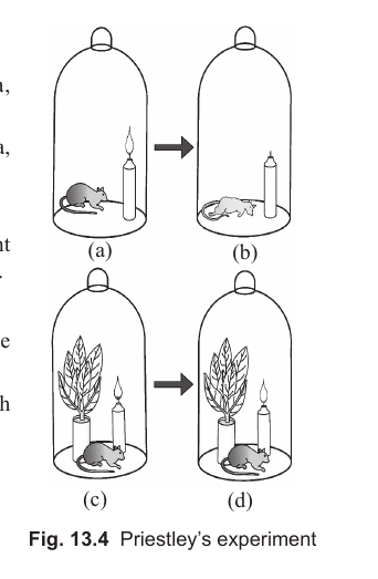
[Figure: Fig. 13.4 Priestley's experiment]
SITE OF PHOTOSYNTHESIS
Chloroplasts were first described by Leeuwenhoek (1679) and are believed to be the site of photosynthesis, as Sachs (1862) found starch grains inside chloroplasts during photosynthesis.
Photosynthesis occurs in the green parts of the plant, mostly in leaves and to a lesser extent in green stems or floral parts. In leaves, photosynthesis occurs particularly in specialized cells called mesophyll cells. These cells contain chloroplasts, which are the actual sites of photosynthesis. Chloroplasts fix CO2 into carbohydrates.
Chloroplasts are green coloured plastids. The entire process of photosynthesis is completed in each chloroplast.
Chloroplasts are double membrane bound organelles. The space limited by the inner membrane of the chloroplast is called the stroma.
A number of organised flattened membranous sacs (called thylakoids) are present in the stroma. Thylakoids are arranged in stacks like piles of coins, called grana.
The internal space of the thylakoid is the lumen. The lumen is the site of oxidation of water and the source of the oxygen evolved during photosynthesis. The lumen also serves as a reservoir for protons, which are pumped across the membrane of the thylakoid during electron transport.
The thylakoids in the chloroplast contain most of the machinery for the photochemical reactions of photosynthesis. They contain pigments for capturing solar energy.
The stroma of the chloroplast contains enzymes required for the synthesis of carbohydrates and proteins.
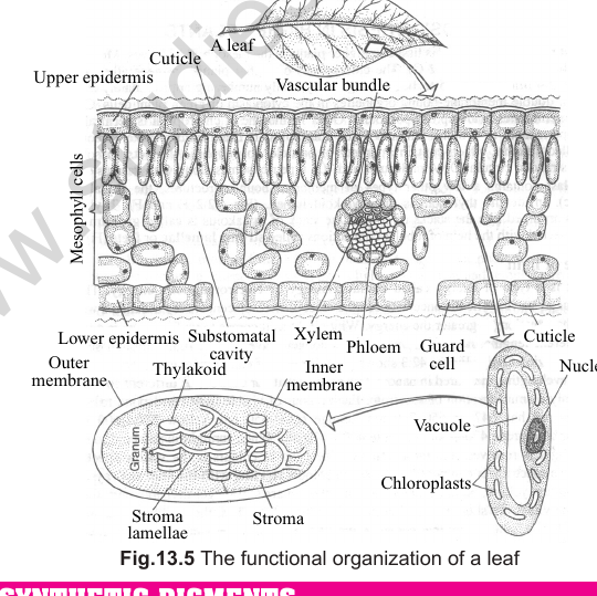
Connecting Concepts: A leaf has an upper epidermis with a cuticle, mesophyll cells containing chloroplasts, a lower epidermis with stomata and guard cells, and vascular bundles with xylem and phloem. Each chloroplast has an outer membrane, an inner membrane, stroma, stroma lamellae and thylakoids.
[Figure: Fig. 13.5 The functional organization of a leaf. Labels include upper epidermis, cuticle, mesophyll cells, chloroplasts, stroma, stroma lamellae, thylakoid, outer membrane, inner membrane, vascular bundle, xylem, phloem, substomatal cavity, lower epidermis, guard cell, nucleus and vacuole]
PHOTOSYNTHETIC PIGMENTS
Pigments are substances that absorb light of specific wavelengths in the visible region, due to the presence of conjugated double bonds in their structures.
Most of the photosynthesis takes place in the blue and red regions of the spectrum.
A photosynthetic unit can be defined as the number of pigment molecules required to effect a photochemical act, that is, the release of a molecule of oxygen. Park and Biggins (1964) gave the term quantasome to photosynthetic units, which is equivalent to 230 chlorophyll molecules. Quantasomes are mainly present in the grana lamellae.
The various contents of a quantasome are:
(i) Chlorophyll a
(ii) Chlorophyll b
(iii) Carotenoids (xanthophylls and carotene)
A chromatographic separation of leaf pigments shows that the colour of leaves is due to four pigments: chlorophyll a (bright or blue-green in the chromatogram), chlorophyll b (yellow-green), xanthophylls (yellow) and carotenes (yellow to yellow-orange). Chlorophyll is the major or principal pigment, and carotenoids are the accessory pigments in photosynthesis.
Chlorophyll
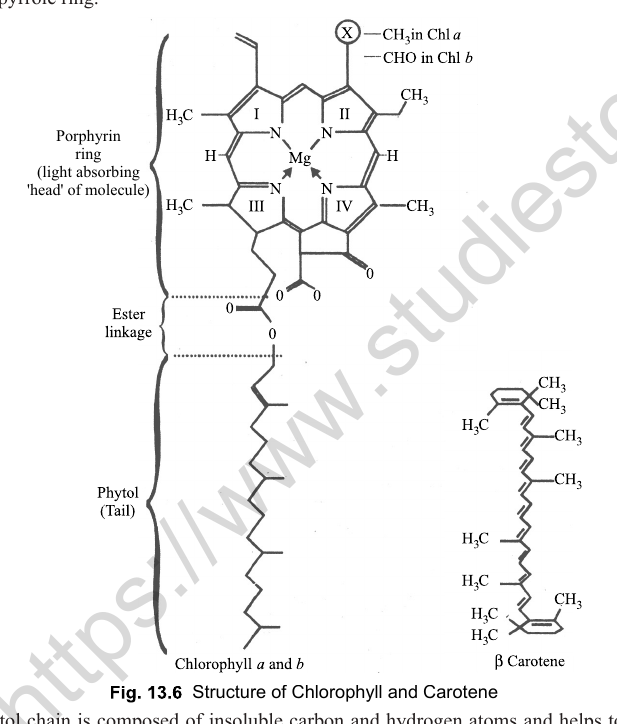
Chlorophyll is the main pigment, useful in photosynthesis. It shows a porphyrin structure. It is a large molecule composed of four five-membered rings called pyrrole rings and a central core of magnesium. A side chain, called the phytol chain, extends from one of the pyrrole rings.
Connecting Concepts: When the central Mg atom is replaced by Fe, the chlorophyll becomes a pigment called 'cytochrome', which is used in photosynthesis (photophosphorylation) and respiration both.
[Figure: Fig. 13.6 Structure of chlorophyll and carotene. The porphyrin ring is the light absorbing head of the molecule.]
The phytol chain is composed of insoluble carbon and hydrogen atoms, and helps to anchor chlorophyll molecules to the thylakoids.
Chlorophyll is a green pigment because it does not absorb green light but reflects it, which gives the leaves their green colour.
In plants, mainly two kinds of chlorophyll (chlorophyll a and chlorophyll b) are present. Chlorophyll a is two or three times more abundant than chlorophyll b and acts as the reaction centre. On absorbing light, the chlorophyll b molecule is excited and transfers its energy to chlorophyll a, which converts the light energy into electrical energy. Thus, chlorophyll a acts as the reaction centre.
Chlorophyll a is bluish-green while chlorophyll b is olive-green. Both are soluble in organic solvents like alcohol and acetone. Chlorophyll a (C55H72O5N4Mg) possesses a -CH3 (methyl group), which is replaced by a -CHO (aldehyde) group in chlorophyll b (C55H70O6N4Mg).
Table 13.1: Differences between chlorophyll a and chlorophyll b
| Characters | Chlorophyll a | Chlorophyll b |
|---|---|---|
| 1. Colour | Appears red in reflected light and bluish green in transmitted light. | Appears brownish red in reflected light and yellow green in transmitted light. |
| 2. Empirical formula | C55H72O5N4Mg | C55H70O6N4Mg |
| 3. Present | Mostly present in PS I. | Mostly present in PS II. |
| 4. Activation | Activated by the red zone of light. | Activated by the blue zone of light. |
| 5. Solubility | Soluble in petroleum ether. | Soluble in methyl alcohol. |
| 6. Group at C-3 of 2nd pyrrole ring | Methyl (-CH3) group. | Aldehyde (-CHO) group. |
Check Point
1. Write one term for the following:
(i) Actual sites of photosynthesis.
(ii) An amorphous, gel-like and enzyme rich material present within the chloroplast.
(iii) Pigment which absorbs light strongly in the blue-violet range.
(iv) Term given by Parks and Biggins.
(v) Pigment mostly present in PS-II.
(vi) Pigment which does not absorb green light but reflects green light.
2. In photosynthesis, which chlorophyll molecule absorbs light energy directly?
Carotenoids
The pigments which absorb light and transmit it to the chlorophyll reaction centre are called carotenoids. They are sometimes called lipochromes, due to their fat-soluble nature. They are lipids and are found in non-green parts of plants. Light is not necessary for their biosynthesis.
Carotenoids mainly absorb violet, indigo and blue wavelengths of the spectrum (maximum absorption occurs in the blue region) in higher plants, and transfer this energy to chlorophyll a. Thus, they act as accessory pigments.
They protect the chlorophyll molecules from photo-oxidation by picking up nascent oxygen and converting it into the harmless molecular stage.
Carotenoids can be classified into two groups, namely carotenes and xanthophylls.
(i) Carotenes: They are orange-red in colour and have the general formula C40H56. They are isolated from carrot. They are found in all groups of plants, from algae to angiosperms. Some of the common carotenes are alpha, beta, gamma and delta carotene, phytotene, lycopene, neurosporene, etc. Lycopene is a red pigment found in ripe tomato and red pepper fruits. Beta-carotene on hydrolysis gives vitamin A, hence carotenes are also called provitamin A. Beta-carotene is an orange-yellow pigment of carrot roots.
(ii) Xanthophylls: They are yellow coloured carotenoids, also called xanthols or carotenols. They contain oxygen along with carbon and hydrogen and have the general formula C40H56O2. Lutein (C40H56O2) is a widely distributed xanthophyll and is responsible for the yellow colour in autumn foliage. Fucoxanthin (C40H52O6) is another important xanthophyll present in Phaeophyceae (brown algae).
Carotenoids act as antenna complexes and harvest light from regions of the spectrum other than that absorbed by chlorophyll a, which is then funnelled into the reaction centre for conversion into electrical energy. The accessory pigments and reaction centres together form a photosystem.
Best photosynthesis takes place in red light, then in blue light, and in green light it is nil.
ABSORPTION AND ACTION SPECTRA
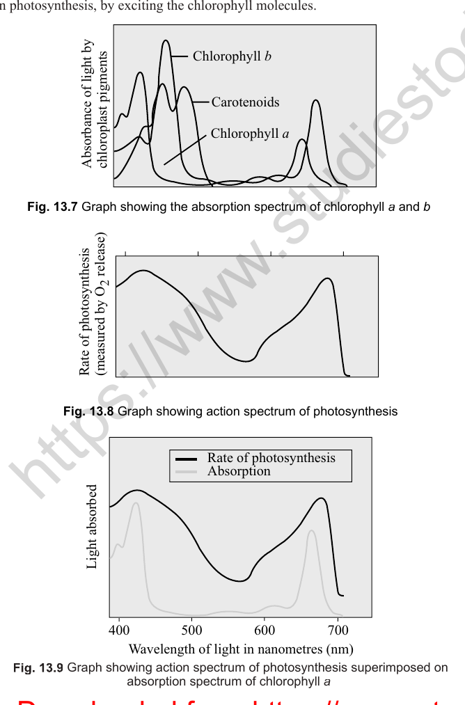
If we study the effect of different wavelengths of light on photosynthesis and plot it on graph paper, taking the wavelength of light on one axis and the rate of photosynthesis on the other, it is called the action spectrum of photosynthesis. The actual rate of photosynthesis, in terms of oxygen evolution or carbon dioxide utilization, is measured as a function of wavelength. Action spectra for photosynthesis and other photobiological processes help identify the pigment involved, because these spectra often closely match the absorption spectrum of the pigment that participates.
If we take an alcoholic or acetone solution of chlorophyll a in a test tube, provide different wavelengths of light to it, and then study the transmitted light, we find that dark bands appear in the red and blue regions of the spectrum (wider in red and narrower in blue). If we plot a graph of wavelength of light against absorption, we get the absorption spectrum.
If the action spectrum of photosynthesis and the absorption spectrum of chlorophyll a solution overlap, it is a clear indication that photosynthesis takes place only at those wavelengths absorbed by chlorophyll a, i.e., only these wavelengths of light participate in photosynthesis, by exciting the chlorophyll molecules.
Connecting Concepts: If a pigment absorbs light energy, the energy may be dissipated as heat, or it may be emitted immediately as a longer wavelength (the phenomenon is called fluorescence), or this energy may trigger a chemical reaction, as in photosynthesis. The first action spectrum of photosynthesis was studied by T. W. Engelmann (1882) using the green alga Spirogyra and oxygen-seeking bacteria.
[Figure: Fig. 13.7 Graph showing the absorption spectrum of chlorophyll a, chlorophyll b and carotenoids. Absorbance is plotted against wavelength of light in nm (400 to 700 nm).]
[Figure: Fig. 13.8 Graph showing the action spectrum of photosynthesis. Rate of photosynthesis is plotted against wavelength of light in nm (400 to 700 nm).]
[Figure: Fig. 13.9 Graph showing the action spectrum of photosynthesis superimposed on the absorption spectrum of chlorophyll a]
MECHANISM OF PHOTOSYNTHESIS
Photosynthesis is an oxidation-reduction process in which water is oxidized and CO2 is reduced to carbohydrates.
The mechanism of photosynthesis demonstrates the existence of two phases:
(i) Light phase (photochemical reaction, light reaction, Hill's reaction or light-dependent reaction).
(ii) Dark phase (biosynthetic phase, Blackman's reaction, light-independent phase or purely chemical reaction).
The reduction of CO2 to carbohydrates needs assimilatory powers, i.e., ATP and NADPH2. The actual reduction of CO2 to carbohydrates is independent of light, i.e., it occurs in the presence or absence of light, but the production of assimilatory powers (ATP and NADPH2) needs light and is light dependent.
During the light stage, energy from sunlight is absorbed and converted to chemical energy, which is stored in ATP and NADPH + H+.
During the dark stage, carbohydrates are synthesized from carbon dioxide using the energy stored in the ATP and NADPH formed in the light-dependent reactions.
Table 13.2: Differences between light reaction and dark reaction
| Characters | Light reaction | Dark reaction |
|---|---|---|
| 1. Light requirement | It is a light-dependent process. | It does not require light. |
| 2. Photosystems | Involves photosystems I and II. | No photosystem is required. |
| 3. Photolysis of water and CO2 | Photolysis of water takes place and O2 is liberated. | Photolysis of water does not take place. CO2 is absorbed. |
| 4. Site | Occurs within the grana of the chloroplast. | Occurs within the stroma of the chloroplast. |
| 5. Products | ATP and NADPH are produced and they are used to drive the dark reaction. | Glucose is produced. Reduced NADP is oxidized. |
Check Point
State true or false.
1. Light phase is also called Blackman's reaction.
2. ATP and NADPH2 are required for the reduction of CO2 to carbohydrates.
3. Light reactions occur within the grana of the chloroplast.
4. Emerson enhancement effect and red drop are the evidences in favour of photorespiration.
5. Quantum requirement in photosynthesis is 8.
6. Reaction centre of PSI is P700.
LIGHT REACTION
The light reaction requires light, and hence it is called the photochemical reaction. It occurs in the grana of the chloroplast. The activities in this reaction are dependent on light. Assimilatory powers (ATP and NADPH2) are mainly produced in this light reaction.
The various events and components of the light phase are: transfer of energy, Emerson effect, two pigment systems, electron transport system, photolysis of water, cyclic and non-cyclic photophosphorylation, and the chemiosmotic hypothesis.
Transfer of energy
When a photon of light energy falls on a chlorophyll molecule, one of the electrons pair from the ground or singlet state passes into a higher energy level called the excited singlet state. It comes back within 10-9 seconds.
The light energy absorbed by the chlorophyll molecule before it comes back to the ground state appears as radiation energy. The radiation given out when the electron returns from the excited singlet state is called fluorescence, and it is temperature independent.
Emerson effect and red drop
R. Emerson and C. M. Lewis (1943) observed that the quantum yield of photosynthesis decreases towards the far red end of the spectrum (680 nm or longer) while determining the quantum yield of photosynthesis in Chlorella using monochromatic light of different wavelengths. Quantum yield is the number of oxygen molecules evolved per light quantum absorbed. This decrease in quantum yield beyond the red region of the spectrum is called the red drop.
Emerson et al. (1957) further observed that the photosynthetic efficiency of light of 680 nm or longer is increased if light of shorter wavelengths (less than 680 nm) is supplied simultaneously. When both short and long wavelengths were given together, the quantum yield of photosynthesis was greater than the total effect when both wavelengths were given separately. This increase in photosynthetic efficiency (or quantum yield) is known as the Emerson effect, or Emerson enhancement effect.
\( E = \frac{\text{Quantum yield in combined beam} - \text{Quantum yield in red beam}}{\text{Quantum yield in far red beam}} \)
The rate or yield of photosynthesis is measured in terms of quantum yield. Quantum requirement is defined as the number of quanta of light required for the evolution of one molecule of O2 in photosynthesis.
Quantum requirement in photosynthesis = 8, i.e., 8 quanta of light are required to evolve one molecule of O2.
Hence, quantum yield = 1 / 8 = 0.125 (i.e., a fraction of 1).
[Figure: Fig. 13.10 Red drop and Emerson's enhancement effect in Chlorella. Quantum yield is plotted against wavelength of light in nm.]
Two pigment system
The discovery of the Emerson effect clearly indicates that the photosynthetic pigments are organised into discrete photochemical light harvesting complexes (LHC), called photosystem I and photosystem II.
The light reaction of photosynthesis involves the participation of two separate pigment systems, i.e., PS I and PS II. These are named in the sequence of their discovery, and not in the sequence in which they function during the light reaction.
The LHCs are made up of hundreds of pigment molecules bound to proteins. Each photosystem has all the pigments (except one molecule of chlorophyll a) forming a light harvesting system, also called antennae. These pigments help to make photosynthesis more efficient by absorbing different wavelengths of light. The single chlorophyll a molecule forms the reaction centre. The reaction centre is different in both photosystems.
Table 13.3: Comparison of photosystem I and photosystem II
| Characters | Photosystem I (PS I) | Photosystem II (PS II) |
|---|---|---|
| 1. Location | Lies on the outer surface of the non-appressed parts of the thylakoids (both grana and stroma thylakoids). | Lies on the inner surface of the appressed parts of the thylakoids (grana thylakoids). |
| 2. Reaction centre | A special chlorophyll molecule called P700. Pigments absorb longer wavelengths of light (above 680 nm). | Reaction centre is P680. Pigments absorb shorter wavelengths of light (below 680 nm). |
| 3. Photophosphorylation | Participates both in cyclic and non-cyclic photophosphorylation. | Participates only in non-cyclic photophosphorylation. |
| 4. Electron source | Receives electrons from photosystem II. | Receives electrons from the photolytic dissociation of water. |
| 5. Main function | Main function is ATP synthesis. | Main functions are ATP synthesis and hydrolysis of water. |
| 6. Photolysis of water | Not related with photolysis of water. Molecular oxygen is not evolved. | As a result of photolysis of water, molecular oxygen is evolved. |
| 7. Pigments | Contains more chlorophyll a, while chlorophyll b and carotenoids are less. | Contains chlorophyll b and some forms of chlorophyll a (such as chlorophyll a 662, 670 and 667). Carotenoid content is higher as compared to PS I. |
| 8. Colour | Light green in colour. | Dark green in colour. |
Electron transport system
The electron transport system was first formulated by Robert Hill (1939). It is a series of electron carriers which pass electrons in a downhill journey. It releases energy at every step, which is used in generating an electrochemical proton gradient that helps in synthesizing ATP.
According to Hill and Bendall, the whole light reaction is a two-step electron transport system. In the first step, photolysis of water occurs, and in the second step, photophosphorylation takes place.
The electron transport chain has two components connected with the two photosystems.
P680 of PS II absorbs light energy, gets excited and transfers its electrons to an electron acceptor, and becomes a strong oxidant. It paves the way for light-dependent splitting of water, called photolysis. Photolysis generates electrons, which are passed on to the electron-deficient P680 for another photoact.
Plastocyanin (a copper-containing soluble protein) transfers electrons to the P700 reaction centre of PS I. On getting excited, P700 hands over electrons to a special electron acceptor molecule, which passes them downhill to an energy-rich molecule. This is called the Z scheme, due to its zig-zag shape.
When the NADP+ reductase complex is not operating, electrons pass to the cyt b-cyt f complex for carrying out cyclic photophosphorylation.
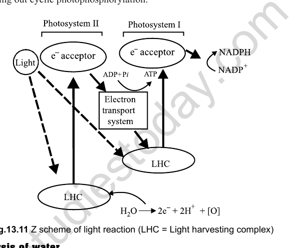
The electron acceptor, on accepting electrons, becomes a strong reducing agent. It donates its electrons to downstream components of the ETC (plastoquinone, cyt b-cyt f complex, plastocyanin).
Check Point
1. A series of electron carriers that pass electrons in a downhill journey is called _____________.
2. Photolysis of water was proposed by __________.
3. Formation of ATP molecules from ADP and H3PO4 in the presence of light and chlorophyll a during the photochemical phase of photosynthesis. (True or False).
4. Non-cyclic involves both PSI and PS II. (True or False).
5. Cyclic phosphorylation is also called Z scheme. (True or False).
6. ATP is synthesized at two steps in __________ phosphorylation.
7. Molybdenum, chloride and calcium ions play an important role in photolysis of water. (True or False).
8. Mechanism of ATP synthesis was given by _________.
[Figure: Fig. 13.11 Z scheme of light reaction (LHC = light harvesting complex)]
Photolysis of water
Splitting up of water and liberation of O2 by chlorophyll in the presence of light and a hydrogen acceptor (e.g., ferricyanide, coenzyme NADP) is called photolysis of water. The Hill reaction, or photolysis of water, was proposed by Robert Hill.
Electron acceptors or hydrogen acceptors that oxidize water are called Hill oxidants. Hill oxidants are ferredoxin, quinone, 2,6-dichlorophenolindophenol (DCPIP) and ferric salts (ferric oxalate).
Experimental proof for the liberation of O2 from H2O was provided by Ruben and Kamen (1941), using radioactive oxygen (O18).
Manganese, chloride and calcium ions play important roles in photolysis of water.
The electrons generated in this process are used to replace the electrons lost by P680. The protons are used for reducing NADP to NADPH. Oxygen is liberated as a by-product of photosynthesis.
2H2O → 4H+ + O2 + 4e-
Photophosphorylation
Arnon showed that during the light reaction, not only is reduced NADP formed and oxygen evolved, but ATP is also formed. This formation of high energy phosphates (ATP) is dependent on light, and hence it is called photophosphorylation.
Photophosphorylation is the process of ATP formation from ADP and inorganic phosphate in the presence of light in chloroplasts.
ADP + H2PO4 → ATP (light, chlorophyll a)
Photophosphorylation occurs in two ways: non-cyclic and cyclic photophosphorylation.
Non-cyclic photophosphorylation
In the light reaction, the formation of ATP from ADP and H3PO4 in the presence of light and chlorophyll a during the non-cyclic transfer of electrons is called non-cyclic photophosphorylation.
This system is dominant in green plants. It involves both PS I and PS II. The flow of electrons is unidirectional. The electrons are not cycled back, and are used in the reduction of NADP to NADPH2. Here H2O is utilized and O2 evolution occurs.
In this chain, high energy electrons released from P680 do not return to P680, but pass through pheophytin, plastoquinone, the cytochrome b6-f complex, plastocyanin (a Cu-containing pigment) and then enter P700. During the transfer of electrons from plastoquinone (PQ) to the cytochrome b6-f complex, ATP is synthesized. Because the high energy electrons released from P680 do not return to P680, and one molecule of ATP is formed, it is called non-cyclic photophosphorylation. ATP is synthesized at only one step.
Non-cyclic photophosphorylation is also known as the Z scheme, because of the shape of the path of electron flow. This was given by Hill and Bendall (1960).
Non-cyclic photophosphorylation, or the Z scheme, is inhibited by CMU and DCMU. (DCMU is a herbicide which kills weeds by inhibiting CO2 fixation, as it is a strong inhibitor of PS II.)
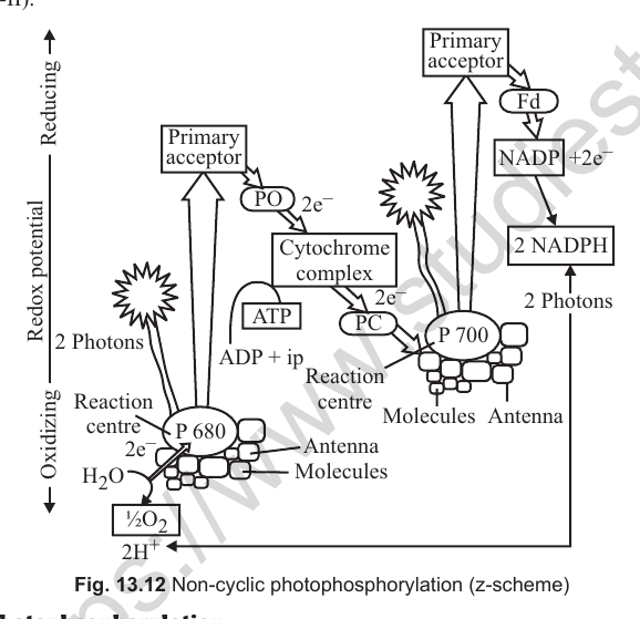
[Figure: Fig. 13.12 Non-cyclic photophosphorylation (Z scheme). It shows the primary acceptor, ferredoxin (Fd), NADP reducing to NADPH, cytochrome complex, plastocyanin (PC), P700 reaction centre with antenna molecules, P680 reaction centre with antenna molecules, 2 photons, H2O splitting to 2H+ and 1/2 O2, ADP + iP giving ATP, on a redox potential scale from oxidizing to reducing.]
Cyclic photophosphorylation
In the light reaction, the formation of ATP from ADP and H3PO4 in the presence of light and chlorophyll a during the cyclic transfer of electrons is called cyclic photophosphorylation.
This system is found dominantly in bacteria. It involves only PS I.
The flow of electrons is cyclic. This process occurs if NADP is not available. When photons activate PS I, a pair of electrons is raised to a higher energy level. They are captured by a primary acceptor, which passes them on to ferredoxin, plastoquinone, the cytochrome complex, plastocyanin, and finally back to the reaction centre of PS I, i.e., P700. At each step of electron transfer, the electrons lose potential energy. Their trip downhill is used by the transport chain to pump H+ across the thylakoid membrane. The proton gradient thus established is responsible for forming 2 molecules of ATP. No reduction of NADP to NADPH + H+ occurs. ATP is synthesized at two steps.
[Figure: Fig. 13.13 Cyclic photophosphorylation. It shows ferredoxin (Fd), plastocyanin (PC), cytochrome complex, primary acceptor, P700 reaction centre with antenna molecules, 2 photons, ADP + iP giving ATP at the 1st and 2nd steps, on a redox potential scale from oxidizing to reducing.]
Table 13.4: Differences between cyclic and non-cyclic photophosphorylation
| Characters | Cyclic photophosphorylation | Non-cyclic photophosphorylation |
|---|---|---|
| 1. Photosystems involved | It involves photosystem I. | It involves both PS I and PS II. |
| 2. Occurrence | It occurs in certain photosynthetic bacteria. | It occurs in higher green plants including blue-green algae. |
| 3. Conditions | It is operative under anaerobic conditions with poor CO2 availability and at low light intensity. | It operates under aerobic conditions with moderate CO2 availability and at optimum light intensity. |
| 4. Photolysis of water | It is not connected with photolysis of water, so no oxygen is evolved. | It is connected with photolysis of water and liberation of oxygen. |
| 5. Path of electrons | Electrons move in a closed circle. Electrons expelled from P700 return to it after passing through different electron acceptors. | Electrons do not move in a closed circle. Electrons expelled by the reaction centre P680 do not return to it. Here water donates electrons to P680. |
| 6. ATP synthesis | In each flow of electrons, 2 molecules of ATP are synthesized. | In each flow of electrons, 1 molecule of ATP is synthesized. |
| 7. NADPH | NADPH is not produced. | NADPH is produced. |
| 8. Effect of DCMU | It is not inhibited by DCMU. | It is inhibited by DCMU. |
Chemiosmotic hypothesis
The chemiosmotic hypothesis was put forward by Mitchell (1961) to explain the mechanism of ATP synthesis. ATP synthesis is linked to the development of a proton gradient across the membranes of the thylakoid.
When electrons are transported through the electron transport system (ETS), protons accumulate inside the thylakoid membranes. The lumen of the thylakoid becomes enriched with H+ ions due to photolytic splitting of water.
The primary acceptor of electrons is located on the outer side of the thylakoid membrane. It transfers its electron to an H-carrier. The carrier removes a proton from the matrix while transporting the electron to the inner side of the membrane. The proton is released into the lumen while the electron passes to the next carrier.
NADP reductase is situated on the outer side of the thylakoid membrane. It obtains electrons from PS I and protons from the matrix to reduce NADP+ to NADPH + H+.
The consequence of these three events is that the concentration of protons decreases in the matrix or stroma region, while their concentration in the thylakoid lumen rises, resulting in a decrease in pH. A proton gradient develops across the thylakoid.
The proton gradient is broken down by the movement of protons through transmembrane channels, F0, of the ATPase. ATPase has two parts, F0 and F1 particles. The rest of the membrane is impermeable to H+.
F0 provides facilitated diffusion to H+ or protons. As protons move to the outer side of the ATPase, they bring about conformational changes in the F1 particle of the ATPase, or coupling factor. The transient F1 particle of the ATPase enzyme forms ATP from ADP and inorganic phosphate.
Therefore, ATP synthesis through chemiosmosis requires a membrane, a proton pump, a proton gradient and an F0-F1 particle or ATPase. One molecule of ATP is formed when 2H+ pass through the ATPase.
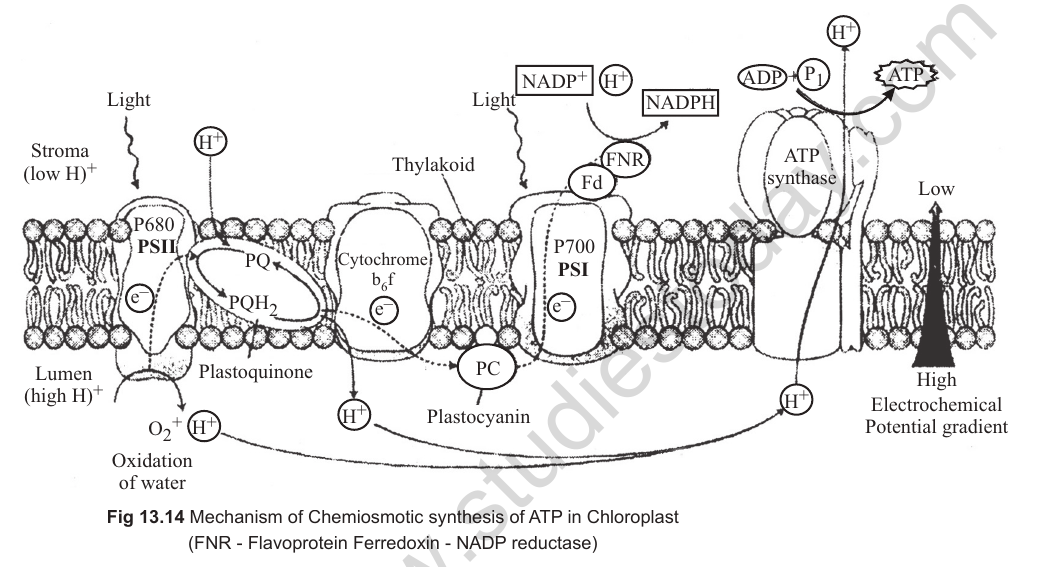
[Figure: Fig. 13.14 Mechanism of chemiosmotic synthesis of ATP in chloroplast (FNR = flavoprotein ferredoxin-NADP reductase). It shows the thylakoid, stroma and lumen with PSII, PSI, plastoquinone, cytochrome b6f, plastocyanin, ferredoxin, FNR, NADP+ + H+ to NADPH + H+, the ATP synthase, and the high electrochemical potential gradient of H+ across the thylakoid membrane.]
Table 13.5: Methods of synthesizing ATP
| Process | Energy source | Site of synthesis |
|---|---|---|
| 1. Photophosphorylation | Sunlight | Chloroplast |
| 2. Substrate level phosphorylation | Reaction not involving oxygen | Cytosol |
| 3. Oxidative phosphorylation | Oxidation with oxygen | Mitochondria |
Flow chart: Mechanism of photosynthesis
Mechanism of photosynthesis is divided into two phases.
Light phase: light dependent; involves photosystems (PS I and II); occurs in the grana of the chloroplast. It involves the following processes:
- Transfer of energy.
- Emerson effect and red drop.
- Two pigment system: Photosystem I (reaction centre is P700) and Photosystem II (reaction centre is P680).
- Electron transport system (formulated by Robert Hill), which is divided into photolysis of water and photophosphorylation. Photophosphorylation occurs in two ways. Cyclic photophosphorylation is found in bacteria, involves only PS I, has a cyclic flow of electrons, and synthesizes ATP in two steps. Non-cyclic photophosphorylation is found in green plants, involves both PS I and PS II, has a unidirectional flow of electrons, and synthesizes ATP at one step only.
- Chemiosmotic hypothesis: mechanism of ATP synthesis given by Mitchell (1961).
Dark phase: light independent; does not involve light; occurs in the stroma of the chloroplast. It involves two main pathways: the Calvin cycle (C3), given by Melvin Calvin in C3 plants, and the dicarboxylic acid cycle (C4), given by Kortschak in sugarcane (or C4 plants).
Check Point
Fill in the blanks
1. Major difference between mesophyll chloroplast and bundle sheath chloroplast is ______.
2. Kranz anatomy is the distinguishing feature of _______ plants.
3. Carbon assimilation in C3 plants was explained by Melvin Calvin in ______ and _______.
4. Three phases of Calvin cycle are ______, ______ and ______.
5. Enzyme involved in carboxylation is _________.
6. _________ are true xerophytic plants.
7. Four examples of C4 plants are ______, ______, ______ and ______.
8. C4 pathway is more efficient than C3 pathway due to the presence of ______.
9. Plants of the family Crassulaceae, Euphorbiaceae and Cactaceae are characterised by the presence of ______.
DARK REACTION
The dark reaction is a thermochemical reaction. It takes place in the stroma of the chloroplast. It is also called CO2 fixation or carbon assimilation.
The dark reaction involves the thermochemical reduction of CO2 to form carbohydrates. This was first established by Blackman (1905), and hence it is also called Blackman reaction. The presence of light is not necessary for the reduction of CO2. However, the dark reaction utilizes the products of the light reaction, i.e., assimilatory powers (ATP and NADPH2), to reduce CO2 into carbohydrates.
There are two main pathways for the biosynthetic or dark phase: the Calvin cycle (C3) and the C4 (dicarboxylic acid) cycle. Plants exhibiting the Calvin cycle and the C4 dicarboxylic acid cycle are respectively called C3 and C4 plants, because the first CO2 product was a 3-carbon organic acid (phosphoglyceric acid, PGA) and a 4-carbon organic acid (oxaloacetic acid, OAA), respectively.
CALVIN CYCLE (C3 CYCLE)
Carbon assimilation in C3 plants was explained by Melvin Calvin et al. (with Benson and Bassham) in Chlorella and Scenedesmus, using the radioisotope 14CO2.
Calvin and Benson discovered the path of carbon in this process. It is known as the C3 cycle, because CO2 reduction is a cyclic process, and the first stable product in this cycle is a 3-carbon compound (3-phosphoglyceric acid or PGA).
The Calvin cycle occurs in all photosynthetic plants, whether they have the C3 or C4 pathway. This pathway occurs in three phases: carboxylation, reduction and regeneration of RuBP.
Carboxylation
• Six molecules of ribulose-1,5-bisphosphate (RuBP) react with six molecules of carbon dioxide to form twelve molecules of 3-phosphoglyceric acid (3-PGA).
• The reaction is catalysed by RuBP carboxylase (located in the stroma).
• RuBisCO has both oxygenation and carboxylation activity.
In this step, CO2 is fixed into a stable organic intermediate.
Reduction
• Twelve molecules of phosphoglyceric acid are converted into twelve molecules of 1,3-diphosphoglycerate, which are then reduced to phosphoglyceraldehyde (PGAL), using ATP and NADPH molecules respectively.
• Two molecules of PGAL are diverted for the synthesis of sugar and then starch.
Regeneration of RuBP
• For the cycle to continue, the primary acceptor of carbon dioxide, i.e., RuBP, has to be regenerated.
• Ten molecules of PGAL, through a series of complex reactions, are converted into six molecules of a 5-carbon compound, RuBP.
• The formation of one molecule of RuBP requires one ATP molecule.
• The reaction is catalysed by the enzyme ribulose bisphosphate carboxylase (RuBisCO).
• PGA, a 3-carbon molecule, is the first stable product of the pathway, hence the name C3 pathway.
• Hence, for every CO2 molecule entering the Calvin cycle, 3 molecules of ATP and 2 molecules of NADPH are required.
Techniques used for studying the steps of the Calvin cycle: radioactive tracer technique using 14C (half-life 5720 years), chromatography and autoradiography. The material used was Chlorella (a unicellular alga) and Scenedesmus (microscopic, unicellular algae that can be easily maintained in the laboratory).
[Figure: Fig. 13.15 Calvin cycle]
C4 CYCLE (DICARBOXYLIC ACID CYCLE)
The C4 acid (dicarboxylic acid) is formed due to carboxylation. It was shown by Kortschak et al. in sugarcane. Kortschak and Hart supplied CO2 to the leaves of sugarcane and found that the first stable product is a 4-carbon (C4) compound, oxaloacetic acid, instead of a 3-carbon compound. The detailed study of this cycle was done by M. D. Hatch and C. R. Slack (1966), so it is called the Hatch and Slack cycle. The stable product in C4 plants is a dicarboxylic substance, hence it is called the dicarboxylic acid cycle or DCA cycle.
C4 plants are true xerophytic plants. They are adapted to hot and dry climates.
The important C4 plants are sugarcane, maize, sorghum, Cyperus rotundus, Digitaria brownii, Amaranthus, etc.
These plants have 'Kranz' (German term meaning halo or wreath) type of leaf anatomy. The vascular bundles in C4 leaves are surrounded by a layer of bundle sheath cells that contain a large number of chloroplasts. The chloroplasts in C4 leaves are dimorphic (two morphologically distinct types). The chloroplasts of bundle sheath cells are larger in size and arranged centripetally. They contain starch grains but lack grana. The mesophyll cells, on the other hand, contain normal chloroplasts. Mesophyll and bundle sheath cells are connected by plasmodesmata. The mesophyll cells perform the C4 cycle, and the cells of the bundle sheath perform the C3 cycle.
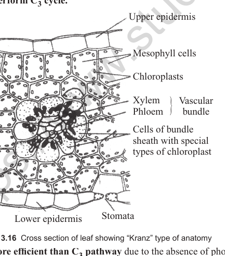
[Figure: Fig. 13.16 Cross section of leaf showing 'Kranz' type of anatomy. It shows upper epidermis, mesophyll cells, lower epidermis, chloroplasts, xylem, phloem, vascular bundle, cells of the bundle sheath with special types of chloroplast, and stomata.]
C4 pathway is more efficient than the C3 pathway due to the absence of photorespiration.
Phosphoenolpyruvate (PEP) is the carbon dioxide acceptor, present in the mesophyll cells. The reaction is catalysed by the enzyme phosphoenolpyruvate carboxylase (PEP carboxylase).
The carbon dioxide liberated is used for the Calvin cycle in the bundle sheath cells.
The first stable product is oxaloacetic acid (OAA), which is a 4-carbon compound.
OAA is converted into malic acid and transported to the bundle sheath cells, where it is decarboxylated into pyruvic acid. Bundle sheath cells are rich in RuBisCO.
Pyruvic acid is transported back to the mesophyll cell, where it is used for the regeneration of PEP, utilizing two molecules of ATP for each pyruvate.
It is an adaptive mechanism to avoid photorespiration. The decarboxylation of malic acid in the bundle sheath cells maintains a high concentration of carbon dioxide near RuBisCO, so that it functions as a carboxylase only and not as an oxygenase.
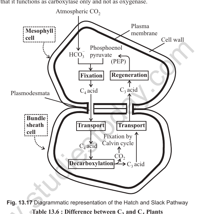
[Figure: Fig. 13.17 Diagrammatic representation of the Hatch and Slack pathway. It shows atmospheric CO2 entering the mesophyll cell, fixation by PEP to C4 acid (OAA), transport of C4 acid to the bundle sheath cell, decarboxylation releasing CO2 for the Calvin cycle, regeneration of PEP from C3 acid (pyruvate), and plasmodesmata connecting the cells. HCO3- is also shown.]
Table 13.6: Differences between C3 and C4 plants
| Characters | C3 plants | C4 plants |
|---|---|---|
| 1. Site of photosynthesis | Occurs in mesophyll cells. | Occurs in mesophyll and bundle sheath cells. |
| 2. Kranz anatomy | Absent. | Present. |
| 3. Primary CO2 acceptor | RuBP. | PEP. |
| 4. First stable product | 3-phosphoglyceric acid (3-PGA), a 3-carbon compound. | Oxaloacetic acid, a 4-carbon compound. |
| 5. Chloroplasts | Of only one type, i.e., granal. | Dimorphic, i.e., granal in the mesophyll cells and agranal in the bundle sheath cells. |
| 6. Photorespiration | Photorespiratory loss is high. | Photorespiration is absent or negligible. |
| 7. CO2 compensation point | High, i.e., 25-100 µl CO2 l-1. | Low, i.e., 0-10 µl CO2 l-1. |
| 8. Temperature optimum for photosynthesis | About 25°C. | About 35°C to 45°C. |
| 9. Efficiency and productivity | Photosynthetically less efficient and productivity is low, e.g., rice, wheat, bean, potato. | Photosynthetically more efficient and productivity is high, e.g., maize, sugarcane, Amaranthus. |
CRASSULACEAN ACID METABOLISM (CAM)
CAM plants are mostly succulent xerophytes, such as plants of the families Crassulaceae, Euphorbiaceae and Cactaceae. The stomata in these plants remain closed during the day, which helps to check transpiration, and open during the cool night for gaseous exchange.
These plants are characterised by the presence of a CAM pathway involving photosynthetic activity at night. This is a xerophytic adaptation, where stomata must remain closed during the day and open during the night to avoid water loss (scotoactive stomata).
They store CO2 during the night in the form of malic acid, in the presence of the enzyme PEP carboxylase. The CO2 stored during the night is used in the Calvin cycle during the day. Succulents re-fix CO2 released during respiration and use it during photosynthesis.
Formation of malic acid during the dark is called acidification, or phase I. Release of CO2 for actual photosynthesis during the day is called de-acidification, or phase II.
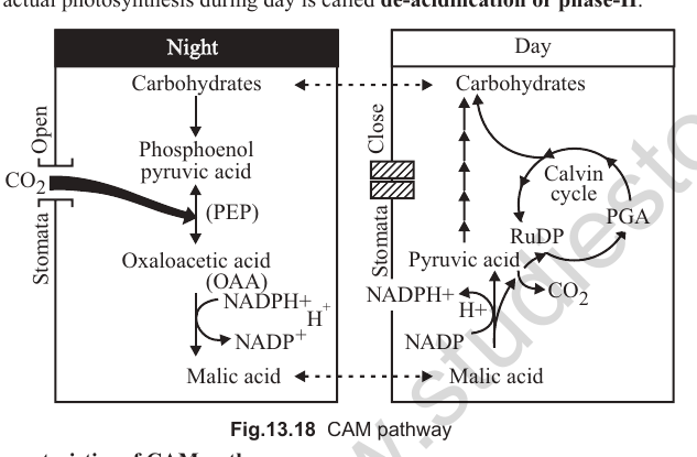
This diurnal change in acidity was first discovered in crassulacean plants, e.g., Bryophyllum. So it is called crassulacean acid metabolism.
[Figure: Fig. 13.18 CAM pathway. During the night, stomata are open, CO2 is fixed by PEP into oxaloacetic acid and then malic acid. During the day, stomata are closed, malic acid is decarboxylated to release CO2 for the Calvin cycle (PGA, RuBP, carbohydrates), with NADPH + H+ regenerated to NADP+.]
Characteristics of CAM pathway
• There is a decrease in pH during the night and an increase in pH during the day.
• CAM plants have enzymes of both the C3 and C4 cycles in mesophyll cells. This metabolism enables CAM plants to survive in xeric habitats. These plants also have the capability of fixing the CO2 lost in respiration.
• Malic acid is stored in the vacuoles during the night, and is decarboxylated to release CO2 during the day.
CO2 compensation point
In photosynthesis, CO2 is utilized in the presence of light to release O2, whereas in respiration, O2 is taken in and CO2 is released. If the light factor is saturating, there will be a certain CO2 concentration at which the rate of photosynthesis is just equal to the rate of respiration. At this point photosynthesis just compensates respiration, or apparent photosynthesis is nil. It is called the CO2 compensation point.
The CO2 compensation point is very low in C4 plants, i.e., 0 to 5 ppm, whereas a high CO2 compensation point is found in C3 plants, i.e., 25 to 100 ppm.
PHOTORESPIRATION
Photorespiration (C2 cycle) is a special type of respiration shown by many green plants when they are exposed to light. It was discovered by Decker and Tio (1959) in tobacco.
Photorespiration is a light-induced CO2 liberation from a C2 compound (glycolic acid) of the dark phase.
It is absent in C4 plants and present in C3 plants. In C4 plants, photorespiration does not occur, because they have a mechanism that increases the concentration of CO2 at the enzyme site. This takes place when the C4 acid from the mesophyll is broken down in the bundle sheath cells to release CO2, which increases the intracellular concentration of CO2. In turn, this ensures that RuBisCO functions as a carboxylase, minimising its oxygenase activity.
There is neither synthesis of sugars nor of ATP. Therefore, photorespiration is a wasteful process.
75% of the carbon lost by oxygenation of RuBP is recovered, and 25% is lost as the release of one molecule of CO2.
Photorespiration is the uptake of O2 and release of CO2 in light, and results from the biosynthesis of glycolate in chloroplasts and the subsequent metabolism of glycolic acid in the same leaf cell. The biochemical mechanism of photorespiration is also called glycolate metabolism.
Loss of energy occurs during this process. Photorespiration involves chloroplasts, peroxisomes and mitochondria.
RuBP carboxylase also catalyses another reaction which interferes with the successful functioning of the Calvin cycle.
RuBisCO is the most abundant enzyme in the world. Its active site can bind both CO2 and O2, hence the name RuBisCO. It has a much greater affinity for CO2 than for O2, so the binding is competitive.
The relative concentrations of O2 and CO2 determine which of the two will bind to the enzyme.
In C3 plants, some O2 does bind to RuBisCO, and hence CO2 fixation is decreased. RuBP, instead of being converted into 2 molecules of PGA, binds with O2 to form one molecule of 3-PGA and one 2-carbon phosphoglycolate, in the pathway called photorespiration.
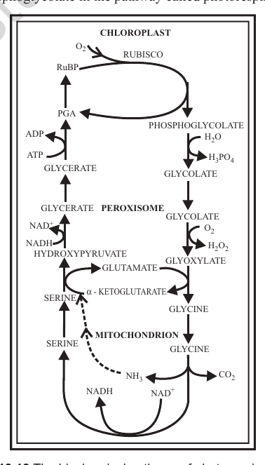
[Figure: Fig. 13.19 The biochemical pathway of photorespiration. It shows chloroplast, RuBisCO, RuBP, O2, PGA, phosphoglycolate, glycerate, glycolate, peroxisome, glyoxylate, glycine, serine, hydroxypyruvate, glutamate, alpha-ketoglutarate, H2O2, mitochondrion, NH3, CO2, NADH and NAD+, ATP, ADP and H3PO4.]
Check Point
1. Name the compound which usually passes into peroxisomes from the chloroplast to form glycolate.
2. Name the cell organelles involved in photorespiration.
3. Give 2 examples of photorespiring plants.
4. Where are the pigments located in photosynthetic bacteria?
5. Give two examples each of bacterial and chemosynthetic photosynthesis.
Biochemical mechanism of photorespiration
(1) Ribulose-1,5-bisphosphate + O2 → 2-phosphoglycolic acid + 3-phosphoglyceric acid (RuBisCO oxygenase)
(2) 2-phosphoglycolic acid + H2O → glycolic acid + phosphoric acid (phosphatase)
(3) Glycolic acid + O2 → glyoxylic acid + H2O2 (glycolic acid oxidase); 2H2O2 → 2H2O + O2 (catalase)
(4) Glyoxylic acid + glutamic acid → glycine + alpha-ketoglutaric acid (glutamate-glyoxylate transaminase)
(5) 2 glycine + H2O + NAD+ → serine + CO2↑ + NH3 + NADH
(6) Serine + glyoxylic acid → glycine + hydroxypyruvic acid
(7) Hydroxypyruvic acid → glyceric acid
(8) Glyceric acid + ATP → 3-phosphoglyceric acid + ADP + phosphate
Importance of photorespiration
Photorespiration is quite different from respiration, as no ATP or NADH are produced.
Photorespiration is closely related to the CO2 compensation point, and occurs only in plants which have a high CO2 compensation point, such as C3 plants.
Photorespiration generally occurs in temperate plants. Some photorespiring plants are rice, bean, wheat and barley.
Inhibitors of glycolic acid oxidase, such as hydroxysulphonates, inhibit the process of photorespiration. Unlike usual mitochondrial respiration, the oxidation of glycolate is not coupled with the formation of ATP molecules. Photorespiration (C2 cycle) is enhanced by bright light, high temperature, high oxygen and low CO2 concentration.
FACTORS AFFECTING PHOTOSYNTHESIS
Photosynthesis is influenced by both external and internal (plant) factors.
Blackman's law of limiting factors (1905): Blackman's law of limiting factors is a modification of Liebig's law of minimum, which states that the rate of a process controlled by several factors is only as rapid as the slowest factor permits. The law states: if a chemical process is affected by more than one factor, its rate will be determined by the factor nearest to its minimal value. It is the factor which directly affects the process if its quantity is changed.
This means that at a given time, only the factor that is limiting among all will determine the rate of photosynthesis. For example, despite the presence of a green leaf and optimal light and CO2 conditions, the plant may not photosynthesize if the temperature is very low. This leaf, given the optimal temperature, will start photosynthesizing.
External factors
Light: Both the quality and intensity of light influence photosynthesis. Light of wavelength between 400 nm and 700 nm is most effective, and this light is called photosynthetically active radiation (PAR).
As the intensity of light increases, the rate of photosynthesis also increases. But at higher light intensity, the rate of photosynthesis decreases, because (i) other factors required for photosynthesis become limiting, and (ii) destruction of chlorophyll occurs.
At low light intensity, the rate of photosynthesis is reduced. There is a linear relationship between incident light and CO2 fixation rates at low light intensities. At higher light intensities, the rate gradually stops showing further increase. Light saturation occurs at about 10% of full sunlight. Hence, except for plants in shade or in dense forests, light is rarely a limiting factor in nature.
Best photosynthesis takes place in red light and in blue light, and in green light it is nil.
[Figure: Fig. 13.20 Effect of light intensity on the rate of photosynthesis]
Temperature: Temperature is also a limiting factor under field conditions, particularly on cool days. The influence of temperature on photosynthesis depends on both light intensity and the availability of carbon dioxide.
The photochemical reactions in the thylakoid membrane are less affected by temperature. But the enzymatic carbon reactions of the C3 cycle or C4 cycle in the stroma are adversely influenced.
C4 plants respond to higher temperatures and show a higher rate of photosynthesis, while C3 plants have a much lower temperature optimum. The temperature optimum for photosynthesis of different plants also depends on the habitat they are adapted to. Tropical plants have a higher temperature optimum than plants adapted to temperate climates.
Overall, the process of photosynthesis is sensitive to high temperature, because:
• at higher temperatures enzymes become inactive.
• affinity of the enzyme RuBisCO for CO2 is also reduced at higher temperature.
• low temperature also inactivates enzymes.
Oxygen: An excess of O2 may become inhibitory to the process. An enhanced supply of O2 increases the rate of respiration, while simultaneously decreasing the rate of photosynthesis, by using common intermediate substances. The concentration of oxygen in the atmosphere is about 21% by volume and seldom fluctuates. O2 is not a limiting factor of photosynthesis. An increase in oxygen concentration decreases photosynthesis, and this phenomenon is called the Warburg effect.
This is due to the competitive inhibition of RuBP carboxylase by increased O2 levels, i.e., O2 competes with CO2 for the active sites of the RuBP carboxylase enzyme. The explanation of this problem lies in the phenomenon of photorespiration. If the amount of oxygen in the atmosphere decreases, photosynthesis will increase in the C3 cycle and show no change in the C4 cycle.
Pollutants and inhibitors: The oxides of nitrogen and hydrocarbons present in smoke react to form peroxyacetyl nitrate (PAN) and ozone. PAN is known to inhibit the Hill reaction. Diquat and paraquat (commonly called viologens) block the transfer of electrons between Q and PQ in PS II. Other inhibitors of photosynthesis are monuron or CMU (chlorophenyl dimethyl urea), diuron or DCMU (dichlorophenyl dimethyl urea), bromacil and atrazine, which have the same mechanism of action as viologens.
At low light intensities, potassium cyanide appears to have no inhibiting effect on photosynthesis.
Minerals: The presence of Mn++ and Cl- is essential for the smooth operation of light reactions (photolysis of water and evolution of oxygen). Mg++, Cu++ and Fe++ ions are important for the synthesis of chlorophyll.
Carbon dioxide concentration: Carbon dioxide is the major limiting factor for photosynthesis. The concentration of CO2 is very low in the atmosphere (between 0.03 and 0.04 per cent). An increase in concentration up to 0.05 per cent can cause an increase in CO2 fixation rates; beyond this level, the effects can become damaging over longer periods.
The C3 and C4 plants respond differently to CO2 concentrations. At low light conditions, neither group responds to high CO2 conditions. At high light intensities, both C3 and C4 plants show an increase in the rates of photosynthesis. C4 plants show saturation at about 360 µl/L, while C3 plants respond to increased CO2 concentration, and saturation is seen only beyond 450 µl/L. Thus, current atmospheric CO2 levels are limiting to C3 plants.
The fact that C3 plants respond to higher CO2 concentrations with increased rates of photosynthesis, leading to higher productivity, has been used for some greenhouse crops such as tomatoes and bell peppers. They are grown in a CO2-enriched atmosphere, which leads to higher yields.
Soil water: Even though water is one of the reactants in the light reaction, the effect of water as a factor is more through its effect on the plant, rather than directly on photosynthesis. Water stress causes the stomata to close, hence reducing CO2 availability. Water stress also makes leaves wilt, thus reducing the surface area of the leaves and their metabolic activity.
Internal factors
Various leaf factors, like leaf age, leaf angle and leaf orientation, affect the rate of photosynthesis. If the leaf undergoes senescence, loss of chlorophyll occurs, which causes deactivation of photosynthetic enzymes and reduces the rate of photosynthesis.
The final product of photosynthesis is sugar, and its accumulation in the cells slows down the process of photosynthesis.
The thickness of the cuticle and epidermis of the leaf, the size and distribution of intercellular spaces, the distribution of stomata, and the development of chlorenchyma and other tissues also affect the rate of photosynthesis.
Connecting Concepts: Factors which increase the rate of photosynthesis are: External factors: high light intensity, red and blue light, intermittent light, optimum temperature (15 to 25°C for C3 plants and 30 to 40°C for C4 plants), higher CO2 concentration, water sufficiency, and sufficient mineral supply (Mn, Mg, Fe). Internal factors: presence of chlorophyll, more palisade parenchyma, maturity of leaf, and rapid translocation of carbohydrates.
Factors which decrease the rate of photosynthesis are: External factors: low intensity of light, continuous supply of light for the same duration, green light, very low and very high temperature, CO2 concentration more than 1%, high oxygen concentration, and water deficiency. Internal factors: absence of chlorophyll, intensive palisade parenchyma, accumulation of end products, and immature and old leaves.
BACTERIAL PHOTOSYNTHESIS
Like green plants, some purple and green sulphur bacteria are capable of synthesizing their organic food in the presence of light and in the absence of O2. This is known as bacterial photosynthesis.
Van Niel was the first to point out these similarities. Oxygen is not liberated in bacteria during photosynthesis. Their photosynthesis is non-oxygenic, because bacteria use H2S in place of water (H2O) as the hydrogen donor. Photosynthetic bacteria are anaerobic. Only one type of pigment system (PS I) is found in bacteria, except in cyanobacteria, which possess both PS I and PS II.
Bacteria have two types of photosynthetic pigments, bacteriochlorophyll and bacterioviridin. Bacteriochlorophyll differs from chlorophyll a in having one pyrrole ring with two hydrogen atoms.
The photosynthetic bacteria fall into three categories:
(1) Green sulphur bacteria: They contain chlorobium chlorophyll, which absorbs 720 to 750 nm (far red light). e.g., Chlorobium.
(2) Purple sulphur bacteria: e.g., Chromatium.
(3) Purple non-sulphur bacteria: e.g., Rhodospirillum, Rhodopseudomonas.
Characteristics of bacterial photosynthesis
(1) No definite chloroplasts, but they contain simple structures having pigments called chromatophores (term coined by Schmitz).
(2) Use longer wavelengths of light (720 to 950 nm).
(3) No utilization of H2O (they use H2S or other reduced organic and inorganic substances).
(4) No evolution of O2.
(5) Photoreductant is NADH2 (not NADPH2).
(6) Only one photoact, and hence one pigment system and one reaction centre, i.e., P890.
(7) Contain chlorobium chlorophyll or bacteriochlorophyll.
(8) Cyclic photophosphorylation is dominant.
(9) It occurs in the presence of light and in the absence of O2.
CHEMOSYNTHESIS
Some forms of bacteria obtain energy by chemosynthesis. This is the process of carbohydrate formation in which organisms use chemical reactions to obtain energy from inorganic compounds.
Such chemoautotrophic bacteria do not require light. They synthesize all their organic cell requirements from CO2, H2O and salts, at the expense of the oxidation of inorganic substances like H2, NO3-, SO4 or carbonate.
Some examples of chemosynthesis are:
(1) Nitrifying bacteria: e.g., Nitrosomonas, Nitrosococcus, Nitrobacter, etc.
(2) Sulphur bacteria: e.g., Beggiatoa, Thiothrix and Thiobacillus.
(3) Iron bacteria: e.g., Ferrobacillus, Leptothrix and Cladothrix.
(4) Hydrogen bacteria: e.g., Bacillus pantotrophus.
(5) Carbon bacteria: e.g., Carboxydomonas, Bacillus oligocarbophilus.

NEET NEET Biology Study Material: Photosynthesis
Comprehensive Study Resources for Photosynthesis
Review targeted study resources for Photosynthesis tailored for NEET learners. Utilizing these structured notes and quick-revision tools ensures complete alignment with current NEET evaluation standards.
Understanding Marking Schemes
Built using official NCERT guidelines for NEET Biology, these materials provide reliable academic support. Integrating past examination questions and step-by-step solutions helps students understand official NEET grading criteria.
Complete Revision for Biology
Maximize your grade potential by utilizing NEET Biology practice papers in tandem with these structured notes. All printable assignments and interactive mock tests on our platform are updated for the 2026 session and available free of charge.
FAQs
Our advanced study package for Photosynthesis includes detailed concepts, diagrams, Mind Maps, and explanation of complex topics to ensure NEET students learn as per syllabus for 2026 exams.
The Mind Maps provided for Photosynthesis act as visual anchors which will help faster recall during high-pressure exams.
Yes, teachers use our NEET Biology resources for lesson planning as they are in simple language and have lot of solved examples.
Yes, You can download the complete, mobile-friendly PDF of the Biology Photosynthesis advanced resources for free.
Yes, our subject matter experts have updated the Photosynthesis material to align with the rationalized NCERT textbooks and have removed deleted topics and added new competency-based questions.