

Download NEET NEET Biology Useful Resources
Explore structured advanced study materials through the NEET Biology Animal Tissues Notes. Tailored for NEET learners, utilizing these Biology resources ensures thorough preparation and strengthens foundational knowledge before final NEET evaluations.
Access NEET Study Guides and Summaries
Access the complete useful resource PDF for NEET Biology below. Regular practice with these targeted academic notes builds familiarity with complex topics and helps secure higher marks in final school evaluations.
These NEET Biology notes on Animal Tissues (Histology) cover the Class 11 chapter on the structure and types of animal tissues, including epithelial, connective, nervous and muscular tissue, along with the histology of organs and the study of cockroach, earthworm and frog as type animals.
Chapter 7: Animal Tissues (Histology) and Cockroach, Earthworm and Frog
Part-I: Animal Tissues (Histology)
Important Terms and Definitions
1. Histology
2. Clasmatocytes
3. Perineurium
4. Mast cells
5. Polycythemia
6. Crenation
7. Sesamoid bone
8. Eosinophilia
9. Neurilemma
10. Nissl granules
11. Glisson's capsule
12. Trichology
Differences To Know
1. Apocrine and holocrine glands
2. Ciliated epithelium and brush bordered epithelium
3. Ligaments and tendons
4. Investing bone and replacing bone
5. Haversian canal and Volkmann's canal
6. Sickle cell anaemia and thalassaemia
7. Stratified and pseudostratified epithelium
8. Granulocytes and agranulocytes
9. Sensory nerves and motor nerves
10. Red pulp and white pulp
11. Integument and skin
Tissues To Know
1. Squamous epithelium
2. Areolar tissue
3. Transitional epithelium
4. Fluid connective tissue
5. Adipose tissue
Structures To Know
1. Sudoriferous glands
2. Ruffini's end bulbs
3. Kupffer cells
4. Pacinian corpuscles
5. Chromatophores
Introduction to Histology
1. The study of tissues is called histology.
2. Marcello Malpighi is the founder of histology.
3. The term 'tissue' was given by Bichat.
4. The term 'histology' was given by Mayer.
Tissue
A tissue is a group of similar cells (of similar embryonic origin) along with intercellular substances, which performs a specific function.
Types of Tissue
There are four major types of tissue:
1. Epithelial tissue
2. Connective tissue
3. Nervous or neural tissue
4. Muscular tissue
At a Glance (Chapter Outline)
- Epithelial tissue: simple epithelium, compound epithelium, glands
- Connective tissue: proper connective tissue, skeletal connective tissue, fluid connective tissue
- Nervous tissue
- Muscular tissue
- Organ histology: liver, spleen, pancreas, gut (alimentary canal), integument (mammalian skin, frog's skin)
- Cockroach: external features, mouth parts, digestive system, excretory system, blood vascular system, respiration, nervous system and sensory organs, reproductive system, development
- Earthworm: external features, body wall and coelom, locomotion, nutrition, respiration, excretory system, blood vascular system, nervous system, reproductive system
- Frog: external morphology, internal morphology and physiology, development and metamorphosis, biological importance
1. Epithelial Tissue
1. Epithelial tissue is ectodermal, mesodermal or endodermal in origin.
2. Its cells are compactly arranged. The intercellular material (matrix) is either absent or negligible.
3. It covers the body surface or lines the body cavity. One surface of this tissue is generally free.
4. The cells rest upon a non-cellular basement membrane, which is secreted by epithelial cells.
5. It is not nourished by blood, so it is non-vascularised.
6. Structures such as cilia, flagella and desmosomes (cell junctions) are usually associated with this tissue.
7. The term 'epithelium' was introduced by Ruysch.
Types of Epithelial Tissue
1. Simple epithelium
2. Compound epithelium: (i) transitional epithelium, (ii) stratified epithelium
Simple Epithelium
Simple epithelium is one cell thick, and can be of three basic types.
(a) Squamous Epithelium
1. It is also called pavement epithelium or tessellated epithelium.
2. The cells resemble floor tiles. The length and breadth of the cells are greater than their thickness.
3. It is generally present in places where diffusion occurs. Examples: alveolar epithelium, inner lining of Bowman's capsule, capillary epithelium (endothelium), coelomic epithelium (peritoneum), pleural membrane (covering of lungs) and eye-lens epithelium.
(b) Cuboidal Epithelium
1. The cells are cubical, with similar length, breadth and height (thickness). Examples: germinal epithelium, follicular epithelium of thyroid gland, lining of DCT, ascending limb (thick) of Henle's loop and collecting tubule of kidney.
(c) Columnar Epithelium
1. The height (thickness) of the cells is greater than their length or breadth.
2. This epithelial lining is generally glandular or secretory. Examples: lining of acini (exocrine part) of pancreas, lining of gall bladder, stratum germinativum of skin, and lining of stomach and intestine.
Specialised Epithelium
(i) Pseudostratified Epithelium
1. The epithelium is single cell thick but appears to be double layered, due to the different heights of the cells and the position of their nuclei.
2. Ciliated epithelium: the majority of cells are ciliated, whereas certain cells in between are non-ciliated. The non-ciliated cells secrete mucus. Example: epithelium of trachea and bronchi.
(ii) Sensory epithelium: these cells are innervated (have neurons) and receive various types of stimuli. Examples: retina, Schneiderian membrane (olfactory) and lining of the membranous labyrinth.
(iii) Brush-bordered epithelium: the cells contain microvilli, which increase the surface area for absorption. The microvilli may be short and regular (example: lining of ileum and PCT) or long and irregular, called stereocilia (example: epididymis).
(iv) Ciliated epithelium: all surface cells are ciliated. The cilia help in movement of sperms in the vas deferens, oviduct or fallopian tube, or in movement of mucus from the trachea into the pharynx.
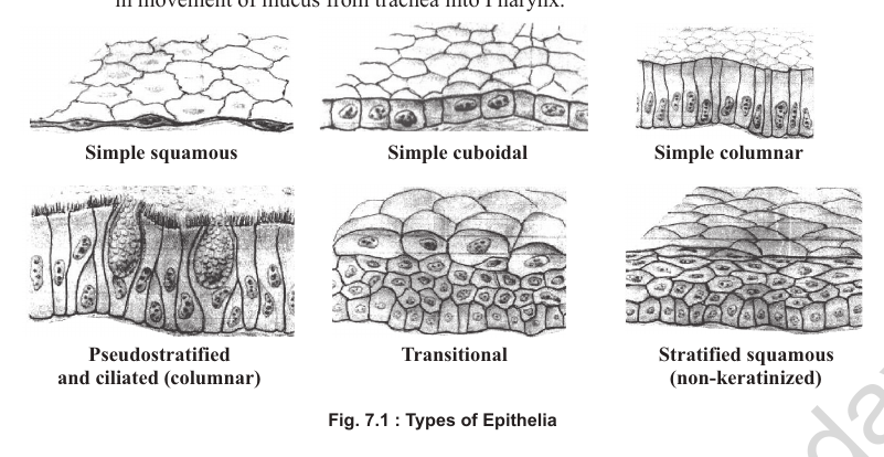
[Figure: Fig. 7.1 Types of epithelia, showing simple squamous, simple cuboidal, simple columnar, pseudostratified and ciliated (columnar), transitional, and stratified squamous (non-keratinised) epithelium]
Compound Epithelium
I. Transitional Epithelium
1. It is 2 to 6 cells thick.
2. Basement membrane is absent.
3. It occurs in areas where stretching is required. Examples: epithelium of urinary bladder, ureter and pelvis.
II. Stratified Epithelium
1. This epithelial layer is more than 6 cells thick.
2. Basement membrane is present but single.
3. The name of the epithelium is according to the cells of the top layer.
(a) Stratified Squamous Epithelium
1. It is the most common stratified epithelium.
2. The cells of the top layer are squamous.
3. It occurs in regions where protection is required, or where there is sufficient wear and tear of the tissue. Examples: buccal cavity (cheek epithelium), skin, pharynx, oesophagus, vaginal epithelium, urethra, conjunctiva and cornea.
(b) Stratified Cuboidal Epithelium
It is present in the ducts of mammary glands and sweat glands.
(c) Stratified Columnar Epithelium
It mainly occurs in embryonic tissue and is poorly developed in adults.
Check Point
Answer the following:
1. (i) Who is the founder of histology? Marcello Malpighi.
(ii) Which epithelium does not have a basement membrane? Transitional epithelium.
(iii) What type of epithelium do the alveoli have? Simple squamous epithelium.
(iv) Which type of glands release their secretion outside the cell without any cell injury? Merocrine glands (eccrine glands).
(v) From which germ layer or layers do the exocrine glands arise? All three germ layers (ectoderm, mesoderm and endoderm), since glands may develop from any of them.
(vi) Which type of epithelium is present in the buccal cavity? Stratified squamous epithelium.
(vii) Which epithelium lines the oviduct? Ciliated columnar epithelium.
(viii) Which part of the respiratory tract has pseudostratified epithelium? Trachea and bronchi.
Glands
1. Glands are epithelial in origin and may therefore develop from any of the three germ layers.
2. They can be exocrine or endocrine. They can be unicellular (example: goblet cells) or multicellular.
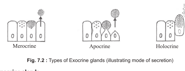
3. Exocrine glands are of three types on the basis of their secretion.
1. Merocrine Glands
1. Also known as eccrine glands.
2. The secretion from these glands is watery.
3. The secretion diffuses out of the cells without cell injury. Examples: salivary glands and sweat glands.
2. Apocrine Glands
1. The secretion in these glands accumulates in the apical part of the cells, and this part is later pinched off. Due to this, the size of the cells is reduced.
2. The cells grow again and accumulate the secretory product. Examples: mammary glands, and sweat glands of the armpit and thigh pit, which produce a brownish pigment with the sweat.
3. The mammary glands are considered to be a modification of apocrine sweat glands.
3. Holocrine Glands
1. The secretion from the cells of these glands is oily.
2. The secretion comes out only after the cells disintegrate. Example: sebaceous glands.
[Figure: Fig. 7.2 Types of exocrine glands, illustrating the mode of secretion: merocrine, apocrine and holocrine]
Cell Junctions
The cells of epithelial and other tissues are held together by various types of cell junctions, which can be grouped into three main categories.
(I) Adhesive Junctions
1. The cells of epithelial tissue, and even cardiac muscle cells, are difficult to separate due to adhesive junctions.
2. These are of two types: (a) adherens junctions and (b) desmosomes.
(a) Adherens Junctions (Zonulae adherens)
1. These are common in epithelial lining of the intestine, where they form a 'belt' encircling the apical portion of cells and binding them to surrounding cells.
2. They are formed by calcium-dependent linkages of cadherin (protein) molecules, which cement the cells and provide a pathway for signals from the cell exterior to the cytoplasm.
(b) Desmosomes (Maculae adherens)
1. These are disc-shaped adhesive junctions found in a variety of tissues, such as skin, uterine cervix and cardiac muscle, which are subjected to mechanical stress.
2. Desmosomes also contain cadherins for binding the cells.
(II) Tight Junctions (Zonulae occludens)
1. In simple or single-layered epithelium the cells are tightly adhered to one another to form a thin cellular sheet.
2. Such junctions seal the extracellular space and block the diffusion of solutes through the paracellular pathway. The adjoining membranes make contact at certain points rather than being fused over a large surface area.
3. Tight junctions act as a barrier to the free diffusion of water and solutes from the extracellular compartment on one side of an epithelial sheet to the other side. They also act as 'fences' that block the diffusion of integral proteins.
4. The proteins that form the structural component of tight junctions are claudin and occludin.
5. Tight junctions make the skin impermeable to water.
6. The junctions between endothelial cells of blood capillaries form the blood-brain barrier, which prevents substances from passing from blood into the brain. This barrier also prevents access of many drugs to the CNS.
(III) Gap Junctions
1. These are specialised for intercellular communication and are composed of the integral membrane protein connexin, which forms molecular pipelines between adjacent cells.
2. Gap junction channels can be opened or closed like gated channels.
3. Stimulation of cardiac or smooth muscle cells involves gap junctions. The impulse from the SAN of the heart flows from one cardiac muscle cell to another through gap junctions, causing the cells to contract in synchrony. Similar gap junctions are responsible for the peristaltic wave in smooth muscles.
4. Gap junctions allow the passage of ATP, cyclic AMP, coenzymes, sugars, phosphates and amino acids.
Plasmodesmata
1. Unlike animal cells, plant cells are separated from one another by a cell wall. Hence, plant cells lack the cell adhesion molecules or specialised cell junctions found in animal cells and tissues.
2. Most plant cells are, however, connected to one another by plasmodesmata (singular: plasmodesma). These are cytoplasmic channels that pass through the cell walls of adjacent cells. They contain a desmotubule derived from the smooth ER of the two cells.
3. Like gap junctions between animal cells, plasmodesmata also serve as sites for cell-to-cell communication.
4. Unlike gap junctions, whose channels have a fixed opening, the plasmodesmata pore can dilate. Plant viruses spread from one cell to another through plasmodesmata by encoding a 'movement protein' that interacts with the wall of the plasmodesmata to increase the diameter of the pore.
2. Connective Tissue
1. It is mesodermal in origin.
2. It binds and supports body parts.
3. The cells are loosely arranged, i.e. the intercellular matrix is well developed.
4. Basement membrane is absent.
5. It is nourished by blood or lymph.
[Figure: Classification of connective tissue. Connective tissue is divided into proper connective tissue, which includes loose connective tissue (areolar, adipose, mucous) and dense connective tissue (dense regular, dense irregular), and specialised connective tissue, which includes skeletal connective tissue (cartilage, bone) and fluid connective tissue (blood, lymph)]
1. Proper Connective Tissue
A. Loose Connective Tissue
(a) Areolar Tissue
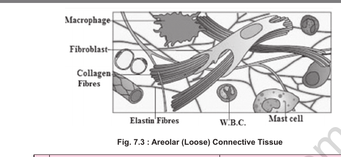
1. It consists of three prominent types of cells: fibroblasts, mast cells and macrophages.
2. Mast cells secrete heparin (an anticoagulant), histamine and serotonin (5-hydroxytryptamine, 5-HT). A major amount of heparin, however, is secreted by the liver.
3. Histamine is a vasodilator, lowers blood pressure, and is involved in allergic reactions.
4. Serotonin is a vasoconstrictor and increases blood pressure.
5. Macrophages, also known as clasmatocytes, are phagocytic cells. They are modified monocytes (WBCs).
6. Fibroblasts produce white and yellow fibres. These cells are the largest cells of areolar tissue.
[Figure: Fig. 7.3 Areolar (loose) connective tissue]
Comparison of White and Yellow Fibres
| White fibres | Yellow fibres |
|---|---|
| Made of collagen protein | Made of elastin protein |
| Unbranched fibres | Branched fibres |
| Present in bundles | Solitary fibres |
| Less resistant to chemicals and produce gelatin on heating | More resistant to chemicals |
| Provide toughness | Provide elasticity |
(b) Adipose Tissue
1. It contains closely packed cells (adipocytes) containing fat globules. Adipocytes are modified fibroblasts.
2. The intercellular matrix is negligible.
3. Adipocytes are of two types: (1) monolocular and (2) multilocular or polylocular.
4. Monolocular cells store white fat, which provides less energy (9 kcal/g).
5. Multilocular cells contain brown fat, which provides 20 times more energy than white fat. Brown fat is present in hibernating animals and newly born human infants.
6. Adipose tissue insulates the body against heat, cushions body organs as a shock absorber, and is a source of reserve food material while occupying minimum space. Examples: the hump of camels, the tail of merino sheep, the blubber of whales and elephants, and the paniculus adiposus (thin subcutaneous layer) in humans.
[Figure: Fig. 7.4 (A) Adipocyte, showing nucleus, cytoplasm and fat globule; (B) Adipose tissue, showing blood vessels and monolocular adipocytes]
(c) Mucous Connective Tissue
1. Also called Wharton's jelly, this tissue has a matrix that is jelly-like (gelatinous), slippery (due to mucopolysaccharides) and semi-solid.
2. Examples: cock's comb, vitreous humour and umbilical cord.
B. Dense (Fibrous) Connective Tissue
(a) Dense Regular Connective Tissue
It contains a large number of protein fibres, and is of the following types:
(i) White Fibrous Connective Tissue
1. It contains collagen fibres and provides strength or toughness to the tissue.
2. Examples: dura mater (outer covering of the brain), scleroid coat of the eye, renal capsule of the kidney, periosteum (covering of bone) and perichondrium (covering of cartilage).
3. The dermis, from which leather is obtained, contains abundant collagen fibres.
4. Tendons, which connect muscles to bones, consist purely of white (collagen) fibres.
(ii) Yellow Fibrous Connective Tissue
1. It contains elastin fibres and provides elasticity to the tissue. Examples: walls of arteries and lungs.
2. Ligaments contain both yellow (mainly) and white fibres and provide bone-to-bone attachment.
3. A sprain is caused by excessive pulling of ligaments.
(b) Dense Irregular Connective Tissue
Fibroblasts are present, and the fibres (mostly white or collagen) are oriented irregularly. Example: dermis part of the skin.
2. Specialised Connective Tissue
A. Skeletal Connective Tissue
Bone and Cartilage
1. This tissue includes bones and cartilages.
2. The study of bones is called osteology, and the study of cartilage is called chondrology.
Comparison of Bone and Cartilage
| Feature | Bone | Cartilage |
|---|---|---|
| Outer covering | White fibrous connective tissue called periosteum | White fibrous connective tissue called perichondrium |
| Forming cells | Osteoblasts | Chondroblasts |
| Protein | Ossein | Chondrin (the sugar in cartilage is chondroitin sulphate) |
| Cells | Osteocytes are solitary | Chondrocytes occur in groups of 2 or 3 |
| Arrangement | Osteocytes are arranged in lamellae | Chondrocytes are scattered in the matrix |
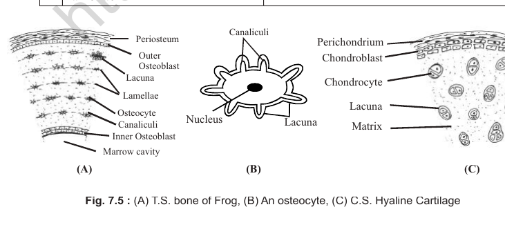
[Figure: Fig. 7.5 (A) T.S. bone of frog, showing periosteum, outer and inner osteoblasts, lacuna, lamellae, osteocyte, canaliculi, marrow cavity; (B) An osteocyte, showing nucleus, cytoplasm and lacuna; (C) C.S. hyaline cartilage, showing perichondrium, chondroblast, chondrocyte, lacuna and matrix]
Bone
Composition of Bone
| Component | Percentage |
|---|---|
| Inorganic material (salts of calcium and magnesium etc.) | 45% |
| Organic material (ossein and collagen etc.) | 35% |
| Water | 20% |
1. Calcium phosphate, in the form of hydroxyapatite, is the largest mineral (salt) and makes up about 80% of the inorganic material. Calcium carbonate is only 12 to 13%.
2. If a bone is kept in dilute HCl for a few days, it is decalcified and only organic material is left. Such a bone becomes flexible and elastic.
3. If a bone is heated to redness, the organic matter burns and only inorganic matter (salt) is left. Such a bone becomes brittle.
4. If a bone is kept in KOH, there is no effect on the organic or inorganic matter. Attached muscles, however, are removed.
Types of Bone
1. Replacing bone (cartilaginous bone): ossification (deposition of bony material) occurs in cartilage, and the cartilage is replaced by bone. Example: most bones of the human and frog body.
2. Investing, dermal or membranous bone: ossification occurs directly in connective tissue, and the bone forms without the development of cartilage. Example: a few bones of the cranium.
3. Sesamoid bone: ossification occurs in a ligament or tendon (both fibrous connective tissue). Example: patella (knee cap), which is formed by ossification of a tendon.
Structure of a Long Bone
1. Each long bone has two parts: (1) epiphysis (articulating end) and (2) diaphysis (shaft).
2. The diaphysis in the centre contains a marrow canal with red or yellow bone marrow.
3. The bone around the marrow canal is compact, whereas at the epiphysis it is spongy, with trabeculae.
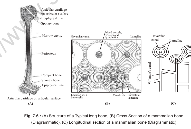
[Figure: Fig. 7.6 (A) Structure of a typical long bone, showing articular cartilage on the articular surface, epiphyseal line, spongy bone, marrow cavity, periosteum and compact bone; (B) Cross section of a mammalian bone (diagrammatic), showing Haversian canal, lamellae, lacunae with bone cells, canaliculi, interstitial lamellae and Volkmann's canal; (C) Longitudinal section of a mammalian bone (diagrammatic), showing Haversian canal, Volkmann's canal, blood vessels, vessels and lymphatics]
Haversian System
1. The compact bone in mammals consists of a large number of Haversian systems. The Haversian system (osteon) is the unit of mammalian bone.
2. It consists of a Haversian canal (longitudinal), circular lamellae, lacunae with osteocytes, and Volkmann's canal, which connects the Haversian canal transversely or obliquely.
3. In non-mammals the Haversian system is lacking, and the unit of bone is also called an osteon. In birds, bones are spongy and pneumatic, without a marrow canal.
Cartilage
1. The matrix of cartilage contains proteoglycans and glycosaminoglycans, including hyaluronic acid and chondroitin sulphate (a sugar).
2. Cartilage is of three types.
1. Hyaline Cartilage
1. It is semitransparent or slightly bluish.
2. Its perichondrium is prominent.
3. Its fibres are not visible. Examples: thyroid, cricoid and arytenoid cartilages of the larynx, hyoid apparatus of frog, endoskeleton of Elasmobranchii and tadpole, tracheal rings, and epiphyseal (articular) cartilage.
4. In frog, deposition of calcium salts in hyaline cartilage forms calcified cartilage. Examples in frog: pubis and suprascapula.
2. Elastic Cartilage
1. It contains yellow fibres.
2. It never forms calcified cartilage.
3. It is elastic in nature. Examples: pinna, epiglottis, tip of the nose and Eustachian tube.
3. Fibrous Cartilage
1. It is the toughest cartilage.
2. The perichondrium is present.
3. Both white and yellow fibres are well developed. Examples: intervertebral discs of mammalian vertebrae, and pubic symphysis in the pelvic girdle. The relaxin hormone softens the pubic symphysis to help in easy birth.
4. The central and softer part of each intervertebral disc is called the nucleus pulposus.
B. Fluid Connective Tissue
This includes blood and lymph.
(a) Blood
1. The study of blood is called haematology.
2. Blood is about 8% of body weight (about 4 litres in a person of 50 kg).
3. Osmotic pressure is equivalent to 7 to 8 atmospheres. pH is 7.4 (slightly alkaline).
4. Specific gravity is 1.003 (slightly heavier than water).
5. Viscosity is 3 to 5 times that of water.
6. Blood consists of (i) plasma and (ii) formed elements.
(i) Plasma
1. Plasma makes up 55% of blood. It consists of about 90% water, 7 to 8% proteins, and 2 to 3% salts, digestive and excretory products etc.
2. The main plasma proteins are albumin, globulin and fibrinogen.
3. Albumin is the most abundant plasma protein (3 to 4%) but has the lowest molecular weight. It maintains the colloidal osmotic pressure of blood.
4. Globulin can be alpha, beta or gamma type, and makes up 2 to 3% of plasma proteins. Alpha and beta globulins bind hormones for transport. Gamma globulins form antibodies.
5. Fibrinogen is about 0.3% and helps in blood clotting.
6. The remaining 2 to 3% contains salts, hormones, digestive end products (amino acids, fatty acids, glycerol etc.), and excretory products such as urea, uric acid and hippuric acid. Bilirubin (a bile pigment) gives plasma its yellow colour.
Normal Values of Some Plasma Components
1. Glucose: about 100 mg per 100 ml.
2. Cholesterol: 120 to 220 mg per 100 ml.
3. Bilirubin: about 0.5 mg per 100 ml.
4. Urea: 20 to 30 mg per 100 ml.
[Figure: Blood composition. Blood divides into plasma (55%) and formed elements (45%), and the formed elements are RBCs, WBCs and platelets]
(ii) Blood Elements (Formed Elements)
1. They constitute 45% of blood and consist of RBCs, WBCs and platelets.
1. Red Blood Corpuscles (RBCs) or Erythrocytes
1. Number of RBCs in males is 5.0 to 5.5 million per mm3; in females it is 4.5 to 5.0 million per mm3.
2. An increase in the number of RBCs is called polycythaemia. It occurs at higher altitudes.
3. A decrease in the number of RBCs or amount of haemoglobin is called erythropenia or anaemia.
4. Erythropoiesis, the production of RBCs, occurs in the yolk sac (in early life), the liver (in middle foetal life) and the bone marrow (in the last stages of foetal development). These are therefore called erythropoietic organs.
5. In early childhood, RBCs are produced in the bone marrow of all bones. At about the age of 20 years, they are produced only in the sternum, ribs, vertebrae and ilium of the hip girdle. Longer bones now serve mainly for support, as they contain inactive yellow (fatty) bone marrow.
6. Erythropoiesis is stimulated by the hormone erythropoietin from the kidney, and by vitamins (folic acid and B12) of the B-complex.
7. The mother cells of RBCs are nucleated erythroblasts.
8. The sequence of development of RBCs is as follows: erythroblast (nucleated, mother cell), normoblast (nucleated RBC), reticulocyte (young RBC), and erythrocyte (mature, non-nucleated RBC).
[Figure: Sequence of development of erythrocytes: Erythroblasts (mother cells, nucleated RBCs) to Normoblasts (nucleated RBCs) to Reticulocytes (young RBCs) to Erythrocytes (mature RBCs, enucleated)]
Features of RBCs
1. RBCs in mammals are non-nucleated (enucleated), circular and biconcave. In camels and llamas, RBCs are oval and biconvex. In lower vertebrates such as frog, RBCs are nucleated. Among mammals, RBCs are smallest in the musk deer.
2. The enucleated condition of mammalian RBCs allows them to accommodate more haemoglobin. The biconcave shape allows faster saturation of haemoglobin.
3. Human RBCs are 7 to 8 micrometres in diameter and about 2 micrometres in thickness.
4. Endoplasmic reticulum, ribosomes, Golgi bodies and mitochondria are absent in mammalian RBCs. Since mitochondria are absent, aerobic respiration is not possible. RBCs therefore respire anaerobically and transport oxygen efficiently.
5. In slow-moving blood, RBCs pile up due to surface tension, like a pile of coins. This is known as the rouleaux phenomenon.
6. The osmotic concentration of the RBC protoplasm is equivalent (isotonic) to a 0.8 to 0.9% NaCl solution. In a hypotonic solution (osmotic concentration below 0.8% NaCl), RBCs swell due to endosmosis. In very dilute solutions or distilled water, they burst. This rupture of RBCs to release haemoglobin is called haemolysis or laking of RBCs.
7. Shrinkage of RBCs in a hypertonic solution is called crenation.
8. The life span of human RBCs is about 120 days (4 months). Worn-out RBCs are destroyed in the spleen and liver. The spleen is therefore also called the graveyard of RBCs.
Composition of RBCs
1. RBCs have very little cytoplasm. Their main component is haemoglobin, a conjugated protein made of a protein (globin) and a non-protein pigment (haem). Its molecular weight is about 67,000 Da.
2. Haem is also present in myoglobin of muscles and in cytochrome c of the respiratory chain.
3. Haemoglobin makes up 34% of wet RBCs and 90% of dry RBCs, by weight.
4. Haemoglobin is about 15 g per 100 ml of blood (about 750 g in a body with 5 litres of blood).
5. One molecule of haemoglobin contains 4 haems and 1 globin. Each haem is formed from 1 porphyrin and 1 iron atom (Fe2+, ferrous form). Therefore one haemoglobin molecule contains 4 Fe2+ and carries 4 molecules of oxygen.
6. Each globin molecule consists of 4 polypeptide chains: 2 alpha chains and 2 beta chains. Each alpha chain has 141 amino acids, and each beta chain has 146 amino acids.
[Figure: Structure of haemoglobin: 1 haem = 1 porphyrin + 1 Fe2+; the globin part is made of four polypeptide chains, each bearing one haem]
Connecting Concepts
1. In chlorophyll, the porphyrin is attached to Mg2+, not Fe2+. The term haem therefore cannot be used for the pigment present in chlorophyll.
2. Leghaemoglobin, different from the haemoglobin above, is present in the root nodules of legumes. Its globin part is synthesised by the plant, while its haem is synthesised by Rhizobium. Like animal haemoglobin, leghaemoglobin has a high affinity for O2 and thus protects the enzyme nitrogenase, which would otherwise be inactivated by O2. In legumes, nitrogenase catalyses the reduction of N2 to NH3.
Anaemia
1. A decrease in the amount of haemoglobin is called anaemia.
Types of Anaemia
1. Nutritional anaemia: caused by deficiency of iron in the food. It is also known as microcytic anaemia, because RBCs become smaller. Copper is essential for the absorption of iron in the body.
2. Megaloblastic anaemia: develops due to deficiency of folic acid and vitamin B12. RBCs do not divide, so their size increases but their number decreases.
3. Pernicious anaemia: caused by deficiency of vitamin B12 alone, and can be fatal. The myelin sheath of neurons is also not formed, and mental abnormality develops. RBCs are larger because the rate of cell division decreases. Such macrocytic or megaloblastic anaemia with mental disorder is injurious and is therefore called pernicious anaemia.
4. Sickle cell anaemia: a hereditary (autosomal recessive) disorder in which the beta chain of globin is affected. At the 6th position of each beta chain, glutamic acid is replaced by valine. This abnormal haemoglobin is called sickle haemoglobin, and the RBCs become sickle shaped. Such RBCs cannot carry sufficient oxygen to the tissues.
5. Thalassaemia: also an autosomal recessive disorder. Either the alpha chain or the beta chain of globin is not synthesised. If the alpha chain is not formed, it is called minor thalassaemia; if the beta chain is not formed, it is called major thalassaemia.
2. White Blood Corpuscles (WBCs) or Leucocytes
1. All WBCs are nucleated.
2. They lack haemoglobin, so they are white.
3. They are wandering cells and move with the help of pseudopodia. They can come out of capillaries through the intercellular spaces of the endothelium. This movement of WBCs out of capillaries is called diapedesis.
4. They form the defence system of the body.
Total Leucocyte Count (TLC)
1. The normal number ranges from 6000 to 9000 per mm3.
2. The ratio between the number of WBCs and RBCs is 1 : 700.
3. An increase in the number of WBCs is called leucocytosis. A decrease is called leucopenia.
Connecting Concepts
In leukaemia, or blood cancer, the number of WBCs increases to more than 25,000 and abnormal corpuscles are released into the blood. A deletion of part of the longer arm of the 22nd autosome (Philadelphia chromosome) is related to this cancer.
Classification of WBCs
WBCs are of two types: agranulocytes and granulocytes.
1. Agranulocytes
1. These cells lack cytoplasmic granules (groups of lysosomes containing various enzymes). They are of two types: (a) lymphocytes and (b) monocytes. They are also called monomorphonuclear leucocytes.
(a) Lymphocytes
1. These cells are of two sizes, 7 to 8 µm and 12 to 15 µm.
2. They have a rounded nucleus.
3. They are formed in lymphatic tissues and bone marrow.
4. Their number is higher in lymph than in blood.
5. They are of two types, T-cells and B-cells (for details refer Chapter 8 in Vol. 2).
6. Their mother cells are called lymphoblasts.
7. B-cells synthesise antibodies and are therefore important for the immune system.
(b) Monocytes
1. They are the largest WBCs, and the largest blood elements in the human body. Their size ranges from 15 to 20 µm.
2. The nucleus is kidney-shaped.
3. They are formed in bone marrow and connective tissue, from monoblasts, their mother cells.
4. They are phagocytic. In connective tissue they become large eating machines called macrophages.
2. Granulocytes
1. These cells contain cytoplasmic granules and are produced from myeloblasts, their mother cells. They range in size from 12 to 15 µm.
2. They are of three types: (a) acidophils, (b) basophils and (c) neutrophils. They are also called polymorphonuclear leucocytes.
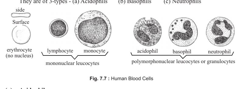
[Figure: Fig. 7.7 Human blood cells, showing erythrocyte (no nucleus), lymphocyte, monocyte, acidophil, basophil and neutrophil]
(a) Acidophils
1. They take up acidic stain (for example eosin) and are therefore also called eosinophils or oxyphils.
2. They have a bilobed nucleus.
3. They produce antitoxins against allergens, the chemicals that cause allergy.
4. An increase in the number of acidophils during allergy or parasitic infection is called eosinophilia.
(b) Basophils
1. They take up basic stain (for example methylene blue).
2. They have an S-shaped, indistinct nucleus.
3. They produce heparin and histamine, and are therefore also known as mast cells of the blood.
(c) Neutrophils
1. They take up neutral stain (for example neutral red).
2. The nucleus of these cells has 2 to 7 lobes (they are polymorphs). The term 'polymorph' can be used for all granulocytes because they have more than one lobed nucleus.
3. They are phagocytic cells. About 5% of neutrophils in females show a Barr body, seen as a drumstick.
Differential Leucocyte Count (DLC)
| WBC type | Percentage of total WBCs |
|---|---|
| Neutrophils | 60 to 65% (maximum in number) |
| Lymphocytes | 20 to 30% |
| Monocytes | 4 to 8% |
| Acidophils | 1 to 4% |
| Basophils | 0 to 1% (minimum in number) |
1. Neutrophils and monocytes are phagocytic and increase during bacterial and viral infection.
2. Acidophils increase during allergy or parasitic infections.
Life Span of Leucocytes
1. Granulocytes (acidophils, basophils and neutrophils) live for 4 to 8 hours.
2. Monocytes live for 10 to 20 hours in blood. In connective tissue they can survive for months.
3. Lymphocytes, as memory cells, may survive for years.
3. Platelets (Thrombocytes)
1. They are the smallest blood elements (cell fragments), 3 to 4 µm in size.
2. They are non-nucleated (enucleated) and irregular in shape.
3. Their mother cells are called megakaryocytes.
4. Their number is 3 to 4 lakh per mm3 (0.3 to 0.4 million per mm3).
5. Their life span is 2 to 5 days.
6. An increase in the number of platelets is called thrombocytosis. A decrease is called thrombocytopenic purpura.
7. Platelets release thromboplastin (thrombokinase) for blood clotting.
Connecting Concepts
WBCs are the only nucleated elements in mammalian blood. In frogs, RBCs and thrombocytes are also nucleated.
(b) Lymph
1. Extracellular fluid (ECF), interstitial fluid or tissue fluid is about 20% of body weight (about 14 litres in a person of 70 kg). It mainly includes lymph and plasma.
2. ECF = lymph (about 11 litres) + plasma (about 3 litres), i.e. about 14 litres.
3. When ECF flows in lymphatic vessels, it is called lymph.
4. Blood has both intracellular fluid (inside blood corpuscles) and extracellular fluid (as plasma).
5. Intracellular fluid (ICF) makes up about 40% of body weight (about 28 litres in a person of 70 kg). Thus ICF is twice the ECF.
6. About 50% of lymph is produced in the liver.
7. Lymph contains more lymphocytes and less protein than blood. It is generally represented as 'blood minus RBCs' or 'plasma plus WBCs'.
8. Lymph contains fibrinogen and can therefore clot like plasma.
9. Lymph differs from plasma in having WBCs, and from serum in having both fibrinogen and WBCs.
10. Lymph helps in the exchange of gases and nutrients between blood and cells or tissues.
11. Fat products are also absorbed through the lacteals of intestinal villi, which carry lymph.
3. Nervous Tissue
1. This tissue is purely ectodermal in origin.
2. Nervous tissue first appeared in Coelenterata (Cnidaria), where the nerve cells are non-polarised.
3. Nervous tissue has the least power of regeneration.
4. The structural and functional unit of nervous tissue is the neuron, or nerve cell. The human nervous system has about 1012 (1 trillion) neurons.
Structure of a Neuron
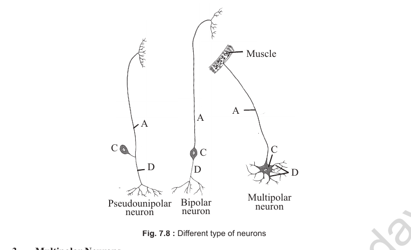
A neuron has three parts: (1) dendrites (dendrons), (2) cyton (cell body or perikaryon) and (3) axon.
1. In polarised fibres, the nerve impulse travels from dendrite to cyton, and from cyton to axon.
Types of Neurons
1. Unipolar or pseudo-unipolar neurons: a single process arises from the cell body and bifurcates to form an axon and a dendrite. Example: neurons of the dorsal root ganglion of the spinal cord.
2. Bipolar neurons: two processes, one dendrite and one axon, arise from the cell body. Examples: neurons of the retina and membranous labyrinth (inner ear).
3. Multipolar neurons: more than two processes arise from the cell body.
[Figure: Types of neurons, showing unipolar, bipolar and multipolar neurons with dendrites (D), cyton, axon (A) and cell body (C) labelled]
4. More than 99% of neurons are multipolar. Such neurons have one axon and two or more dendrites.
[Figure: Fig. 7.8 Different types of neurons, showing pseudounipolar neuron, bipolar neuron and multipolar neuron]
Neuroglia
1. Neurons are surrounded by supporting cells, or packing cells, called neuroglia. The number of glial cells is 30 to 40 times the number of neurons, but they are so small that together they make up only half of the volume of neural tissue.
2. Glial cells around neurons of the peripheral nervous system are called Schwann cells.
3. Glial cells of the central nervous system are of three types:
(i) Astrocytes: star-shaped cells that form the blood-brain barrier and transport nutrients from blood to the brain (CNS).
(ii) Microglia: phagocytic cells that remove debris from nervous tissue.
(iii) Oligodendrocytes: secrete the myelin sheath around many nerve fibres together.
Structure of a Neuron
1. The cyton (cell body) contains groups of ribosomes and rough endoplasmic reticulum, called Nissl granules. Nissl granules are also present in dendrites but absent in the axon. They help in protein synthesis. Their presence is a characteristic feature of neurons.
2. The centrosome is absent in the cyton, so neurons cannot divide.
3. The cytoplasm and cell membrane of the axon are called axoplasm and axolemma respectively.
4. Long fibres (axons or dendrites) may be grey (non-myelinated) or white (myelinated). In the PNS, the myelin sheath is secreted by Schwann cells. Myelinated fibres are also known as medullated fibres.
5. The covering formed by Schwann cells around an axon is called the neurilemma.
6. Because the myelin sheath around an axon in myelinated fibres is discontinuous, fibres are differentiated into nodes and internodes. At nodes (nodes of Ranvier) the myelin sheath is absent but the neurilemma is present.
[Figure: Fig. 7.9 Structure of a multipolar neuron, showing cyton, axon hillock, dendrites, Nissl's granules, axoplasm, Schwann cell, myelin sheath, internode, node of Ranvier, neurilemma, terminal buttons, oligodendrocyte (glia cell), and the C.N.S. and P.N.S. regions]
Ganglia and Nerves
1. A ganglion is a group of cytons (cell bodies). In the brain (CNS), such groups of cytons are called nuclei.
2. A nerve is mainly a group of axons. In the brain, such groups of axons are called tracts.
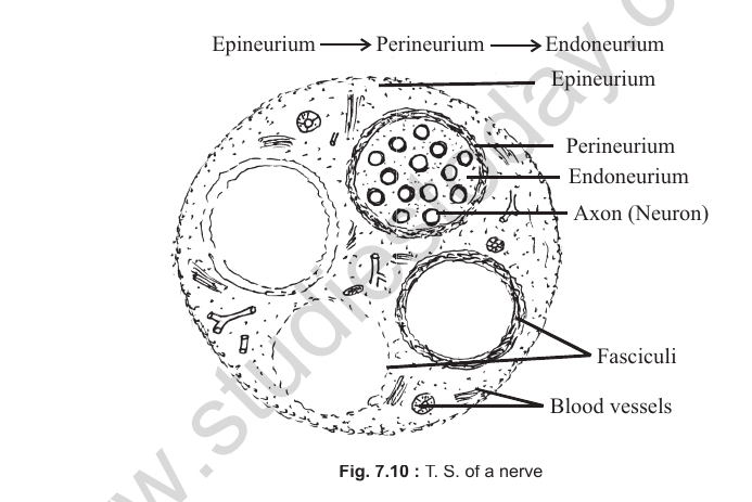
Structure of a Nerve
1. The outermost connective tissue covering of a nerve is the epineurium. Several bundles of neurons, called nerve fasciculi, are embedded in the epineurium.
2. Each nerve fasciculus has a connective tissue covering called the perineurium.
3. Each individual neuron is surrounded by a connective tissue covering called the endoneurium.
4. From outer to inner, the connective tissue coatings of a nerve are: epineurium, perineurium and endoneurium.
[Figure: Fig. 7.10 T.S. of a nerve, showing epineurium, perineurium, endoneurium, axon (neuron), fasciculi and blood vessels]
Types of Nerves
On the basis of their nature or function, nerves are of three types.
1. Sensory nerves: carry impulses from receptors (skin, retina etc.) to the CNS. Example: optic nerve.
2. Motor nerves: carry impulses from the CNS to effector organs (muscles or glands). Example: trochlear nerve.
3. Mixed nerves: some fibres carry impulses towards the CNS and others carry them away from it. Example: vagus nerve.
[Figure: Fig. 7.11 Types of nerves (D: dendrites, C: cyton, A: axon, N: nucleus, S: Schwann cell), showing sensory nerve, motor nerve and mixed nerve]
4. Muscular Tissue
1. Muscular tissue is mesodermal in origin (exception: iris muscles, which are ectodermal).
2. It forms 40 to 50% of the body weight in humans.
3. The total number of muscles in the human body is 639.
Important Muscles of the Human Body
1. The largest or biggest muscle is the gluteus maximus (hip muscle).
2. The longest muscle is the sartorius (thigh muscle).
3. The strongest muscle is the masseter (jaw muscle).
4. The longest smooth muscle is the rectus abdominis.
5. The shortest muscle is the stapedial muscle.
Special or Important Muscles
1. Auricular muscles: in the pinna (not in the auricles).
2. Papillary muscles: in the ventricles of the heart (not in the pupil).
3. Rectus muscles: in the eyeball (not in the rectum).
4. Gastrocnemius muscle: in the shank (calf) region (not in the stomach).
5. Intercostal muscles: between the ribs.
6. Biceps and triceps: in the upper arm.
7. Quadriceps: in the thigh region.
8. Segmental muscles (vestigial in humans): on either side of the vertebral column.
9. Pectoralis major and pectoralis minor: flight muscles in birds, or chest muscles in mammals.
Structure of Muscle Fibre
1. The structural unit of muscular tissue is the muscle fibre, or muscle cell. The functional unit is different.
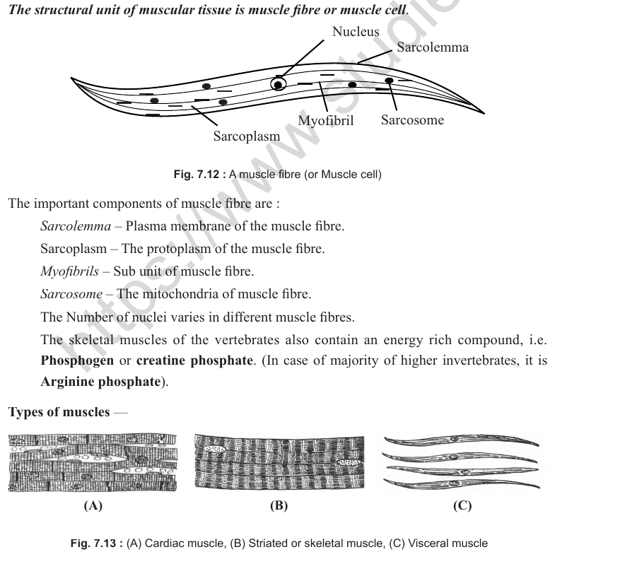
[Figure: Fig. 7.12 A muscle fibre (or muscle cell), showing nucleus, sarcolemma, myofibril, sarcosome and sarcoplasm]
Components of a Muscle Fibre
1. Sarcolemma: plasma membrane of the muscle fibre.
2. Sarcoplasm: protoplasm of the muscle fibre.
3. Myofibrils: subunits of the muscle fibre.
4. Sarcosomes: mitochondria of the muscle fibre.
1. The number of nuclei varies in different muscle fibres.
2. Skeletal muscles of vertebrates contain an energy-rich compound called phosphogen or creatine phosphate. In most higher invertebrates, it is arginine phosphate.
Types of Muscles
[Figure: Fig. 7.13 (A) Cardiac muscle, (B) Striated or skeletal muscle, (C) Visceral muscle]
| Feature | Cardiac muscle | Skeletal muscle | Visceral muscle |
|---|---|---|---|
| Location | Present in the myocardium of the heart | Attached to the skeleton, except the diaphragm and tongue muscles | Present in visceral organs like gut, blood vessels and urinary bladder |
| Sarcolemma | Ill defined | Well defined | Ill defined |
| Shape of fibres | Cylindrical but branched, with intercalated discs at the junctions | Cylindrical, unbranched and in bundles | Fusiform or spindle shaped, unbranched and solitary |
| Striations | Faintly striated | Striated (dark and light bands present) | Unstriated (dark and light bands absent), so called smooth muscles |
| Nature | Involuntary | Voluntary | Involuntary |
| Speed of contraction | Slow but rhythmic | Quick | Slow |
| Nuclei | Fibres with 1 to 2 nuclei | Fibres multinucleated | Fibres uninucleated |
| Fatigue | Does not develop | Develops | Does not develop |
Antagonistic Sets of Skeletal Muscles
1. Flexor and extensor muscles: the flexor (example: biceps) bends the arm at the elbow, and the extensor (example: triceps) straightens it.
2. Sphincter and dilator muscles: a sphincter reduces, while a dilator increases, the diameter of an opening (example: circular and radial muscles of the iris).
3. Adductor and abductor muscles: adductors draw limbs towards the body axis, while abductors take them away.
4. Levator and depressor muscles: a levator lifts a body part vertically upward, whereas a depressor brings it down.
5. Protractor and retractor muscles: a protractor moves organs forward from their normal position, while a retractor brings them back (example: muscles of the lower jaw for to-and-fro movement).
6. Pronator and supinator: a pronator turns the palm backward and downward, whereas a supinator turns it forward and upward.
Skeletal Muscle Fibres
In birds and mammals (homeotherms), these fibres are of two functional types.
| Red muscle fibres* | White muscle fibres |
|---|---|
| Thin fibres | Thick fibres |
| Myoglobin present | Myoglobin absent |
| Many mitochondria | Few mitochondria |
| Contraction slow | Contraction fast |
| Fatigue develops slowly | Fatigue develops early |
| Examples: flight muscles in kite and back muscles in human | Examples: flight muscles in house sparrow and eyelid muscles in human |
* Since these muscle fibres have plenty of mitochondria, they use large amounts of oxygen (stored in them) to produce ATP, and are therefore also known as aerobic muscles.
Smooth Muscle Fibres
In birds and mammals (warm-blooded animals), these fibres are also of two functional types.
| Multi-unit type | Single-unit type |
|---|---|
| All the muscle fibres contract independently as separate units. | All muscle fibres contract together as a single unit. |
| Examples: muscle fibres of hair roots and the wall of blood vessels | Examples: muscle fibres of the urinary bladder, gut and uterus |
Organ Histology
1. Liver
1. The liver is the largest gland of the body and weighs 1 to 2 kg.
2. It has the maximum power of regeneration (compensatory regeneration).
3. It is endodermal in origin and lies on the right side of the abdominal cavity.
4. In humans, the liver has four lobes: left lobe, right lobe, quadrate lobe and caudate lobe. In frog and rabbit it has 3 and 5 lobes respectively. The gall bladder is attached to the right lobe of the liver.
5. The structural and functional unit of the liver is the lobule.
6. Each lobule is surrounded by a connective tissue covering called Glisson's capsule (in humans, Glisson's capsule is not well developed).
7. At the centre of each lobule is a branch of the hepatic vein, also known as the intralobular vein.
8. Between adjacent lobules are interlobular ducts or portal canals, containing branches of the hepatic artery, lymphatic vessels, bile duct and hepatic portal vein.
9. Hepatic cells (hepatocytes) are arranged in sheets, each 2 cells thick. Between the sheets are blood lacunae and sinuses (sinusoids).
10. Phagocytic Kupffer cells lie in Glisson's capsule and at the border of the sinuses.
[Figure: Fig. 7.15 T.S. of a liver lobule, showing polygonal lobule, portal canal or interlobular duct, liver cords (usually one or two cells thick), sinusoids (small sinuses), central vein (branch of hepatic vein) or intralobular vein, hepatic artery, bile duct and hepatic portal vein]
[Figure: Fig. 7.14 T.S. of ileum, showing the layers of the mucosa (villi with lacteal, smooth muscle fibres, goblet cells, columnar epithelium, mucous membrane, lamina propria, crypts of Lieberkühn or intestinal glands, Paneth cells, muscularis mucosa and blood vessel), submucosa (blood vessel, connective tissue and sympathetic ganglion of Auerbach's plexus), muscularis externa (circular muscle layer and longitudinal muscle layer) and serosa]
Functions of the Liver
1. Phagocytosis: Kupffer cells are phagocytic and remove pathogens and toxic material from the blood.
2. Excretion: the liver is not an excretory organ, but urea, uric acid, creatinine etc. are synthesised in the liver. Bilirubin, an excretory product, is also formed in the liver.
3. Storage: it stores vitamins A, D and B12, iron (as ferritin), blood and glycogen etc.
4. Haemopoiesis: in the foetus, the liver is a haemopoietic organ and produces RBCs, WBCs and platelets.
5. Production: it produces bile juice, which is concentrated in the gall bladder, and heparin, an anticoagulant. It produces many plasma proteins, such as albumin, prothrombin and angiotensinogen. About 50% of the body's lymph is produced in the liver. It also synthesises fatty acids, cholesterol and phospholipids.
6. It is the seat of metabolic reactions such as glycogenesis, glycogenolysis, gluconeogenesis, deamination and the Cori cycle. It is an important site for beta-oxidation of fatty acids and for the degradation of cholesterol to form cholic acid of bile.
7. It is the busiest organ of the body. It receives both oxygenated and deoxygenated blood. Blood supply to the liver in humans is about 1500 ml per minute (1000 ml from the hepatic portal vein and 500 ml from the hepatic artery).
[Figure: Fig. 7.15 C.S. human liver]
2. Spleen
1. It is mesodermal in origin.
2. It lies close to the fundic stomach, on the left side of the abdominal cavity. In frog it lies close to the large intestine.
3. It is the largest lymphatic organ (about 15 cm in diameter and 150 g in weight).
4. Unlike other lymphatic organs, it is not differentiated into cortex and medulla. Lymphatic sinuses and lymphatic vessels are also absent.
5. It consists of an outer red pulp (containing RBCs) and an inner white pulp (lacking RBCs). Nodules (germinal centres) in the white pulp synthesise lymphocytes.
Functions of the Spleen
1. Biological filter: it removes pathogens such as bacteria and foreign material from the blood. Its phagocytic cells belong to the reticulo-endothelial system.
2. Blood bank: it stores blood in frog. In humans, most blood is stored in the liver.
3. Haemopoietic organ: in the human foetus and adult frog, blood elements are manufactured in the spleen. It also stores RBCs.
4. Graveyard of RBCs: worn-out RBCs, whose life span is complete, are destroyed here.
5. Immunological function: it is the centre for lymphocyte production. Lymphocytes produce plasma cells and hence antibodies. It plays an important role in immunity.
3. Pancreas
1. Like the liver, it is endodermal in origin.
2. It is the second largest digestive gland in the body, after the liver (about 15 cm in size).
3. It lies in the coil of the duodenum in the abdominal cavity.
4. It is a heterocrine (mixed) gland, containing both exocrine and endocrine parts.
5. On the basis of structure, it is a compound tubulo-alveolar gland.
6. Its exocrine part consists of many acini (singular: acinus), which produce pancreatic juice. The endocrine part, the islets of Langerhans, is discussed in the chapter on hormonal coordination.
7. The pancreatic duct joins the common bile duct before opening into the duodenum, forming the ampulla of Vater, which has the sphincter of Oddi at the common opening.
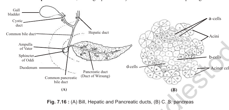
[Figure: Fig. 7.16 (A) Bile, hepatic and pancreatic ducts, showing gall bladder, cystic duct, hepatic duct, common bile duct, ampulla of Vater, sphincter of Oddi, duodenum, pancreatic duct (duct of Wirsung) and common pancreatic bile duct; (B) C.S. pancreas, showing acini, alpha cells, beta cells, delta cells and acinar cells]
Classification of Exocrine Glands on the Basis of Structure
Exocrine glands are of two broad categories: simple glands and compound glands.
(i) Simple Glands
In simple glands the duct (non-secretory part) is unbranched.
1. Simple unbranched glands: the secretory part is also unbranched.
(i) Tubular: uncoiled, example crypts of Lieberkühn; coiled, example sweat glands.
(ii) Alveolar: example mucous and poison glands in frog's skin.
2. Simple branched glands: the secretory part is branched.
(i) Tubular: example gastric glands.
(ii) Alveolar: example sebaceous glands and meibomian or tarsal glands.
(ii) Compound Glands
In compound glands the duct (non-secretory part) is branched.
1. Tubular: example non-lactating mammary glands and Brunner's glands.
2. Tubulo-alveolar: example lactating mammary glands, parotid (salivary) gland and pancreas.
3. Alveolar: example submaxillary salivary glands.
[Figure: Fig. 7.17 Types of exocrine glands, showing simple coiled tubular, simple branched tubular, simple alveolar, simple branched alveolar, compound tubular, compound alveolar and compound tubulo-alveolar glands]
4. Gut (Alimentary Canal)
The gut wall has four coatings, from inner to outer.
1. Tunica mucosa: differentiated into epithelium, lamina propria and muscularis mucosae. It contains digestive glands. The epithelium of most of the gut is simple columnar. The lamina propria contains lymphatic tissue (GALT, gut-associated lymphatic tissue) and cells such as lymphocytes, plasma cells and eosinophils. It lacks blood vessels and nerves.
2. Tunica submucosa: connective tissue with blood vessels, nerve fibres and nerve plexuses (example: Meissner's plexus).
3. Tunica muscularis: usually an inner layer of circular muscles and an outer layer of longitudinal muscles. Auerbach's plexuses are usually present between these two layers of smooth muscle.
4. Tunica externa: a thin layer of connective tissue covered by visceral epithelium, or serosa (formed from coelomic mesoderm or peritoneum), and attached to mesenteries.
[Figure: Fig. 7.18 General plan of the alimentary canal, showing epithelium, lamina propria and mucosa, muscularis mucosae, submucosa with Meissner's plexus, muscularis externa with inner circular and outer longitudinal muscle layers, Auerbach's plexus, and serosa]
Oesophagus
1. The epithelium or mucous membrane of the tunica mucosa is formed by stratified squamous cells.
2. The tunica submucosa is thin.
3. The tunica muscularis has voluntary (striated) muscles in the upper one-third, both voluntary and involuntary muscles in the middle one-third, and involuntary (smooth) muscles in the lower one-third.
Stomach
1. When the stomach is empty, the tunica mucosa forms longitudinal folds called rugae.
2. The gastric glands in the tunica mucosa are simple, tubular and branched. The neck of each gland contains mucus-secreting cells. The body of each gland has chief (zymogen) cells and parietal (oxyntic) cells. Cells at the bottom are argentaffin (enterochromaffin) cells, which are endocrine cells of the stomach.
3. Chief cells produce digestive enzymes.
4. Parietal cells secrete the intrinsic factor of Castle and HCl. (For details, see the chapter on Animal Nutrition.)
5. The tunica muscularis of the stomach contains inner oblique, middle circular (thickest) and outer longitudinal layers of smooth muscle.
[Figure: Fig. 7.19 (A) T.S. stomach, showing mucosa with gastric pits, lamina propria, cardiac glands, muscularis mucosa, adipose tissue, blood vessels, submucosa, circular muscle layer, longitudinal and oblique muscle layers, serosa; (B) Fundic (gastric) gland, showing gastric pit, mucoid neck cells, chief or peptic cells (lower third, secreting pepsinogen and dilute hydrochloric acid) and parietal or oxyntic cells]
Small Intestine
1. It is characterised by villi of various shapes.
2. The tunica mucosa consists of four types of cells:
(1) Enterocytes or absorptive cells: have microvilli forming a brush-bordered epithelium, which increases the surface area for absorption. They also secrete digestive enzymes.
(2) Goblet cells: single-celled, egg-shaped glands that secrete mucus.
(3) Paneth cells: secrete digestive enzymes that digest the walls of bacteria.
(4) Argentaffin cells: endocrine cells.
Duodenum
1. The lamina propria is thick and contains lymphatic nodules called Peyer's patches.
2. Crypts of Lieberkühn are present in the mucosa.
3. Brunner's glands, the characteristic mucus-secreting glands of the duodenum, are embedded in the submucosa. These glands open into the crypts of Lieberkühn.
4. The tunica mucosa is thrown into leaf-shaped villi.
5. The tunica muscularis does not have oblique muscles.
6. Goblet cells are few in number.
Ileum
1. Its villi are tubular.
2. Goblet cells are more numerous.
3. Brunner's glands are absent.
4. Crypts of Lieberkühn, with abundant Paneth cells, are present.
5. Peyer's patches are present in the lamina propria.
6. The tunica mucosa is thrown into circular folds called plicae circulares, or folds of Kerckring.
Large Intestine
1. Villi are absent in the large intestine.
2. Colon: crypts of Lieberkühn are present. Goblet cells are innumerable. Paneth cells are absent. The longitudinal muscles of the tunica muscularis form three longitudinal bands called teniae coli, which make the colon sacculated.
3. Anal canal: the tunica muscularis is formed by striated (voluntary) muscles. Crypts of Lieberkühn and villi are absent. The mucous membrane is thrown into longitudinal folds called rectal columns of Morgagni.
5. Integument
1. The integument is the part of the body wall and consists of the skin and its derivatives (glands and exoskeleton).
2. The skin is ectomesodermal in origin. Its epidermis is ectodermal, and its dermis is mesodermal.
Mammalian Skin
1. Epidermis
1. The epidermis is stratified squamous epithelium. From inner to outer, it consists of the following layers:
(i) Stratum germinativum or stratum Malpighii (innermost)
(ii) Stratum spinosum (innermost to outer)
(iii) Stratum granulosum
(iv) Stratum lucidum
(v) Stratum corneum (outermost)
1. Stratum germinativum (stratum Malpighii): simple columnar epithelium. It is the only meristematic layer of the epidermis (mitotic divisions are faster at night). All other layers, cutaneous glands and the exoskeleton develop from this layer. Melanocytes, the pigment-bearing cells, lie between these cells.
2. Stratum spinosum: a prickly layer of polyhedral cells bearing minute spine-like structures.
3. Stratum granulosum: polygonal cells containing granules of keratohyalin protein.
4. Stratum lucidum: cells are non-nucleated due to degeneration of nuclei. These cells contain eleidin (a transparent protein and precursor of keratin), which makes the layer lustrous. It is prominent and thick in the heel.
5. Stratum corneum: consists of flat, dead, non-nucleated squamous cells containing keratin (keratinised). The dead cells make the skin germ-proof, and keratin is important for waterproofing.
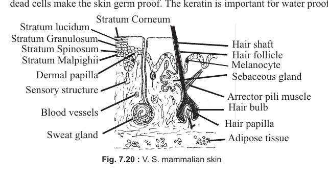
[Figure: Fig. 7.20 V.S. mammalian skin, showing stratum corneum, stratum lucidum, stratum granulosum, stratum spinosum, stratum Malpighii, dermal papilla, sensory structure, blood vessels, sweat gland, hair shaft, hair follicle, melanocyte, sebaceous gland, arrector pili muscle, hair bulb, hair papilla and adipose tissue]
Derivatives of the Skin
1. Sweat Glands
1. Also known as sudoriferous glands.
2. Structurally they are simple, unbranched or branched, coiled and tubular.
3. They are mainly merocrine, or apocrine (in the axilla and pubis). They produce a watery secretion, which diffuses out of the cells without cell injury.
4. Sweating and its evaporation help to cool the body.
5. In rabbits, sweat glands are concentrated on the lips; in deer, on the base of the tail; in dogs and cats, on the paws. In humans, they are most numerous on the palm and sole, and are absent on the lips and glans penis.
2. Sebaceous Glands
1. These glands secrete sebum, which is germicidal due to the presence of lysozymes.
2. They are holocrine glands and release an oily secretion into hair follicles.
3. Structurally they are simple, branched and alveolar.
4. They are absent on the sole and palm.
5. Their cells synthesise vitamin D with the help of UV radiation.
3. Exoskeleton
1. Mammals have various forms of exoskeleton, such as hoofs, horns, nails or claws, scales or spines, and hair.
2. All of these develop from the stratum germinativum of the epidermis and are therefore ectodermal in origin. Antlers are bony structures and not skin derivatives; they are formed from mesoderm.
3. The outer layer of the exoskeleton is keratinised and contains dead cells.
4. Mammalian hair, which is ectodermal, is covered with a cuticle. It is differentiated into an outer cortex and an inner medulla. The pigment melanin is present in the cortex.
5. The hair coat of mammals is called pelage. Because human hair lacks a medulla, it is hollow or tubular. Hair is absent on the palm, sole, lips, glans penis and teats.
6. The study of hair is called trichology.
7. Greying of hair occurs due to degeneration of melanin.
8. Contraction of the arrector pili muscles causes hair erection (goose flesh).
9. Each hair grows from the base of the shaft (hair root), which is called the hair bulb. The hair
papilla receives blood and nerve supply.
2. Dermis
1. The dermis is differentiated into an outer papillary layer and an inner reticular layer. The papillary layer is compact, with finger-like projections called papillae. The reticular layer consists of loose connective tissue, which merges with the subcutaneous fat.
2. Below the dermis is a thin layer of fat, called paniculus adiposus.
3. Leather is produced from the dermis of mammals.
Sensory Structures of the Skin
Most sensory structures in the skin are encapsulated dendrites embedded in the dermis. All of them are ectodermal in origin.
1. Meissner's corpuscles: sensory for touch (thigmoreceptors).
2. Merkel's discs: sensory for touch or soft pressure.
3. Pacinian corpuscles: sensory for pressure (baroreceptors).
4. Krause's end bulbs: sensory for cold (frigidoreceptors).
5. End bulbs of Ruffini: sensory for warmth (caloreceptors).
6. Free nerve endings: sensory for pain (algesireceptors).
Because the skin performs many functions, such as protection, temperature regulation, sensation and minimising water loss, it is called the 'Jack of all trades'.
Frog's Skin
1. The epidermis (ectodermal) is thin and has only two layers: an inner stratum Malpighii (stratum germinativum) and an outer stratum corneum.
2. The stratum Malpighii is simple columnar epithelium. Because of its cylindrical cells, it is also called stratum cylindricum. It is the only meristematic layer of the skin.
3. The stratum corneum consists of stratified squamous epithelium.
4. The exoskeleton is absent in frog.
5. The dermis (mesodermal) is differentiated into an outer stratum spongiosum and an inner stratum compactum.
6. The stratum spongiosum contains loose connective tissue and pigment-bearing cells called chromatophores. Chromatophores are of three types:
(i) Melanophores: contain blue, black or brown pigments.
(ii) Lipophores or xanthophores: contain red, orange or yellow pigments.
(iii) Guanophores or iridocytes: colourless or transparent.
7. Because of their colour-changing ability (metachrosis), frogs resemble their background (camouflage).
8. The stratum compactum has closely packed white fibres, and also contains nerve fibres and blood vessels.
9. The frog's skin can be easily removed, as it is not fused with the musculature. Lymph-filled spaces lie between the skin and musculature. Such skin cannot yield leather.
Glands of Frog's Skin
Frog skin has two types of glands. Both develop from the stratum Malpighii and are therefore ectodermal in origin.
1. Mucous glands: simple alveolar, distributed all over the body. They secrete mucus, which keeps the skin moist for cutaneous respiration. The secretion also makes the skin slippery, helping the frog escape predators.
2. Poison glands: simple alveolar, mainly concentrated on the shoulders. In toads these glands together form the parotid glands on the shoulders.
3. Frogs can absorb water through the skin but cannot regulate their body temperature. Sweat glands are absent. Vitamin D cannot be synthesised in the skin, and subcutaneous fat storage is absent.
[Figure: Fig. 7.21 V.S. frog's skin, showing stratum corneum, stratum Malpighii, chromatophores, blood vessels, lymph spaces, stratum spongiosum, stratum compactum, musculature, poison gland and mucous glands]
Check Point (Part I: Tissues)
Distinguish between the following
1. Ligaments and tendons: ligaments join bone to bone and contain mainly yellow (elastin) fibres; tendons join muscles to bones and consist purely of white (collagen) fibres.
2. Investing bone and cartilaginous (replacing) bone: investing bone forms by ossification directly in connective tissue without a cartilage model; replacing bone forms when ossification replaces a cartilage model.
3. Hyaline cartilage and fibrous cartilage: hyaline cartilage is semitransparent with invisible fibres and a prominent perichondrium; fibrous cartilage is the toughest, with well-developed white and yellow fibres.
4. Bone and cartilage: bone has a hard matrix of ossein and mineral salts (calcium phosphate), with Haversian systems and solitary osteocytes; cartilage has a flexible chondrin matrix, chondrocytes in groups, and no Haversian systems.
5. Plasma and lymph: plasma contains more proteins (including fibrinogen) and RBCs; lymph contains fewer proteins, no RBCs, and more lymphocytes.
6. RBCs and WBCs: RBCs are non-nucleated (in mammals), contain haemoglobin and carry oxygen; WBCs are nucleated, lack haemoglobin and form the body's defence system.
7. Chief cells and oxyntic cells: chief cells secrete digestive enzymes (pepsinogen); oxyntic (parietal) cells secrete HCl and the intrinsic factor of Castle.
Fill in the blanks
1. Cuboidal cells form the epithelium of thyroid follicles.
2. The term 'tissue' was coined by Bichat.
3. A compound epithelium that is two or more cells thick is called stratified epithelium.
4. Where wear and tear is high, the type of epithelium is stratified squamous epithelium.
5. Mammary glands are a modification of apocrine sweat glands.
6. Histologically, blood is a connective tissue.
7. Elastic cartilage occurs in the epiglottis and pinna.
8. Sesamoid bones are formed by the ossification of tendons.
9. An increase in the number of RBCs is called polycythaemia.
10. Monocytes are the largest blood element in humans.
Name the following structures
1. Graveyard of RBCs: spleen.
2. Digestive gland that is also an endocrine gland: pancreas.
3. Part of the gut that has Brunner's glands: duodenum.
4. Caloreceptors in the skin: end bulbs of Ruffini.
5. 'Jack of all trades': skin.
6. A WBC with a 2 to 7 lobed nucleus: neutrophil.
7. Blood element that shows the rouleaux phenomenon: erythrocyte (RBC).
8. Bone containing a Haversian system: compact bone of a mammalian long bone (replacing bone).
9. Blood element that shows diapedesis: leucocyte (WBC).
10. Blood cell associated with eosinophilia: acidophil (eosinophil).
Part-II: Cockroach, Earthworm and Frog
While going through this part, pay special attention to the following.
Terms and Definitions
1. Typhlosole
2. Holonephridia
3. Nephrostome
4. Phaosome
5. Mosaic image
6. Ecdysis
7. Paurometabola
8. Metachrosis
9. Anal style and anal cerci
10. Ocelli and ommatidia
11. Aspiration and inspiration
12. Blood and haemolymph
13. Apposition and superposition (image)
14. Prostomium and peristomium
Structures to Know
1. Elytra
2. Notum
3. Arolium
4. Stomodeal valve
5. Peritrophic membrane
6. Alary muscles
7. Corpora allata
8. Gonapophyses
9. Bidder's canal
10. Corpora bigemina
Cockroach
Taxonomic Position
Phylum: Arthropoda
Class: Insecta
Order: Dictyoptera
Genus: Periplaneta
Species: americana
1. There are two main types of cockroach in India, Periplaneta and Blatta.
2. The term Periplaneta was given by Burmeister, and the term Blatta was given by Linnaeus.
3. The most common cockroach in India is Periplaneta americana. Blatta orientalis is found in the oriental region but is less common in India.
Differences Between Blatta orientalis and Periplaneta americana
| Feature | Blatta orientalis | Periplaneta americana |
|---|---|---|
| Distribution | Oriental and Indian cockroach | American cockroach, but cosmopolitan in distribution |
| Colour | Black, so called black beetle | Dark brown |
| Wings | In males the wings cover half of the body, whereas in females they are rudimentary | Wings are larger and cover the whole of the abdomen |
| Flight | Cannot fly | Can fly |
| Arolium (pulvillus) | Absent; legs do not have arolium for moving on smooth surfaces | Present at the terminal end of each leg |
| Diploid chromosome number (2n) | 48 | 34 |
Body Structure
1. Like other insects, the body of the cockroach is differentiated into three parts: head, thorax and abdomen.
Head
1. The head consists of six fused segments and is directed downwards (hypognathous).
2. The exoskeletal plates on the head are the vertex (epicranium), frons, gena and clypeus. The vertex has an inverted Y-shaped epicranial suture.
3. The antennae (one pair) are many-segmented and are differentiated into three regions: scape, pedicel and flagellum.
4. There is one pair of compound eyes, one on each side of the head. Each eye is sessile and contains about 2000 visual units (ommatidia).
5. There is one pair of simple eyes (ocelli). They are vestigial and do not form images.
6. The mouthparts are of the cutting and chewing type (mandibulate). They consist of the upper lip (labrum), lower lip (labium), mandibles, maxillae and hypopharynx.
7. The hypopharynx (tongue), mandibles and labrum are unjointed structures, whereas the maxillae and labium are jointed. Jointed structures are based on the biramous plan.
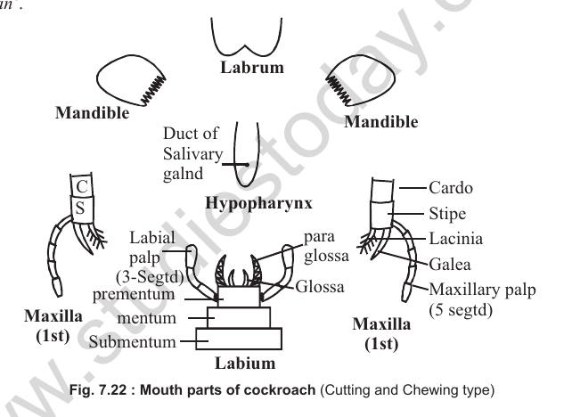
[Figure: Fig. 7.22 Mouthparts of cockroach (cutting and chewing type), showing labrum, mandible, duct of salivary gland, hypopharynx, labial palp (3-segmented), prementum, mentum, submentum, glossa, paraglossa, maxilla (1st) with cardo, stipe, lacinia, galea and maxillary palp (5-segmented), and labium]
8. Each maxilla (first maxilla) consists of two podomeres, the cardo and the stipe. Two rami arise from the stipe: the maxillary palp and the lacinia-galea. The maxillary palp is 5-jointed and is sensory for smell (olfactory receptor).
9. The labium is formed by the fusion of two second maxillae. Each half of the labium has three podomeres: prementum, mentum and submentum. Its two rami are the outer labial palp and the inner glossa-paraglossa. The labial palp is three-segmented and is sensory for taste (gustatoreceptor). The glossa and paraglossa together are called the ligula.
10. Lingula is a living fossil and belongs to a minor phylum, Brachiopoda.
[Figure: Fig. 7.23 External features of Periplaneta americana (diagrammatic), showing epicranial suture, vertex, antenna, compound eye, ocellus, frons, clypeus, labrum, gena, neck, forewings (elytra or tegmina), hind wings (membranous), 3 pairs of walking legs, 10-segmented abdomen and anal cercus]
Thorax
1. The thorax is differentiated into prothorax, mesothorax and metathorax.
2. The exoskeleton of each segment is called a sclerite. It consists of a dorsal plate (tergum or notum), a ventral plate (sternum) and lateral plates (pleuron).
3. There are two pairs of wings. The first pair is attached to the mesothorax. It is leathery and protective, called tegmina or elytra. The second pair is attached to the metathorax. It is membranous and is used in flight.
4. Each thoracic segment has one pair of legs. Because there are three pairs (six in all), insects are called hexapods.
[Figure: Structure of a cockroach leg, showing coxa, trochanter, femur, tibia with tibial spines, and tarsus]
5. Each leg is differentiated, from proximal to distal end, into five regions: coxa, trochanter, femur, tibia and tarsus. The coxa is attached to the pleuron. The tibia bears tibial spines and is the longest part of the leg. The tarsus has five tarsomeres. The last tarsomere carries a pretarsus with an arolium and two claws.
[Figure: Fig. 7.24 Leg of cockroach, showing coxa, trochanter, femur, tibia with tibial spines, 5-tarsomere tarsus, pretarsus, arolium (pulvillus) and claw]
Abdomen
1. The abdomen consists of 10 segments.
2. One pair of 15-segmented anal cerci arise from the 10th segment and perceive ground-borne vibrations.
3. In males there is one pair of unsegmented anal styles arising from the 9th segment. The male abdomen is narrower than the female abdomen.
4. In females, anal styles are absent and the abdomen is broader. Thus sexual dimorphism is prominent in cockroach.
[Figure: Fig. 7.25 Abdomen of cockroach (ventral view). In the male, 9 sterna are visible, with the anal cercus from the 10th segment and anal style from the 9th segment. In the female, 7 sterna are visible, with the anal cercus from the 10th segment]
Digestive System
1. In cockroach the alimentary canal is well differentiated and consists of three regions: foregut (stomodaeum), midgut (mesenteron) and hindgut (proctodaeum).
[Figure: Fig. 7.26 Alimentary canal of cockroach, showing salivary gland, reservoir, crop, gizzard, hepatic caeca (8), mesenteron (mid gut), ileum, rectum, anus, pharynx, oesophagus and anal cercus]
Foregut (Stomodaeum)
1. The stomodaeum includes the mouth, buccal cavity, oesophagus, crop and gizzard. It is ectodermal in origin.
2. There is one pair of salivary glands in cockroach. Each gland is bilobed. Saliva is stored in a reservoir before it is released onto the hypopharynx in the buccal cavity.
3. The crop mainly stores food material.
4. The gizzard is an organ for mastication. It consists of six cuticular plates alternating with six muscular pads of longitudinal muscles.
5. The valve between the gizzard and midgut is called the stomodaeal valve. It allows only fine particles of food to pass into the midgut.
Midgut (Mesenteron)
1. It is endodermal in origin.
2. The anterior part of the midgut bears four pairs of hepatic caeca (eight in all).
Check Point
Answer the following
1. (i) Who proposed the term 'Blatta' for cockroach? Linnaeus.
(ii) How many segments constitute the head? Six (fused) segments.
(iii) What is mosaic image? An image formed by the compound eye, made up of many small images from different ommatidia.
(iv) What is the visual unit in the compound eye of cockroach? Ommatidium.
(v) Which is the longest component of the leg? Tibia.
(vi) How many sterna are visible in the female cockroach? Seven.
(vii) What is the number of hepatic caeca? Eight (four pairs).
(viii) Which part of the gut is called the proctodaeum? The hindgut.
(ix) What is the scientific term for moulting? Ecdysis.
Midgut Secretions
1. The lining of the midgut secretes the peritrophic membrane, which protects the soft lining of the midgut from injury by spiny food. It is a temporary membrane, removed after food passes into the hindgut.
2. Digestive enzymes are secreted by the hepatic caeca and the lining of the midgut.
Hindgut (Proctodaeum)
1. Like the foregut, it is ectodermal in origin.
2. At the junction of the midgut and hindgut there are 60 to 150 blind Malpighian tubules for excretion.
3. The hindgut is differentiated into ileum, colon and rectum. The rectum stores undigested waste. It also contains six rectal pads.
Food and Digestion
1. Cockroach is omnivorous and feeds on every type of food material, such as paper, leather and kitchen waste.
2. Saliva contains amylase for the digestion of starch.
3. Enzymes secreted by the hepatic caeca and midgut are proteolytic, lipolytic and sucrolytic in nature.
4. Digested food is absorbed through the midgut and hepatic caeca. Hepatic caeca increase the surface area for absorption.
5. In the hindgut, only water and minerals are absorbed.
6. Undigested waste passes out through the anus from the rectum.
Excretory System
1. Primary excretory organs: Malpighian tubules.
2. Accessory excretory organs: fat bodies, skin and reproductive organs.
Malpighian Tubules
1. They are blind tubes between the midgut and hindgut.
2. Their number ranges from 60 to 150.
3. They take excretory matter from the haemocoel and pour it into the midgut, from where it passes out with faecal matter.
4. Malpighian tubules can be compared with the septal nephridia of the earthworm, as both are adapted for water conservation.
5. The major excretory product of cockroach is uric acid, so it is uricotelic.
Accessory Excretory Organs
(a) Fat body
1. It is single and multilobular, and is present around the visceral organs.
2. It consists of four types of cells: trophocytes, mycetocytes, urate cells and oenocytes.
3. Trophocytes store reserve food material. Mycetocytes contain bacteria that help in the synthesis of glycogen, amino acids and vitamins. Urate cells form uric acid. Oenocytes help in moulting.
4. The fat body is analogous to the liver of vertebrates.
(b) Skin and reproductive organs
Excretory matter from the skin is removed through moulting (ecdysis). Uric acid is also removed through the covering of the spermatophore.
Blood Vascular System
1. The blood vascular system of cockroach is open type. Capillaries are absent and the haemocoel is well developed.
2. The body cavity (haemocoel) is divided into three sinuses by two perforated diaphragms. The dorsal sinus around the heart is the pericardial sinus; the ventral sinus around the ventral nerve cord is the perineural sinus; and the middle sinus around the visceral organs is the perivisceral sinus.
3. The coelom (schizocoel) is reduced in cockroach.
4. Since the blood pigment is absent, the blood of cockroach is called haemolymph. Oxygen is carried directly to the tissues through the tracheal system.
[Figure: Heart and alary muscles of cockroach, showing anterior aorta, heart, ostia (valves open and valves closed), alary muscles and segments 10th and 11th]
[Figure: Fig. 7.27 (A) Blood vascular system of cockroach, showing 13-chambered heart, pericardial sinus, perivisceral sinus, perineural sinus, ventral nerve cord, dorsal and ventral perforated diaphragm, alary muscles, ostia, and anterior aorta; (B) Systole and diastole phases in the working of the heart]
Heart
1. The cockroach has a single, mid-dorsal, 13-chambered heart (three chambers in the thoracic region and ten in the abdominal region), present in the pericardial sinus.
2. Except for the last chamber, all chambers have one pair of ostia each (12 pairs in total). Each ostium has valves.
3. The heart is suspended in the pericardial sinus by 12 pairs of alary muscles.
4. The chambers of the heart pulsate with rhythmic diastole and systole. During diastole the ostia open and blood enters the heart. During systole the ostia close and blood is pushed from posterior to anterior through the dorsal aorta.
5. Deoxygenated blood from the perivisceral and perineural sinuses passes into the pericardial sinus through the perforated diaphragm.
6. The function of blood (haemolymph) is only to transport excretory and nutritive materials. Blood has no role in respiration or gas transport.
Respiratory System
1. The respiratory system consists of tracheal tubes, which are ectodermal in origin.
2. Each tracheal tube is lined internally with cuticle, called intima, which prevents the tubes from collapsing. Both the tracheal tube of cockroach and the trachea of humans have non-collapsible walls.
3. There are six longitudinal tracheal ducts: two dorsal, two ventral and two lateral.
4. The lateral longitudinal ducts open to the outside through spiracles. Cockroach has 10 pairs of spiracles: two pairs in the thorax and eight pairs in the abdomen.
5. The thoracic spiracles lie on the pleuron between the prothorax and mesothorax, and between the mesothorax and metathorax. The first pair of abdominal spiracles lies on the tergum; the other seven pairs lie on the pleura.
6. Each spiracle opens into a chamber called the atrium. From the atrium, the tracheal tube branches into fine tracheae and then tracheoles. Tracheoles have no intima lining.
Process of Respiration
(a) Resting stage
1. Abdominal movements are absent and oxygen requirement is low. All 10 pairs of spiracles remain open during both inspiration and expiration.
2. Tissue fluid is drawn into the tracheoles, and oxygen diffuses into the tissues after dissolving in the tissue fluid.
(b) Active stage
1. Abdominal movements become active and oxygen requirement increases.
2. Tissue fluid is not drawn into the tracheoles. Oxygen passes directly to the tissues.
3. During inspiration all 10 pairs of spiracles remain open. During expiration all spiracles except the first and third pairs are closed. Thus the first pair of thoracic and the first pair of abdominal spiracles always remain open.
Nervous System
All three types of nervous system, CNS, PNS and ANS, are present in cockroach.
Central Nervous System (CNS)
1. It consists of a nerve ring and a ganglionated, double (paired) ventral nerve cord.
2. The nerve ring is formed by a dorsal supra-oesophageal ganglion, a ventral sub-oesophageal ganglion and a pair of circum-oesophageal connectives.
3. Each ganglion is formed by the fusion of three pairs of (cephalic) ganglia. The supra-oesophageal ganglion forms the brain of cockroach.
4. The ventral nerve cord is paired and solid, but unlike the earthworm, it is neither fused nor enclosed in a common sheath. It contains nine pairs of ganglia (three pairs in the thoracic region and six pairs in the abdominal region). The sixth abdominal ganglion, present in the 7th segment, is the largest (representing many fused ganglia) and supplies nerves to the muscles of the remaining segments.
PNS and ANS
1. Nerves arising from the brain and the ganglia of the ventral nerve cord form the peripheral nervous system (PNS).
2. The autonomic nervous system (ANS) is also known as the visceral nervous system or stomatogastric nervous system, because it mainly supplies the anterior part of the alimentary canal.
3. Important ganglia of the ANS are the frontal ganglion, the ingluvial ganglion on the crop, and the proventricular ganglion on the gizzard.
Endocrine System
Cockroach has three types of endocrine glands.
1. Corpora Cardiaca
1. It is a paired gland. Each gland lies below the brain on either side of the ventral nerve cord.
2. Neurohormones produced by the brain accumulate in the corpora cardiaca before being released into the body. These hormones regulate metabolism, including heart beat, breathing mechanism and peristalsis.
2. Corpora Allata
1. These bodies (one pair) are attached to the corpora cardiaca, close to the brain, on either side of the ventral nerve cord.
2. They produce juvenile hormone (JH), also called neotenin. This hormone maintains juvenile (nymphal or larval) characters and prevents metamorphosis, the change of nymph into adult or larva into pupa.
3. Prothoracic Gland
1. It is a single, lobular gland present on the dorsal side of the prothoracic segment.
2. It secretes the moulting hormone, called ecdysone, which is steroid in nature. It controls ecdysis, or moulting, which is mainly meant for growth. Hence ecdysone is also known as the growth hormone.
Sensory Organs
1. The sensory structures of cockroach include one pair of compound eyes, one pair of antennae and one pair of anal cerci.
2. The anal cerci remain in contact with the ground and perceive ground-borne vibrations.
Compound Eyes
1. The functional unit of the compound eye is the ommatidium. There are about 2000 ommatidia in each compound eye of cockroach.
2. Each compound eye is differentiated into an outer dioptrical region and an inner receptor region.
3. Each ommatidium on the eye surface is represented by a hexagonal corneal facet.
4. The dioptrical region consists of a crystalline cone, surrounded by four cone cells, and is separated from adjacent ommatidia by iris pigment.
5. The receptor region consists of a transversely striated rhabdome, surrounded by seven retinal cells, and is separated from adjacent ommatidia by retinal pigment.
6. Nerve fibres from each ommatidium form the optic ganglion and pass to the brain through the optic nerve.
[Figure: Fig. 7.28 (A) Compound eye in section (diagrammatic), showing corneal facet, corneagen cell, iris pigment, cone cells (4), crystalline cone, diopterical region, receptor region, retinal cells (7), rhabdome, retinal pigment, basement membrane, nerve fibre, optic ganglion and optic nerve; (B) Two ommatidia]
Image Formation
1. In compound eyes, the image is formed in pieces. Such an image is called a mosaic image. Each compound eye forms as many pieces of the image as there are ommatidia facing the object.
2. In insects there are two types of images: apposition image and superposition image.
Apposition Image
1. This image is formed in diurnal insects (active during the day), for example butterflies, bees and houseflies.
2. As there is bright light outside, only the direct rays form the image on the rhabdome. The pigmented sheath, being contractile, elongates in such cases, and oblique rays are prevented from forming the image.
3. Such an image is sharp and clear, as there is no overlapping of images.
Superposition Image
1. This image is formed in nocturnal insects (active at night), for example mosquitoes and moths.
2. As there is dim light around, both direct and oblique rays form the image on the rhabdome. The pigmented sheaths shorten to allow oblique rays to reach the rhabdome.
3. Because of overlapping, the image formed is blurred.
4. Although cockroach is nocturnal, it forms an apposition image because the pigmented sheaths in its eye are non-contractile.
Check Point
1. What is the function of:
(i) Elytra in cockroach: they are leathery and protect the membranous hind wings.
(ii) Peritrophic membrane in cockroach: it protects the soft lining of the midgut from injury by spiny food.
(iii) Alary muscles: they suspend the heart in the pericardial sinus.
(iv) Juvenile hormone in insects: it maintains juvenile characters and prevents metamorphosis.
(v) Collaterial gland in cockroach: it is a female accessory gland that secretes the material that forms the ootheca (egg case).
(vi) Anal cerci in abdomen: they perceive ground-borne vibrations.
(vii) Maxillary palp: it is sensory for smell (olfactory receptor).
(viii) Arolium in Periplaneta: it is a pad at the tip of each leg that helps the insect walk on smooth surfaces.
(ix) Gizzard in cockroach: it is an organ for mastication (grinding of food).
(x) Sclerite in abdomen: the exoskeletal plate of each segment, which protects the body.
2. How can you differentiate between:
(i) Male and female cockroach: the male has a narrower abdomen, a pair of anal styles from the 9th segment, and 9 visible sterna; the female has a broader abdomen, no anal styles, and 7 visible sterna.
(ii) Simple and compound eyes: compound eyes have many ommatidia and form mosaic images; simple eyes (ocelli) have a single lens, do not form images, and detect light intensity.
(iii) Anal style and anal cerci: anal styles are unsegmented, found only in males, and arise from the 9th segment; anal cerci are segmented, found in both sexes, and arise from the 10th segment.
(iv) Females of Blatta and Periplaneta: in Blatta orientalis the wings of the female are rudimentary and it cannot fly, whereas in Periplaneta americana the wings are well developed and it can fly.
(v) Superposition image and apposition image: apposition images are formed in diurnal insects and are sharp and clear; superposition images are formed in nocturnal insects and are blurred due to overlapping.
(vi) Blood and haemolymph: blood of vertebrates contains a respiratory pigment (haemoglobin) in RBCs and carries gases; haemolymph has no pigment and transports only nutritive and excretory materials.
(vii) Mushroom gland and phallic gland: the mushroom gland secretes a fluid that hardens into a spermatophore; the phallic (conglobate) gland releases pheromones as a sex attractant.
(viii) Seminal vesicle and spermatheca: the seminal vesicle is a male organ that stores sperms; the spermatheca is a female organ that stores sperms received during mating.
Reproductive System
1. Sexual dimorphism is prominent in cockroach, so males and females can be distinguished externally. The male has a narrower abdomen and a pair of anal styles arising from the 9th segment. Nine sterna are visible in the male, whereas only seven are visible in the female.
1. Male Reproductive System
1. It consists of the following structures:
(i) Testes: one pair
(ii) Vas deferens: one pair
(iii) Ejaculatory duct: single
(iv) Seminal vesicles: one pair
(v) Male gonopore: single
(vi) Genital chamber: single
(vii) Gonapophyses: three
(viii) Reproductive glands: mushroom glands and conglobate (phallic) gland
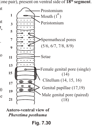
2. Each testis is trilobed and lies in the abdomen, in segments 4, 5 and 6.
3. Two vasa deferentia, arising from the testes, join to form a common ejaculatory duct.
4. Two seminal vesicles at the base of the ejaculatory duct store sperms.
5. The genital chamber has three phallomeres, or gonapophyses, that handle the spermatophore and help in the transfer of sperms.
6. The mushroom gland, also known as the utricular or uricose gland, is a bunch of tubules that secrete a fluid which hardens to form a spermatophore.
7. The conglobate or phallic gland is single, opens into the genital chamber, and releases pheromones as a sex attractant.
[Figure: Fig. 7.29 Reproductive systems of cockroach, showing male: testis, mushroom gland, vas deferens, seminal vesicle, ejaculatory duct, conglobate gland, gonapophyses (phallomere, 3), genital chamber, anal style (from 9th segment) and male gonopore; female: germarium, ovary (with 8 ovarioles), oviduct, common oviduct (vagina), spermatheca (1-bilobed), genital chamber, brood pouch, oothecal chamber, collaterial gland, gonapophyses (6) and female gonopore]
2. Female Reproductive System
1. It consists of the following structures:
(i) Ovaries: one pair
(ii) Oviducts: one pair
(iii) Vagina: single
(iv) Spermatheca: single
5. Gonapophyses: three pairs.
6. Female gonopore: single.
7. Brood pouch: single.
8. Collaterial glands: one pair.
1. Each ovary contains eight ovarioles. Each ovariole is differentiated into a distal germarium and a proximal vitellarium. Eggs mature towards the vitellarium, where yolk deposition is completed.
2. The two oviducts join to form a common oviduct, or vagina, which opens into the brood pouch through a single gonopore.
3. A single but bilobed spermatheca lies near the female gonopore. It stores sperms after mating.
4. The gonapophyses help in handling the spermatophore to transfer sperms into the spermatheca, and later remove it from the female body.
5. The brood pouch is formed by the 7th, 8th and 9th segments. It is differentiated into a proximal genital chamber and a distal oothecal chamber.
6. Of the two collaterial glands, the left one is larger. Both open into the brood pouch. The secretion of the collaterial glands forms the ootheca.
Breeding Season and Fertilisation
1. In cockroach the breeding season is March to September.
2. The spermatophore is formed from the secretion of the mushroom gland around sperms in the ejaculatory duct. The lining of the ejaculatory duct produces a second covering around the spermatophore. Later, when the spermatophore passes out of the male's body, the secretion of the conglobate gland forms a third covering around it.
3. Eight eggs are released from each ovary, one from each ovariole, so 16 eggs in all are released into the genital chamber.
4. During mating, the spermatophore is transferred into the female body with the help of the male gonapophyses. The female gonapophyses fix the spermatophore against the spermathecal opening. All sperms from the spermatophore are transferred into the spermatheca. The empty spermatophore is then thrown out by the female gonapophyses.
5. Fertilisation of the eggs occurs in the genital chamber, by sperms released from the spermatheca.
6. The secretion of the collaterial glands forms an ootheca around the fertilised eggs. All eggs are arranged in two rows of eight. Ootheca formation occurs in the oothecal chamber of the brood pouch.
7. The female cockroach places the ootheca in a dark and warm region. This placement is called oviposition.
Development
1. Cockroach is a Paurometabola insect, showing gradual metamorphosis.
2. Nymphs (16 in number) hatch from the ootheca. Nymphs are immature, smaller in size and wingless.
3. They undergo five to six moultings, each stage called a stadium, to form the adult.
Earthworm
Taxonomic Position
Phylum: Annelida
Class: Oligochaeta
Order: Opisthopora (Haplotaxida)
Genus: Pheretima
Species: posthuma
External Features
1. The body is metamerically segmented, with 100 to 120 segments (metameres), each separated by an intersegmental groove. The number of segments remains fixed throughout life: new segments are not added, and old ones are not removed.
2. The earthworm is burrowing (fossorial).
3. The skin is light brown due to the porphyrin pigment derived from chlorophyll, which the young animal takes in with the soil.
4. The dorsal side can be recognised by a prominent dorsal blood vessel. The ventral side contains two pairs of prominent genital papillae, one pair each on segments 17 and 19.
5. The first segment, called the peristomium, bears the mouth on the ventral side. The hood-like projection of the peristomium is called the prostomium.
6. The male genital apertures (gonopores) are one pair, on the ventral side of the 18th segment.
7. The female genital aperture is single, on the mid-ventral side of the 14th segment.
8. Spermathecal pores are four pairs, in the intersegmental grooves of segments 5/6, 6/7, 7/8 and 8/9.
9. The anal opening is in the last segment.
[Figure: Fig. 7.30 Antero-ventral view of Pheretima posthuma, showing prostomium, mouth (1st), peristomium, spermathecal pores (5/6, 6/7, 7/8, 8/9), setae, female genital pore (single, 14th segment), clitellum (14, 15, 16), genital papillae (17, 19) and male genital pore (paired, 18th segment)]
10. The body of the earthworm is differentiated into three regions:
(1) Pre-clitellar region (1st to 13th segments)
(2) Clitellar region (14th, 15th and 16th segments)
(3) Post-clitellar region (17th to the last segment)
11. Because the total number of segments ranges from 100 to 120, the number of segments in the post-clitellar region may differ between earthworms. The numbers of segments in the pre-clitellar and clitellar regions are fixed.
Study of the earthworm is not included in the NEET syllabus.
Septa
1. Septa are the partitions between adjacent segments, lying in the intersegmental grooves.
2. Septa are absent from segments 1 to 4. The continuous coelomic fluid in these segments makes them turgid, which helps in burrowing.
3. Septa are also absent between the 9th and 10th segments.
4. In the remaining segments (up to the 14th) the septa are non-porous.
5. From the 14th or 15th segment onwards, all septa are porous, and the coelomic fluid, which acts as a hydrostatic skeleton, is interconnected.
Body Wall and Coelom
1. The body wall consists of four layers: cuticle, epidermis, circular muscles and longitudinal muscles.
2. The earthworm is eucoelomate, with a schizocoel lined by somatic (parietal) mesoderm on the outside and visceral (splanchnic) mesoderm on the inside.
3. The coelom contains coelomic fluid, which acts as a hydrostatic skeleton. This fluid oozes out of the body through dorsal pores in the intersegmental grooves, one per segment, from the 12/13th groove to the last segment.
Locomotion
1. Locomotion is carried out by the body wall musculature (circular and longitudinal muscles).
2. Setae are chitinous structures present in setigerous sacs, produced from formative cells. They are embedded in the body wall and can be moved by protractor and retractor muscles.
3. Locomotion depends on coelomic fluid (the hydrostatic skeleton) and the setae.
4. First, the circular muscles of the anterior part contract, and that part becomes longer and thinner. Next, the longitudinal muscles contract, and the diameter of the region increases. To prevent the anterior segments from retracting, the setae are anchored in the ground.
5. This alternate contraction of circular and longitudinal muscles spreads posteriorly, and the whole body moves forward.
6. This process of locomotion, called the 'thinning and thickening process', was described by Gray and Lissman.
Nutrition
1. The earthworm is soil-eating (geophagous), detritivorous or omnivorous.
2. The alimentary canal is complete and highly specialised. It consists of the following parts: mouth and buccal cavity, pharynx, oesophagus, gizzard, stomach and intestine.
[Figure: Fig. 7.31 Alimentary canal of earthworm, showing peristomium (1st segment), mouth, pharynx (3rd and 4th segments), oesophagus, gizzard (8th and 9th segments), stomach (9th to 14th segments), intestine (from 15th segment), intestinal or hepatic caeca (26th to 22nd/23rd) and typhlosole]
7. The pharynx (3rd and 4th segments) is suctorial and is divided into two chambers, a dorsal chamber and a ventral chamber. The pharyngeal gland, or salivary gland, lies on the dorsal side of the pharynx and secretes saliva into the dorsal chamber. The chromophil cells of the pharyngeal gland are analogous to the chief cells of the vertebrate stomach, as they secrete proteolytic enzymes, like pepsin, which digest food in an acidic medium in the presence of humus acid of the soil.
8. The gizzard (8th and 9th segments) is a masticatory organ. It cuts down food material by muscular movements. It has a cuticular lining, and its longitudinal muscles are absent.
9. The stomach extends from the 9th to the 14th segment. In earthworms other than Pheretima posthuma, calciferous glands in the stomach neutralise the acidic medium.
10. The intestine is the longest and widest part of the alimentary canal, extending from the 15th segment to the last. It digests and absorbs food. One pair of intestinal caeca (hepatic caeca) arise from the 26th segment and extend forward up to the 22nd or 23rd segment. These caeca secrete amylase for starch digestion. Other enzymes secreted from the intestinal lining are cellulase, chitinase, lipase and trypsin.
Regions of the Intestine
1. Pre-typhlosolar region (15th to 26th segments)
2. Typhlosolar region (27th to the last 25 segments)
3. Post-typhlosolar region (the last 25 segments)
The post-typhlosolar region, also known as the rectum, stores undigested waste, which is removed from the gut as ball-shaped castings.
Connecting Concepts
The typhlosole is a dorsal infolding of the major part of the intestine. It increases the surface area for absorption and is analogous to the villi of the vertebrate ileum.
Respiration
1. The earthworm respires through its moist skin (cutaneous respiration). The skin is kept moist by coelomic fluid oozing out through the dorsal pores.
2. Because of its cylindrical shape, the surface area to volume ratio is high in the earthworm.
3. There is no specialised respiratory structure in the body.
Excretory System
1. The excretory structures of the earthworm are nephridia (metanephridia). They are ectodermal in origin and are of three types:
(1) Integumentary nephridia
(2) Pharyngeal nephridia
(3) Septal nephridia
Connecting Concepts
In the larvae of polychaetes, the nephridia are simple and are called protonephridia; they have no nephrostome. In the earthworm they are called metanephridia and do have a nephrostome. In integumentary and pharyngeal nephridia, the nephrostome has become secondarily absent. Septal nephridia are truly metanephridia.
1. Integumentary Nephridia
1. They are present in the skin (integument) from the 3rd segment to the last.
2. They are most numerous in the clitellar region (14th, 15th and 16th segments). The term 'forest of nephridia' is used for these in the clitellar region, where their number ranges from 2000 to 2500 per segment.
3. In other segments they number 200 to 250 per segment.
4. They are the simplest nephridia and have no nephrostome.
5. They take excretory matter from the blood and pour it outside the body through nephridiopores. Hence they are called exonephric or exteronephric nephridia.
2. Pharyngeal Nephridia
1. They are present near the pharynx, in the 4th, 5th and 6th segments only.
2. There is one pair of tufts (bunches) per segment. Each tuft has about 100 nephridia, so there are about 200 nephridia per segment.
3. Of the three types, pharyngeal nephridia are the least numerous.
4. They also lack a nephrostome and are therefore simple in structure.
5. Like integumentary nephridia, they take excretory matter from the blood.
6. They pour the excretory matter into the pharynx through nephridial ducts. Hence they are called endonephric or enteronephric nephridia.
3. Septal Nephridia
1. They are attached to the intersegmental septa, from the 15/16th septum to the last.
2. They are called typical nephridia or holonephridia because they are complete and contain a nephrostome.
3. They are present in the number of 80 to 100 per segment.
4. They collect excretory matter from the blood as well as from the coelom (through the nephrostome).
5. The excretory matter is poured through a septal excretory canal and the supra-intestinal duct into the intestine.
6. Such nephridia are also called endonephric or enteronephric nephridia, since the excretory matter is discharged into the gut.
[Figure: Fig. 7.32 Excretory structures in Pheretima, showing pharynx, pharyngeal nephridia and their duct, integumentary nephridia, septal nephridia, septal excretory canal and supra-intestinal duct]
7. Both pharyngeal and septal nephridia pour excretory products into the gut for water absorption, and are therefore adapted for water conservation.
Parts of a Septal Nephridium
1. Nephrostome
2. Twisted lobe (proximal and distal limbs)
3. Straight lobe
4. Terminal duct
Route of Excretory Product through Septal Nephridia
Nephrostome → proximal limb → distal limb → straight lobe → distal limb → proximal limb → terminal duct
1. The excretory product of the earthworm is urea, so it is ureotelic. During the rainy season, when water is plentiful, the major excretory product becomes ammonia.
2. Chloragogen cells, present in the visceral mesoderm of the gut and in the coelomic fluid, synthesise urea and other excretory materials, including chloragogue formed by the metabolism of haem.
3. Chloragogen cells also store reserve food material, and are therefore analogous to the vertebrate liver.
Blood Vascular System (BVS)
1. The BVS of the earthworm is closed type.
2. Haemoglobin is dissolved in plasma, and RBCs are absent. WBCs or phagocytic cells are present.
3. Blood is formed in blood glands in the 4th, 5th and 6th segments. Pharyngeal nephridia and blood glands occur together in the same segments (4th, 5th and 6th).
4. Lymph glands, which produce phagocytic cells and release them into the coelomic fluid, are present as one pair per segment from the 26th segment to the last. They are dorso-lateral in position.
5. The BVS of the earthworm can be studied in two parts.
1. BVS in the First 13 Segments
It consists of the following prominent structures: (i) dorsal blood vessel (DBV), (ii) supra-oesophageal blood vessel (SOBV), (iii) ventral blood vessel (VBV), (iv) lateral oesophageal blood vessels (LOBV), (v) hearts, and (vi) anterior loops.
(i) Dorsal Blood Vessel
1. It is single (unpaired) and is the prominent blood vessel on the mid-dorsal side of the body.
2. It is distributive in nature and supplies blood to the anterior part of the gut: buccal cavity, pharynx, oesophagus, stomach and gizzard.
3. It has one pair of valves per segment, and is comparable to the veins of vertebrates or to the heart of cockroach.
4. Blood flows from posterior to anterior.
5. It is connected to the ventral blood vessel through two pairs of false hearts and two pairs of true hearts.
(ii) Supra-Oesophageal Blood Vessel
1. It is single (unpaired) and present only in the 9th to 13th segments.
2. It is collective in nature and collects blood from the anterior part of the alimentary canal.
3. Valves are absent.
4. It receives blood from the LOBV through the anterior loops.
(iii) Ventral Blood Vessel
1. It is single (unpaired) and lies on the mid-ventral side of the body.
2. It is distributive in nature and supplies blood to the nephridia, body wall, septa and reproductive organs.
3. Blood flows from anterior to posterior.
4. Valves are absent.
5. It receives blood from the dorsal blood vessel through the false and true hearts.
(iv) Lateral Oesophageal Blood Vessel
1. It is paired and lateral in position.
2. It is collective in nature and collects blood from the skin, excretory organs, reproductive organs and so on.
3. Blood flows from anterior to posterior.
4. Valves are absent.
(v) Hearts
1. There are four pairs of hearts (eight in all) in the earthworm.
2. They are of two types:
(a) Lateral hearts (false hearts): two pairs, one pair each in the 7th and 9th segments. They connect the dorsal blood vessel to the ventral blood vessel. Blood flows from dorsal to ventral. Each lateral heart has four pairs of valves.
(b) Lateral oesophageal hearts (true hearts): two pairs, one pair each in the 12th and 13th segments. They connect the dorsal blood vessel and the supra-oesophageal blood vessel to the ventral blood vessel. Blood flows from dorsal to ventral. Each heart has three pairs of valves, one pair at each opening of the dorsal, ventral and supra-oesophageal blood vessels.
(vi) Anterior Loops
1. There are two pairs of anterior loops (four in all), present in the 10th and 11th segments.
2. They are non-pulsating.
3. They connect the supra-oesophageal blood vessel to the lateral oesophageal blood vessels.
4. Blood flows from the LOBV to the SOBV (ventral to dorsal). Valves are absent.
[Figure: Fig. 7.33 Blood vascular system of Pheretima, showing DBV (dorsal blood vessel), VBV (ventral blood vessel), SOBV (supra-oesophageal blood vessel), LOBV (lateral oesophageal blood vessel), CV (commissural vessel), false hearts, true hearts, anterior loops, intestine, intestinal blood vessels, SNBV (subneural blood vessel) and segments 6 to 17]
2. BVS After the 13th Segment
From the 14th segment to the last, the blood vascular system consists of the following important vessels: (i) dorsal blood vessel (DBV), (ii) ventral blood vessel (VBV), (iii) subneural blood vessel (SNBV), (iv) commissural vessels and (v) intestinal vessels.
1. Dorsal blood vessel: after the 13th segment it becomes collective in nature, collecting blood from the alimentary canal, excretory organs and reproductive organs. Since it collects blood from about 100 segments (14th to last), it is mainly collective in nature. The supra-oesophageal blood vessel is absent after the 13th segment.
2. Ventral blood vessel: it is the same throughout the body.
3. Subneural blood vessel: the two LOBVs unite after the 13th segment to form a single vessel that runs posteriorly below the ventral nerve cord. It is connected to the dorsal blood vessel through commissural vessels.
4. Commissural vessels: two (one pair) per segment, present from the 15th segment to the last. They connect the dorsal blood vessel to the subneural blood vessel. Blood flows from ventral to dorsal.
5. Intestinal vessels: two pairs per segment. They are collective in nature and pour blood into the dorsal blood vessel.
Nervous System
The earthworm has a central nervous system (CNS), a peripheral nervous system (PNS) and an autonomic nervous system (ANS).
1. The central nervous system consists of a nerve ring and a paired ventral nerve cord.
2. The nerve ring is formed by supra-pharyngeal ganglia, circum-pharyngeal connectives and sub-pharyngeal ganglia. It lies around the pharynx.
3. The supra-pharyngeal ganglia form the brain of the earthworm.
4. The ventral nerve cord is paired but fused to appear single, and is enclosed in a common sheath. It is ganglionated, with one pair of (fused) ganglia per segment.
5. Above the ventral nerve cord lie four giant nerve fibres of large diameter, which conduct impulses faster.
Sensory Structures
1. Sensory structures in the skin are thigmoreceptors.
2. The dorsal side of the anterior segments, particularly the peristomium and prostomium, contains photoreceptor cells. Each photoreceptor cell has a lens-like structure called a phaosome. These cells do not form an image, but detect the presence of light.
Reproductive System
1. The earthworm is bisexual (monoecious) or hermaphrodite.
2. Both male and female reproductive systems are present on the antero-ventral side of the body.
Male Reproductive System
1. Testes: two pairs (10th and 11th segments).
2. Testes sacs: two (10th and 11th segments).
3. Vasa deferentia: two pairs.
4. Seminal vesicles: two pairs (11th and 12th segments).
5. Prostate gland: one pair (17th to 20th segments).
6. Genital apertures: one pair (18th segment).
1. Testes lie in the testes sacs. They produce spermatocytes, which enter the seminal vesicles.
2. Sperms mature in the seminal vesicles, not in the testes.
3. Sperms pass from the seminal vesicles into the vasa deferentia through the spermatic funnel.
4. Sperms, together with the secretion of the prostate gland, are released through the male genital apertures as spermatic fluid.
5. Accessory glands, which secrete an adhesive substance, open into the genital papillae of the 17th and 19th segments.
Female Reproductive System
1. Ovaries: one pair (13th segment).
2. Oviducts: one pair.
3. Genital aperture: single (14th segment).
4. Spermathecae: four pairs (6th, 7th, 8th and 9th segments).
1. Each ovary consists of a number of ovarioles and is attached to the septum of the 12th/13th segment.
2. Eggs pass through the oviducal funnel and oviduct, and are released through the single genital aperture in the 14th segment.
3. Each spermatheca is differentiated into an outer ampulla and an inner diverticulum. Spermathecae open into the intersegmental grooves of the 5/6, 6/7, 7/8 and 8/9 segments. In Pheretima, sperms of other earthworms are stored in the diverticulum of the spermathecae.
[Figure: Fig. 7.34 Reproductive system of Pheretima, showing spermathecae (6, 7, 8 and 9), testes (10 and 11), testes sac (10 and 11), seminal vesicle (11 and 12), ovary (13), female genital pore (14), vasa deferentia, prostate gland (17 to 20), male genital pore (18), and genital papillae (17 and 19)]
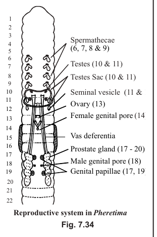
Breeding and Fertilisation
1. Breeding occurs before dawn, during the rainy season.
2. The earthworm is protandrous, and self-fertilisation does not occur. Fertilisation is cross and reciprocal.
3. Two earthworms attach to each other on the antero-ventral side with the help of their genital papillae. The male genital apertures of one earthworm face the first pair of spermathecal openings (5/6 segment) of the other.
4. During mating, sperms of one earthworm are transferred into the diverticulum of the spermathecae of the other, and vice versa.
5. Eggs are released through the female genital aperture in the 14th segment (the anterior clitellar region).
6. The epithelium of the clitellum secretes a cocoon. This cocoon contains unfertilised eggs.
[Figure: Fig. 7.35 Clitellum secretes cocoon, showing sperms, and the cocoon with fertilised eggs]
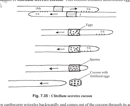
7. The earthworm then wriggles backwards and comes out of the cocoon through its anterior end. When the cocoon passes the openings of the spermathecae, sperms are released over the eggs, and the eggs are fertilised inside the cocoon. Thus fertilisation is external but occurs in the cocoon.
8. Epidermal glands also secrete albumin, which is stored in the cocoon as reserve food for the developing embryo.
Development
1. Development in the earthworm is direct, that is, without a larval stage.
2. Of 2 to 20 embryos, usually 4 mature in the cocoon, and the rest are used as food.
Economic Importance
1. The earliest use of the earthworm was in Unani and Ayurvedic medicine, for the treatment of gout.
2. Earthworms are used as bait for fishing.
3. They are natural ploughmen and make the soil porous.
4. They enrich the soil by adding excretory and faecal matter.
5. Vermicompost is in high demand these days.
Frog
Taxonomic Position
Phylum: Chordata
Class: Amphibia
Order: Anura
Genus: Rana
Species: tigrina
1. Rana tigrina is commonly called the Indian bull-frog.
2. It is amphibious. It is found in ponds and shallow water bodies, and also on land.
3. Its close relative, the toad (Bufo melanostictus), is largely terrestrial.
4. It is cold-blooded (poikilothermic). It shows aestivation (summer sleep) and hibernation (winter sleep) in extreme temperatures.
5. Its body colouration shows camouflage. Its colour-changing ability is called metachrosis.
6. It is insectivorous or carnivorous.
External Morphology
1. The body of the frog is streamlined, well adapted for swimming, leaping and burrowing.
2. The skin is slimy and moist, without an exoskeleton or scales. It contains mucous and poison glands, the latter concentrated on the shoulders. Chromatophores in the skin help in camouflage.
3. The eyes are placed dorso-laterally on the head. The frog has three eyelids: upper, lower and a third eyelid (nictitating membrane), which covers the eyeball during swimming.
4. The external ear is absent. The tympanum (ear drum) lies near the eyes.
5. The body is divided into head and trunk. The neck is absent. The trunk bears one pair of shorter forelimbs and one pair of longer hind limbs.
6. Each hand has four fingers (digits), and each foot has five toes (digits). The pollex, or thumb, is absent in the frog.
Internal Morphology (Anatomy) and Physiology
(a) Digestive System
1. Because the frog is carnivorous, its alimentary canal is short. The posterior-most part of the gut is the cloaca. Urine also passes out through the cloacal opening.
2. Uvula is absent, so the buccal cavity and pharynx are not differentiated. The bucco-pharynx contains numerous maxillary teeth. The teeth of frog are homodont (similar in type), acrodont (fused with the jaw bones) and polyphyodont (many sets of teeth). Teeth are absent in the lower jaw. Besides the upper jaw, teeth are also present on the vomer bone of the olfactory capsule, in the roof of the bucco-pharynx.
3. The teeth are not used for cutting food, but to prevent the escape of prey.
4. The tongue of the frog is bifid at the tip and free behind. It is used for capturing prey (insects).
(b) Respiratory System
1. The frog respires through the skin (cutaneous respiration), the lining of the bucco-pharynx, and the lungs (pulmonary respiration). During aestivation and hibernation, cutaneous respiration is the only mode of respiration.
2. The lungs are hollow, non-lobular and of the positive-pressure type. Pulmonary respiration has three phases: aspiration, inspiration and expiration. During aspiration, the sternohyoid
muscles of the lower jaw contract and air enters the bucco-pharynx through the nostrils. During inspiration the lower jaw is raised (due to contraction of the petrohyoid muscles) and the nostrils are closed, so air is forced into the lungs. The lungs are positive-pressure lungs. During expiration, air leaves the body through the nostrils.
(c) Blood Vascular System
1. The BVS of frog is closed type and has double circulation, though it is not as efficient as in birds and mammals.
2. The heart is three-chambered, with two atria and one ventricle.
3. On the dorsal side of the heart is a triangular structure, the sinus venosus, which receives deoxygenated blood from two precavals and one postcaval vein. It pours the blood into the right atrium.
4. On the ventral side of the heart is a prominent structure, the conus arteriosus, which arises from the ventricle. The conus arteriosus is differentiated into a proximal pylangium (bulbous arteriosus) and a distal synangium (ventral aorta). The pylangium contains cardiac muscle and is part of the heart. It contains a fold-like spiral valve, which divides it into a right ventral chamber, the cavum aorticum, and a left dorsal chamber, the cavum pulmocutaneum.
5. Each branch of the ventral aorta contains three arches: pulmocutaneous (dorsal), systemic (middle) and common carotid (ventral).
6. The SA node is present in the wall of the sinus venosus, not in the right atrium as in mammals.
7. Since there is a single ventricle (no interventricular septum), the bicuspid and tricuspid valves are not differentiated, and blood from the two atria mixes in the ventricle.
8. The AV node and bundle of His are absent in frog.
9. There are two types of portal systems in the frog: the hepatic portal system (between gut and liver) and the renal portal system (between posterior abdominal parts and kidney).
[Figure: Fig. 7.36 Heart of frog (dorsal, ventral and frontal sections), showing pulmonary veins, aortic trunks, carotid (ventral), systemic and pulmocutaneous (dorsal) arches, anterior vena cavae or precavals, right atrium, left atrium, sinus venosus, conus arteriosus, bulbous arteriosus (BA), ventral aorta (VA), spiral valve, semilunar valves, ventricle, atrio-ventricular valves and posterior vena cavae or postcaval (BA-bulbous arteriosus or pylangium, VA-ventral aorta or synangium)]
Renal Portal System
1. The femoral vein, arising from the outer thigh region, bifurcates after entering the abdomen to form an outer renal portal vein and an inner pelvic vein.
2. The renal portal vein ends in the kidney. The pelvic veins of the two sides join to form the anterior abdominal vein, which finally ends in the liver.
3. The sciatic vein, arising from the inner thigh region, joins the renal portal vein and pours its blood into it.
[Figure: Renal portal system of frog, showing the liver, anterior abdominal vein, left kidney and right kidney]
[Figure: Fig. 7.37 Renal portal system of frog, showing femoral vein, sciatic vein, pelvic vein, renal portal vein, anterior abdominal vein, liver, right kidney and left kidney]
(d) Nervous System
1. The frog has all three types of nervous system: CNS (brain and spinal cord), PNS (cranial and spinal nerves) and ANS (sympathetic and parasympathetic nervous systems).
2. The frog has 10 pairs of cranial nerves and 10 pairs of spinal nerves. In Rana tigrina, however, there are 9 pairs of spinal nerves.
3. In the brain, the cerebral hemispheres are less developed than in mammals. The corpus callosum, which connects the two cerebral hemispheres in mammals, is absent in frog.
4. The mid-brain consists of two optic lobes (corpora bigemina). Both lobes are hollow, with a cavity called the optocoel.
5. The hind brain consists of the cerebellum and medulla oblongata. The pons is absent in frog.
(e) Sensory Organs
1. The eyeball of frog is cartilaginous. Colour vision is lacking, because there are no cones in the retina.
2. In place of the lacrimal and meibomian glands, frog has Harderian glands.
3. The sensory part for hearing in the internal ear of frog is the pars basilaris and pars lagena. The cochlea and organ of Corti are absent. The round window (fenestra rotunda) between the middle and internal ear is also absent.
4. In the olfactory capsule, the septomaxillary bone is the smallest bone of the frog's body.
Endoskeleton
Skull
The skull has two parts: (i) neurocranium and (ii) splanchnocranium.
[Figure: Fig. 7.38 (A) Skull, dorsal view, showing septomaxillary, premaxilla, premaxillary teeth, internal nares, external naris, nasal, cartilaginous nasal capsule or olfactory chamber, vomer, vomerine teeth, maxilla, maxillary teeth, frontoparietals, orbital fossa, palatine foramen for vein and nerve, pterygoid, squamosal, prootic, auditory capsule, quadratojugal, quadrate cartilage, fenestra ovalis, columella, exoccipital, occipital condyle, foramen magnum, sphenethmoid diamond-shaped area, parasphenoid, foramina for II, V, VI and VII nerves; (B) Skull, ventral view]
(i) Neurocranium
The neurocranium forms the cranium (enclosing the brain) and the sensory capsules (enclosing the sense organs).
(A) Cranium is the brain box and is mainly differentiated into:
1. Occipital region: formed by two exoccipitals, which enclose the foramen magnum. Each exoccipital has one occipital condyle, so the skull is dicondylic.
2. Frontal and parietal regions: fused to form the fronto-parietal bone (dorsally) and the parasphenoid (ventrally). The parasphenoid is unpaired and is an inverted T-shaped bone. Between the fronto-parietal and the parasphenoid, in the anterior part, lies another unpaired bone, the sphenethmoid, of which only a diamond-shaped area is visible.
(B) Sensory Capsules include the optic capsule, auditory capsule and olfactory capsule.
1. Optic capsule: has no specific bone.
2. Auditory capsule: includes the columella auris (in the middle ear) and the prootic (around the internal ear or membranous labyrinth).
3. Olfactory capsule: consists of the nasal (dorsal), vomer (ventral), which bears vomerine teeth, and septomaxillary (anterior). The septomaxillary is the smallest bone in the frog's body.
(ii) Splanchnocranium forms the visceral skeleton: the lower jaw, upper jaw, suspensorium and hyoid apparatus.
(A) Upper Jaw
1. Each half (ramus) of the upper jaw consists of six bones: three on the outer margin (premaxilla, maxilla and quadratojugal), two on the inner margin (palatine and pterygoid), and one on the dorsal side (squamosal).
2. The premaxilla and maxilla bear teeth (acrodont, homodont and polyphyodont type).
3. The quadratojugal is comma-shaped (quadrate is cartilage and jugal is bone). The palatine is rod-shaped and the pterygoid is tri-radiate. The squamosal is also tri-radiate but hammer-shaped.
(B) Lower Jaw
1. Each half (ramus) of the lower jaw consists of three bones (angulosplenial, dentary and mentomeckelian) and one cartilage (Meckel's cartilage). The two halves are attached at the mentomeckelian. Teeth are absent in the lower jaw of frog.
2. The lower jaw articulates with the quadratojugal, through the angulosplenial, and forms the autostylic type of suspensorium. The quadratojugal is called the bone of the suspensorium.
3. The occipitals, sphenethmoid, mentomeckelian and auditory capsule are cartilaginous or replacing bones. The rest of the skull bones are investing or dermal bones.
[Figure: Fig. 7.39 Lower jaw, showing dentary, angulosplenial, coronary process, mentomeckelian and articular facet for quadrate]
(C) Hyoid Apparatus
1. It consists of the body of hyoid, anterior cornua, posterior cornua and alar processes. The whole hyoid apparatus, except the posterior cornua, is cartilaginous.
2. The posterior cornua are bony (replacing bone) and guard the glottis opening.
Vertebral Column
Total number of vertebrae = 10.
1. Cervical: one (1st vertebra).
2. Thoracic: six (2nd to 7th vertebrae).
3. Lumbar: one (8th vertebra).
4. Sacral: one (9th vertebra).
5. Caudal: one (10th vertebra).
1. 1st vertebra (atlas): ring-shaped, with a reduced centrum. Prezygapophyses and transverse processes are absent. The anterior side, towards the skull, has two facets for the occipital condyles (the skull is dicondylic).
2. 2nd to 7th vertebrae (typical vertebrae): centrum is procoelous, pre- and post-zygapophyses are present, and transverse processes are well developed.
3. 8th vertebra: centrum is amphicoelous. Its other features are similar to a typical vertebra.
4. 9th vertebra: centrum is acoelous, with a double convexity on the posterior side.
5. 10th vertebra (urostyle): centrum is rod-shaped. It is sword-shaped, formed by the fusion of several caudal vertebrae of the tadpole. It is the longest vertebra, with two concavities on the anterior side that fit the two convexities of the 9th vertebra.
Sternum
The sternum has four components, from anterior to posterior: episternum (cartilaginous), omosternum (inverted Y-shaped and bony), mesosternum (rod-shaped and bony) and xiphisternum (cartilaginous).
Girdles
Pectoral Girdle
1. The pectoral girdle is arc-shaped and mainly consists of replacing bones. Its two halves are fused with the sternum to protect the lungs and heart. Ribs are absent in frog.
[Figure: Fig. 7.40 Pectoral girdle and sternum, showing clavicle, episternum, suprascapula, omosternum, scapula, precoracoid, coracoid, glenoid cavity, mesosternum, epicoracoid and xiphisternum]
1. Each half of the pectoral girdle consists of:
(i) Coracoid
(ii) Scapula: the universal bone of the pectoral girdle, with an acromion process.
(iii) Glenoid cavity: between the coracoid and scapula, for articulation with the head of the humerus.
(iv) Clavicle: the collar bone, an investing bone.
(v) Suprascapula: calcified cartilage.
Pelvic Girdle
1. Each half, called the innominate bone, consists of the ilium (the longest), ischium and pubis. The pubis is the smallest component and is of calcified cartilage.
2. The acetabulum (for articulation with the head of the femur) is formed by all three bones.
3. The obturator foramen is absent.
[Figure: Fig. 7.41 (A) Pelvic girdle, showing facet for 9th vertebra, ilium, iliac crest, articular surface for urostyle, acetabulum, ischium and pubis; (B) Innominate bone, showing ilium, iliac crest, acetabulum, ischium and pubis]
Limb Bones
(1) Fore Limbs
Each fore limb has 23 bones.
1. Humerus (1): characterised by a deltoid ridge.
2. Radio-ulna (1): the radius and ulna are fused. The ulna is characterised by an olecranon process.
3. Carpals (6): arranged in two rows of three each.
4. Metacarpals (5).
5. Phalanges (10): the thumb (pollex) is absent. Phalangeal formula is 0-2-2-3-3.
(2) Hind Limbs
Each hind limb has 25 bones.
1. Femur (1): stretched, S-shaped. The trochanter is absent.
2. Tibio-fibula (1): the longest bone of the frog's body, with a nutrient foramen in the middle. The tibia has a small cnemial crest on its anterior side.
3. Tarsals (4): arranged in two rows. The proximal tarsals are well developed and are called astragalus or tibiale (thin and curved) and calcaneum or fibulare (thick and straight).
4. Metatarsals (5).
5. Phalanges (14): the hallux, or great toe, is present. Phalangeal formula is 2-2-3-4-3, and the ring finger is the longest.
6. The pivotal joint is absent in frog, since the axis vertebra (odontoid process) is lacking.
[Figure: Fig. 7.42 Endoskeleton of frog (dorsal view), showing maxilla, premaxillae, nasal, orbital fossa, frontoparietals, squamosal, atlas, typical vertebrae, 8th and 9th vertebrae, urostyle, suprascapula, radio-ulna, humerus, carpals, metacarpals, phalanges, femur, tibio-fibula, calcar, astragalus, calcaneum, distal tarsals and metatarsals]
Excretory System
1. The kidneys of frog are mesonephros. They are not differentiated into cortex and medulla.
2. Each kidney has four to five pairs of renal arteries and veins.
3. The nephrons (uriniferous tubules) are not differentiated into PCT, DCT and loop of Henle.
4. The major excretory product of frog is urea (ureotelic).
Reproductive System
Sexual dimorphism in frog is prominent during the breeding season. The male frog differs from the female in the following features.
1. The male has a nuptial pad at the base of the first finger (actually the second finger, as the pollex is absent).
2. Males have vocal sacs for amplifying sound.
3. The colour pattern of the male is different from that of the female.
[Figure: Reproductive system of frog, showing fat body, vasa efferentia and ostium, and fat bodies and oviduct]
[Figure: Fig. 7.43 Urinogenital system in frog. A: male, showing right testis, left testis, adrenal gland, kidney, urinogenital duct, rectum, urinary bladder, cloaca and cloacal aperture; B: female, showing kidney, adrenal gland, ureter, ovary, ovisac, rectum, urinary bladder, cloaca and cloacal aperture]
Male Reproductive System
1. The testes are attached to the kidney by a fold of peritoneum called the mesorchium.
2. Sperms from the testes pass through 10 to 12 vasa efferentia and are released into Bidder's canal, a collecting duct of the kidney. This is because the vas deferens is absent in frog.
3. The mesonephric (Wolffian) duct of the kidney is therefore also known as the urinogenital duct. Faecal matter, urine and sperms are all released through the cloacal opening.
Female Reproductive System
1. The ovaries lie close to the kidney but, unlike the testes, are not functionally connected to it.
2. The oviducts open separately into the cloaca.
Development
1. The frog is a spontaneous or seasonal ovulator, meaning ovulation occurs according to the season. (The rabbit is an induced or reflex ovulator, in which copulation induces ovulation.) Frogs lay 2000 to 4000 eggs in a spawn.
2. The eggs of frog are mesolecithal and telolecithal. The animal hemisphere is pigmented due to melanin, and shows camouflage. The eggs are at the secondary oocyte stage, with one polar body.
3. Each egg is covered by an inner non-living vitelline membrane and an outer jelly, which is secreted by the oviducts.
4. The jelly is protective (a shock absorber), distasteful, adhesive and a non-conductor of heat. It is light in weight and keeps the eggs floating on the water surface, to ensure sufficient oxygen and sunlight. The jelly also contains fertilizin, which helps sperms attach to the egg.
5. Copulation in frog occurs without a copulatory organ in the male, and is called amplexus.
6. Fertilisation is external, in water. After fertilisation, movement of melanin particles forms a grey crescent opposite the point of sperm entry. The first cleavage passes through the middle of the grey crescent, establishing bilateral symmetry.
7. Cleavage is unequal holoblastic. The first and second cleavages, however, are equal holoblastic.
8. Development is indirect, and the larva is the tadpole.
[Figure: Fig. 7.44 Fertilised egg of frog, showing polar bodies, animal hemisphere, vegetal hemisphere, grey crescent, jelly coats (3) and vitelline membrane]
The important stages of development are:
Zygote → Morula → Blastula → Gastrula → Neurula → Tail-bud stage → Gill bud stage → External gill stage → Internal gill stage (mature tadpole).
[Figure: Fig. 7.45 (A) Blastula, showing (a) entire view and (b) sagittal section with animal hemisphere and vegetal hemisphere; (B) Gastrula, showing lateral blastopore lip, gastral mesoderm, yolk plug and archenteron]
[Figure: Fig. 7.46 Neurula, showing neurocoel, neurenteric canal, hindgut, proctodeum, rectum, midgut, notochord, foregut, stomodaeum, optic vesicle, head mesenchyme, oral evagination, olfactory pit, mandibular arch, oral sucker or cement gland, heart mesenchyme, liver diverticulum, yolk, rudiment of eye, external gills, myotomes, tail fin, gill clefts and cloacal aperture]
[Figure: Fig. 7.47 (A) External gill stage, showing eye, 3 external gills, myotomes, olfactory pit, mouth with horny jaws, oral sucker, opercular fold, tail fin and cloacal aperture; (B) Tadpole, showing eye, olfactory pit, mouth with horny jaws, myotomes, tail fin, hindlimb, operculum, spiracle and cloacal aperture]
1. A mature tadpole has a pronephros kidney, a cartilaginous skeleton, and respires through the skin and four pairs of internal gills.
2. Its blood vascular system is fish-like, and its heart is two-chambered.
3. Its alimentary canal is coiled like a spring. It is herbivorous.
4. The fore limbs and hind limbs are developed, but the fore limbs are not visible because they are covered by the operculum over the gills.
5. The excretory product is ammonia.
Metamorphosis
1. Metamorphosis occurs in the presence of the hormone thyroxine. In deficiency of thyroxine or iodine, the tadpole increases in size and does not undergo metamorphosis.
2. The emergence of the fore limbs out of the operculum is the first sign of metamorphosis.
3. The nervous system is least affected, and the blood vascular system is most affected, during metamorphosis.
4. Important destructive or regressive changes in metamorphosis are: (i) shedding of horny jaws and teeth, (ii) resorption of the tail by autolysis, and (iii) disappearance of the cloacal tube, lateral line organs, operculum, spiracle and internal gills.
Biological Importance of Frog
1. It is used in laboratories for teaching and research.
2. Being insectivorous, it protects crops from insect pests and saves expenditure on insecticides.
3. Its muscular legs are used as food and in pickles.
4. Froglets or tadpoles can be used as fish bait.
5. The frog is an important link in the ecosystem and helps maintain ecological balance.
Free NEET Study Guides: NEET Biology Animal Tissues
Core Study Kit: NEET Biology Animal Tissues
Access comprehensive study material for Animal Tissues, including revision notes, concept maps, and high-probability questions. These resources are designed in alignment with the latest 2026 NEET syllabus for NEET Biology to support effective exam preparation.
NCERT-Aligned Solutions for Animal Tissues
Each resource draws directly from authorized textbooks to maintain academic accuracy. Evaluating solved examples allows NEET students to master the formal presentation and answer-writing standards expected in upcoming school exams.
Next Steps in Your Exam Preparation
Wrap up your chapter revision by testing your knowledge against standard objective question formats. Explore our full library of free, up-to-date sample papers and online assessments to secure top grades in your NEET examinations.
FAQs
Our advanced study package for Animal Tissues includes detailed concepts, diagrams, Mind Maps, and explanation of complex topics to ensure NEET students learn as per syllabus for 2026 exams.
The Mind Maps provided for Animal Tissues act as visual anchors which will help faster recall during high-pressure exams.
Yes, teachers use our NEET Biology resources for lesson planning as they are in simple language and have lot of solved examples.
Yes, You can download the complete, mobile-friendly PDF of the Biology Animal Tissues advanced resources for free.
Yes, our subject matter experts have updated the Animal Tissues material to align with the rationalized NCERT textbooks and have removed deleted topics and added new competency-based questions.