

Official NEET Study Materials for NEET Biology
Review targeted academic resources with the NEET Biology Molecular Basis of Inheritance Notes. Built according to official NEET standards for the 2026-27 term, these downloadable NEET Biology study materials support effective daily practice and deeper conceptual understanding for Molecular Basis of Inheritance.
Advanced Resources for Biology
Access the complete useful resource PDF for NEET Biology below. Regular practice with these targeted academic notes builds familiarity with complex topics and helps secure higher marks in final school evaluations.
Molecular basis of inheritance explains how genetic information is stored, copied, expressed and regulated at the molecular level. This NEET Biology notes page for Class 12 covers the structure of nucleic acids (DNA and RNA), the double helix and packaging of DNA, the experiments that proved DNA as the genetic material, DNA replication, transcription, the genetic code, translation, regulation of gene expression and the Human Genome Project.
Terms and Definitions to Know
1. Melting of DNA
2. Annealing of DNA
3. Ribozyme
4. Solenoid model
5. DNA proof reading
6. Monocistronic and polycistronic gene
7. Split gene
8. RNA splicing
9. RNA editing
10. Wobble position
11. Junk DNA
12. VNTRs (variable number tandem repeats)
13. Southern blotting
14. DNA typing
Topics Covered
1. Nucleic acids (DNA and RNA)
2. Chemical structure of nucleic acid
3. DNA as genetic material, including Griffith's experiment, Hershey-Chase experiment and bacterial transformation
4. Replication
5. Transcription
6. Genetic code and tRNA as an adapter molecule
7. Translation
8. Regulation of gene expression
9. Human Genome Project
Differences to Note
1. Differences between A, B, C, D and Z-DNA.
2. Differences between prokaryotic and eukaryotic transcription.
Nucleic Acids (DNA and RNA)
1. Nucleic acids are present in all cells and viruses.
2. They are polymers of nucleotides and hence macromolecules. The structural unit of a nucleic acid is the nucleotide.
3. They are informational molecules of universal occurrence.
Chemical Structure of Nucleic Acid
On hydrolysis, a nucleic acid yields a pentose sugar, two types of heterocyclic nitrogenous bases (purines and pyrimidines) and phosphoric acid.
| Nucleic Acid | Purines | Pyrimidines |
|---|---|---|
| DNA | Adenine and Guanine | Cytosine and Thymine |
| RNA | Adenine and Guanine | Cytosine and Uracil |
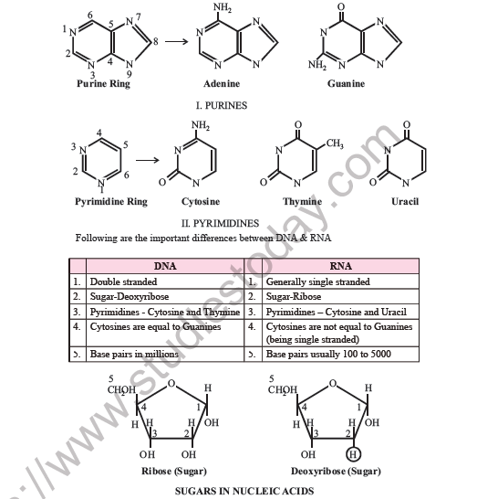
Purines and Pyrimidines
1. Purines (two rings): Adenine and Guanine.
2. Pyrimidines (one ring): Cytosine, Thymine and Uracil. Thymine is found in DNA, and uracil is found in RNA.
Ring structures and numbering
1. Purine ring: a six-membered ring fused to a five-membered ring, numbered N1, C2, N3, C4, C5, C6, N7, C8, N9.
2. Adenine: purine with an NH2 group at C6.
3. Guanine: purine with an O (keto) group at C6 and an NH2 group at C2.
4. Pyrimidine ring: a single six-membered ring numbered N1, C2, N3, C4, C5, C6.
5. Cytosine: pyrimidine with an NH2 group at C4 and an O at C2.
6. Thymine: pyrimidine with O at C2 and C4, and a CH3 group at C5.
7. Uracil: pyrimidine with O at C2 and C4 (no methyl group).
8. Sugar rings: the pentose ring has an O in the ring and carbons numbered 1' to 4'. Carbon 5' is the CH2OH group attached to C4'. Ribose has an OH on C2'; deoxyribose has H in place of that OH.
Differences Between DNA and RNA
| Feature | DNA | RNA |
|---|---|---|
| Strands | Double stranded | Generally single stranded |
| Sugar | Deoxyribose | Ribose |
| Pyrimidines | Cytosine and Thymine | Cytosine and Uracil |
| Base equality | Cytosines are equal to guanines | Cytosines are not equal to guanines (being single stranded) |
| Size | Base pairs in millions | Base pairs usually 100 to 5000 |
Sugars in Nucleic Acids
[Figure: Ribose (sugar) and deoxyribose (sugar) rings numbered 1 to 5, with a 5-CH2OH group at the top. Ribose has an OH group on carbon 2; deoxyribose has H in place of that OH group]
Deoxyribonucleic Acid (DNA)
1. DNA is mainly located in the nucleus.
2. A small amount of DNA is also present in mitochondria and chloroplasts.
3. Mitochondrial DNA (mtDNA) is circular and contains about 16,500 nucleotides. It has information for the synthesis of 2 rRNA and 22 tRNA molecules. It is inherited from the mother through the female gamete. It has a low mutation rate but does not show genetic recombination.
Nucleosides
A combination of a pentose sugar with a nitrogenous base (purine or pyrimidine) is called a nucleoside.
1. Nucleosides of purines: Adenine gives adenosine; guanine gives guanosine.
2. Nucleosides of pyrimidines: Cytosine gives cytidine; thymine (in DNA) gives thymidine; uracil (in RNA) gives uridine.
Nucleotides
1. A phosphate ester of a nucleoside is called a nucleotide. Each nucleotide consists of a nitrogenous base, a pentose sugar and one or more phosphate groups.
2. Nucleotides of purines: Adenosine with one phosphate gives adenosine monophosphate (AMP), also called adenylic acid. Adenosine with two phosphates gives ADP. Adenosine with three phosphates gives ATP. Guanosine with one phosphate gives guanosine monophosphate (GMP), also called guanylic acid.
3. Nucleotides of pyrimidines: Cytidine with one phosphate gives cytidine monophosphate (CMP), also called cytidylic acid. Cytidine with two phosphates gives CDP, and with three phosphates gives CTP. Thymidine with one phosphate gives thymidine monophosphate (thymidylic acid). Uridine with one phosphate gives uridine monophosphate (uridylic acid).
4. In deoxyribose nucleotides the names are preceded by "d", for example dAMP (deoxyadenosine monophosphate) and dTDP (deoxythymidine diphosphate).
ATP (adenosine triphosphate) is also a nucleotide. It contains one adenine base, one ribose sugar and three phosphate groups. It is an energy-rich compound and is called the energy currency of the cell. Its second and third phosphate bonds are energy-rich bonds, each releasing 31 kJ of energy per mole.
Polynucleotide Chain
1. Nucleotides are joined by phosphodiester linkages between the 5' carbon and the 3' carbon of the pentose sugars. The carbons of the pentose sugar are numbered 1', 2', 3' and so on, to distinguish them from the carbons of the nitrogenous bases.
2. The chain of a nucleic acid is written from the 5' end to the 3' end, from left to right.
3. The nucleotides are acids and are negatively charged at neutral pH.
[Figure: Schematic representation of a polynucleotide chain, with the 5' end at the phosphate and the 3' end at the sugar, and bases attached to the sugars; shown in short form as a line with the bases written along it]
DNA double helix and antiparallel strands (labels of the figure)
1. DNA double helical structure: sugar-phosphate backbone, two antiparallel strands, nitrogenous bases, minor groove, major groove. One complete turn has 10 base pairs = 3.4 nm. The diameter is 2 nm.
2. Two antiparallel strands with nitrogenous bases: base pairs joined by hydrogen bonds (T=A, G≡C, C≡G). One strand runs 5' to 3' and the other 3' to 5'. The 5' end and the 3' end are marked. Sugars are linked by phosphodiester bonds, shown as P.
3. Representation in short form: two chains of sugar and P, one running 5'→3' and the other 3'→5'.
4. Key: P = phosphodiester linkage; A = adenine (purine); T = thymine (pyrimidine); G = guanine (purine); C = cytosine (pyrimidine).
Chargaff's Rules
1. Edwin Chargaff reported that the amount of adenine is equal to thymine (A = T), and the amount of guanine is equal to cytosine (G = C).
2. The total number of purines is equal to the total number of pyrimidines (A + G = T + C).
3. The base composition of DNA varies from species to species, so the AT/GC ratio also varies. The AT/GC ratio is 1.52 in humans and 0.93 in E. coli.
Double Helical Structure of DNA
1. To explain base equivalence (A/T, G/C) and other properties of DNA, Watson and Crick (1953), using X-ray diffraction studies, proposed the double helical structure of DNA.
2. The structure has two right-handed helical polynucleotide chains (strands) wound around a central axis, like a spiral staircase.
3. The two strands are antiparallel, that is, the 5' end of one strand lies opposite the 3' end of the other.
4. The sugar-phosphate backbone forms the rails or sides of the ladder, and the bases are held inside like the steps (rungs) of the staircase by hydrogen bonds.
5. Between A and T there are two hydrogen bonds (A = T). Between C and G there are three hydrogen bonds (C ≡ G). The C ≡ G pair is more stable than the A = T pair.
6. The diameter of the double helix is 2 nm (20 Å).
7. The distance from one base pair to the next (rise) is 0.34 nm (3.4 Å).
8. There are 10 base pairs in one complete turn (pitch) of B-DNA. The pitch is therefore 3.4 nm (34 Å).
Types of DNA
On the basis of the number of base pairs per turn and the handedness of the helix, DNA exists in more than 12 forms. The main forms are:
| Type | Type of helix | Properties |
|---|---|---|
| A-DNA | Right handed | 11 base pairs per turn; formed under artificial conditions |
| B-DNA | Right handed | 10 base pairs per turn; the most stable and most common form, described by Watson and Crick |
| C-DNA (9½ base pairs per turn) | Right handed | Formed under artificial conditions |
| D-DNA (8 base pairs per turn) | Right handed | Formed under artificial conditions |
| Z-DNA | Left handed | 12 base pairs per turn (6 dinucleotides); repeating unit is a dinucleotide; sugar-phosphate rail is zig-zag, hence named Z-DNA; pitch 45 Å; diameter 18 Å |
Denaturation and Annealing of DNA
1. On heating, the two strands of DNA separate from each other. This is called melting, or denaturation.
2. On cooling, the strands hybridise again. This is called annealing, or renaturation.
3. The temperature at which the two strands separate completely is the melting temperature (Tm), which is specific for each sequence.
4. A DNA sample with a higher Tm has more C ≡ G pairs, because these have three hydrogen bonds and are harder to break.
5. Separated or denatured DNA strands absorb more light than the intact double strand. This increased absorption is called the hyperchromic effect. It is used to show whether DNA is single or double stranded.
Ribonucleic Acid (RNA)
1. RNA has a single helical structure and is mainly of three types: mRNA, rRNA and tRNA.
2. RNA is double stranded in reoviruses, wound tumour virus, rice dwarf virus and mycophages.
Messenger RNA (mRNA)
1. mRNA makes up about 5% of the total RNA.
2. Its base sequence is complementary to one DNA strand.
3. It has more than 1500 nucleotides and is the longest RNA when the length of the polynucleotide is considered.
4. It has the shortest life, about 1 to 4 minutes.
5. Its length varies according to the length of the polynucleotide used. It generally exists for only a short time.
Ribosomal RNA (rRNA)
1. rRNA makes up about 80% of the total RNA.
2. It is synthesised in the nucleolar organiser region (NOR) of the DNA. Its base sequence is similar in all organisms.
3. It has the longest life, several days, and is the most stable RNA.
4. Ribozyme, an RNA that acts as an enzyme, is also a type of rRNA.
Transfer RNA (tRNA)
1. tRNA makes up about 15% of the total RNA.
2. It is the smallest RNA, with an average of 80 nucleotides per molecule.
3. More than 20 different tRNAs may be present in a cell.
4. It is easily soluble, so it is also called soluble RNA (sRNA).
5. It transfers amino acids from the cytoplasm to the ribosomal machinery.
6. It folds on itself to form a clover-leaf structure. Unusual bases such as dihydrouracil, pseudouridine and hypoxanthine (inosine) cause the single strand to coil into an L shape or clover shape.
DNA Packaging in Prokaryotes
1. The DNA of bacteria such as E. coli is a circular, double-stranded molecule with 4.6 million base pairs.
2. The circular DNA is packaged into the nucleoid, where it is organised into loops or domains bound to a central protein.
3. The scaffold DNA is supercoiled, that is, twisted upon itself, and is complexed with 7 DNA-binding proteins like HU, HLP and HNS. HLP is a histone-like basic protein.
C-Value
The haploid DNA content of an organism is called its C-value.
The Nobel Prize for artificial synthesis of DNA (gene), or DNA synthesis in vitro, was given to A. Kornberg (1959).
The Nobel Prize for artificial synthesis of RNA was given to S. Ochoa (1959).
DNA Packaging in Eukaryotes
1. DNA is organised into a beaded structure called the nucleosome.
2. Histones are a set of positively charged basic proteins rich in the amino acids lysine and arginine.
3. Eight histone molecules form a histone octamer. Negatively charged DNA is wrapped around the positively charged octamer to form a nucleosome.
4. There are five classes of histone: H1, H2A, H2B, H3 and H4. H1, H2A and H2B are rich in lysine, and H3 and H4 are rich in arginine.
5. H3 and H4 are the most conserved histones, and H1 is the least conserved.
6. The histone octamer has two molecules each of H2A, H2B, H3 and H4.
7. DNA makes 1.8 left-handed turns around the octamer. A typical nucleosome contains about 200 bp of DNA, of which about 146 nucleotide pairs are wrapped around the octamer.
8. Nucleosomes are joined by linker DNA, which is associated with the H1 protein.
9. Winding around the nucleosome shortens the DNA about seven-fold.
10. Nucleosomes form chromatin in the form of beads on a string.
11. Chromatin condenses or supercoils at metaphase to form chromosomes. This packaging needs an additional set of proteins called NHC (non-histone chromosomal) proteins. NHC are high molecular weight proteins rich in the amino acids tyrosine and tryptophan.
12. The DNA packaging is best explained by the solenoid model.
[Figure: Fig. 6.1 Structure of nucleosome, showing a nucleosome bead of about 10 nm with 8 histone molecules and 146 nucleotide pairs of DNA, and linker DNA]
[Figure: Fig. 6.2 Bead on a string structure of chromatin, showing nucleosome, wrapped DNA, histone octamer and linker DNA]
Order of Packaging
DNA → histone octamer → nucleosome → 6 nucleosomes → solenoid → 7 solenoids → 30 nm fibre → chromatin → protein scaffold or nuclear matrix → radial loops → packaging → chromosome.
Types of Chromatin
| Feature | Euchromatin | Heterochromatin |
|---|---|---|
| Transcription | Transcriptionally active | Transcriptionally inactive |
| Packing | Loosely packed, rich in gene concentration | Densely packed |
| Staining | Lightly stained | Darkly stained |
DNA as the Genetic Material
Several experiments proved that DNA is the genetic material.
Griffith's Experiment (1928)
1. Frederick Griffith, a British medical officer, described the phenomenon of bacterial transformation. He worked with Streptococcus pneumoniae, the bacterium that causes pneumonia, which he used to infect mice.
2. The bacteria form two types of colonies on a culture plate: S (smooth, due to a mucous polysaccharide capsule that makes it infective) and R (rough, with no mucous polysaccharide).
Steps of the experiment:
1. S strain (virulent) injected into mice: the mice die.
2. R strain (non-virulent) injected into mice: the mice survive.
3. Heat-killed S strain injected into mice: the mice survive.
4. Heat-killed S strain plus live R strain injected into mice: the mice die.
Conclusion: Some material from the heat-killed S strain transformed the R strain, making it virulent and enabling it to make the smooth polysaccharide. This shows that genetic material passed from the S strain to the R strain.
[Figure: Griffith's experiment of bacterial transformation, showing four mouse experiments with S strain, R strain and heat-killed S strain]
Avery, MacLeod and McCarty (1933-34)
1. Avery, Colin MacLeod and Maclyn McCarty revealed the chemical nature of the transforming substance to be DNA.
2. They purified DNA, RNA and protein from heat-killed S strain and tested which could transform R cells into S cells.
3. Protease (protein-digesting enzyme) and RNase (RNA-digesting enzyme) had no effect on transformation, whereas digestion of DNA with DNase would not cause transformation. This showed that the transforming genetic material is DNA.
Hershey-Chase Experiment (1952)
1. Alfred Hershey and Martha Chase worked with the bacteriophage T2, which infects the bacterium E. coli.
2. Some viruses were grown on a medium containing radioactive sulphur (which labels protein) and some on a medium containing radioactive phosphorus (which labels DNA).
3. Radioactive sulphur-labelled viruses were allowed to infect E. coli, and the bacteria showed no radioactivity.
4. Radioactive phosphorus-labelled viruses infected E. coli, and the bacteria showed radioactivity.
Conclusion: Protein did not enter the bacteria, but DNA from the virus entered the bacteria as the genetic material. Hershey and Chase therefore showed that DNA is the genetic material of bacteriophage.
[Figure: Fig. 6.3 The Hershey-Chase experiment]
Labels of the figure: bacteriophage with radioactive (35S) labelled protein capsule, and bacteriophage with radioactive (32P) labelled DNA. Steps: 1. Infection, 2. Blending, 3. Centrifugation. Result for 35S: no radioactive 35S detected in cells, radioactive 35S detected in supernatant. Result for 32P: radioactive 32P detected in cells, no radioactivity detected in supernatant.
Properties of Genetic Material
Genetic material must fulfil these criteria:
1. It must be stably inheritable.
2. It must be chemically and structurally stable.
3. It must be stably replicable, that is, it must undergo replication.
4. It must undergo evolution through mutation, recombination and variation.
5. It must follow Mendel's laws of inheritance.
Why DNA is More Stable Than RNA
1. Base pairing, stacking and complementarity make DNA structurally and chemically stable.
2. Denaturation means separation of the two DNA strands on heating. Renaturation means restoring the double strands when conditions are restored.
3. The 2' OH group present at every nucleotide of RNA is a reactive group that is prone to nucleophilic attack. This makes RNA labile and unstable.
4. The presence of thymine instead of uracil makes DNA additionally stable and helps in DNA repair, which makes it suitable as genetic material.
5. DNA is preferred for storage of genetic information, and RNA is more suitable for transmission of genetic information.
RNA as the First Genetic Material
From an evolutionary point of view, RNA is thought to be the first genetic material because:
(i) It is catalytic as a ribozyme, for example in post-transcriptional tRNA modifications.
(ii) For biocatalysis, RNA was later replaced by protein enzymes, which are more stable, efficient and able to catalyse a wide variety of reactions.
(iii) Single-stranded RNA could have given rise to double-stranded DNA, which is more stable and less susceptible to mutation.
Central Dogma of Molecular Biology
1. Francis Crick (1958) proposed the central dogma, which states that information flows in one direction, from DNA to RNA and then to protein.
2. The flow is: DNA → (replication) → DNA; DNA → (transcription) → mRNA; mRNA → (translation) → polypeptide (protein).
3. Temin reported that retroviruses reverse this flow of information inside host cells, from RNA to DNA. This process is catalysed by the enzyme reverse transcriptase.
DNA Replication
1. The formation of a new DNA strand from an old DNA template is called DNA replication, or DNA duplication. It occurs just before cell division.
2. Replication is semiconservative: each new DNA molecule has one parental strand and one newly formed strand.
3. Taylor (1957) experimentally proved semiconservative replication in chromosomes of Vicia faba (broad bean) using tritiated thymidine.
4. Meselson and Stahl also proved it in E. coli, using the following steps:
(a) E. coli was grown for many generations in a medium containing N15, which was incorporated as a nitrogen base in the newly synthesised DNA.
(b) The bacteria were then transferred to a normal N14 medium.
(c) DNA was extracted at intervals and separated by CsCl (cesium chloride) density gradient centrifugation.
(d) After one generation, all the DNA was hybrid, with one heavy N15 strand and one light N14 strand.
(e) After two generations, DNA appeared as hybrid and light types. This confirmed semiconservative replication.
[Figure: Fig. 6.4 Semiconservative DNA replication, and Fig. 6.5 Meselson and Stahl's experiment, showing heavy, hybrid and light DNA bands after successive generations in CsCl density gradient]
Labels of the figure: 15N-DNA (heavy) → after 20 min, Generation I: 15N-DNA / 14N-DNA hybrid → after 40 min, Generation II: 14N-DNA (light) and hybrid DNA. The centrifuge tubes (separation of DNA by centrifugation, in the direction of the gravitational force) show one band for heavy (15N15N), one band for hybrid (14N15N), then light (14N14N) and hybrid (14N15N) bands.
Mechanism of DNA Replication
1. DNA polymerase enzymes are responsible for template-directed polymerisation of deoxyribonucleotide triphosphates. In prokaryotes there are three types:
(i) DNA polymerase I: DNA repair after replication and 5' to 3' polymerisation activity. It removes mismatched or wrong nucleotides and replaces them using the intact strand as template. The gap is then sealed by DNA ligase.
(ii) DNA polymerase II: 5' to 3' synthesis and 3' to 5' exonuclease activity.
(iii) DNA polymerase III: DNA synthesis in the 5' to 3' direction, with 3' to 5' and 5' to 3' exonuclease activity.
2. The removal of a mismatched nucleotide by the exonuclease activity of polymerases is called proofreading.
3. In eukaryotes five DNA polymerases have been identified: alpha, beta, gamma, delta and epsilon.
4. DNA repair is carried out when mutation, UV exposure or mismatching escapes proofreading. An endonuclease makes a nick near the damaged region, and DNA polymerase I removes the wrong nucleotides.
5. A more complex enzyme, the primosome, is required in phage φX174 and some other prokaryotic systems.
Requirements for Replication
1. All four deoxyribonucleoside triphosphates (dATP, dGTP, dTTP and dCTP) and Mg2+.
2. A DNA template to be copied.
3. An RNA primer with a free 3' OH end, which can be extended by DNA polymerase.
4. DNA replication occurs during the S phase of the cell cycle.
Steps of Replication
1. The DNA double helix is unwound by the enzyme helicase, and the single-stranded DNA is stabilised by SSB (single-stranded binding) proteins.
2. DNA topoisomerase allows the helix to unwind without causing extensive rotation of the chromosome.
3. Replication begins at a specific site called the origin of replication (ori), which provides the recognition site for the enzymes and the attachment site for the RNA primer.
4. The RNA primer is essential, because DNA polymerase cannot add nucleotides without it. The primer is synthesised at the 5' end of the new DNA by the enzyme primase.
5. The RNA primer is later removed. The gap is filled by DNA polymerase I in prokaryotes and by DNA polymerase beta in eukaryotes.
6. In a bacterial chromosome, replication starts at a single origin and proceeds in both directions, so it is bidirectional. The region being replicated forms a replication bubble, or replication eye, with replicating forks moving in opposite directions.
7. The double strand is antiparallel. DNA polymerase works only in the 5' to 3' direction.
8. The leading strand is synthesised continuously, in the 5' to 3' direction, on the template strand running 3' to 5'.
9. The lagging strand is synthesised discontinuously, in short pieces of about 1000 nucleotides called Okazaki fragments, on the template strand running 5' to 3'. The fragments are joined by DNA ligase.
[Figure: Fig. 6.6 Replicating fork, showing template DNA, continuous synthesis of the leading strand, and discontinuous synthesis of the lagging strand with Okazaki fragments]
Connecting Concepts on Replication
1. Bacterial and viral DNA has a single origin of replication and works as one replicating unit, called a replicon. Eukaryotic DNA has many origins and is multireplicon.
2. Some viruses have a primosome requirement; phage φX174 is an example.
3. Uracil is found in RNA, while thymine is found in DNA, which makes DNA more stable.
Transcription
1. The process of copying genetic information from DNA to RNA is called transcription.
2. At any one time, only one DNA strand is transcribed into RNA.
3. The DNA strand with polarity 3' to 5' acts as the template strand, and the strand with polarity 5' to 3' is the coding strand. For example: template strand 3'-ATGCATGCATGCATGC-5' and coding strand 5'-TACGTACGTACGTACG-3'.
4. A transcription unit is the segment of DNA between the sites of initiation and termination of transcription. It has three regions: a promoter, where RNA polymerase binds; a structural gene, which undergoes transcription; and a terminator, where transcription ends.
5. Transcription requires RNA polymerase, a DNA template, all four ribonucleoside triphosphates (ATP, GTP, CTP and UTP), divalent metal ions Mg2+ or Mn2+ as cofactors, and rho (ρ) factor in some cases.
6. RNA polymerase has five polypeptides: sigma (σ), beta (β), beta prime (β'), alpha (α) and omega (ω). The β, β', α and ω polypeptides together form the core enzyme. The sigma factor recognises the promoter, while the core enzyme carries out transcription.
[Figure: Fig. 6.7 Schematic structure of a transcription unit, showing promoter, transcription start site, structural gene, template strand, coding strand and terminator]
Process of Transcription
1. Initiation: RNA polymerase recognises a specific promoter site on the DNA upstream of the gene. It unwinds the DNA locally, recognising the -35 sequence.
Check Point
Fill in the blanks
1. Prokaryotic DNA is circular, double stranded and packaged into the nucleoid.
2. In eukaryotes, DNA is organised into a beaded structure called the nucleosome.
3. DNA packaging in eukaryotes is best explained by the solenoid model.
4. When heat-killed S strain of Streptococcus pneumoniae is injected into mice, the mice survive.
5. Non-histone chromosomal protein is high molecular weight and rich in tyrosine and tryptophan amino acids.
Write the correct value or number
(i) Pitch of B-DNA: 34 Å (3.4 nm).
(ii) Base pairs per turn in A-DNA: 11.
(iii) Percentage of r-RNA: about 80% of total RNA.
(iv) AT/GC ratio in human: 1.52.
State True or False
1. RNA is more primitive than DNA: True.
2. Other than protein, RNA also acts as an enzyme: True.
3. DNA can give rise to RNA and vice versa: True (transcription and reverse transcription).
4. Presence of uracil instead of thymine makes DNA stable: False. The presence of thymine makes DNA stable.
5. Replication of DNA is semiconservative and it is experimentally proved: True.
6. DNA polymerase-II polymerises in the 5'-3' direction and has 3'-5' exonuclease activity: True.
7. DNA polymerase-I synthesises the DNA chain in the 5'-3' direction and has 5'-3' exonuclease activity: True.
8. An RNA primer is needed to replicate DNA: True.
Transcription (Continued)
Elongation
1. The core enzyme contains the polymerisation catalytic unit within the β subunit.
2. The first nucleotide in an RNA transcript is usually pppG or pppA.
3. RNA polymerase synthesises RNA in the 5' to 3' direction, using ATP, CTP, GTP and UTP as precursors.
4. The transcription bubble is the region of unwound DNA undergoing transcription. The RNA transcript forms a transient DNA-RNA hybrid and pulls away from the template as transcription proceeds. The DNA is wound ahead of the bubble and rewinds after the transcription complex passes.
Termination
1. Transcription continues until a termination sequence is reached. This is a GC-rich or palindromic region. The DNA of this palindrome is self-complementary and base pairs internally to form a hairpin structure.
2. The termination sequence lies downstream, towards the 3' end of the coding strand.
3. The rho (ρ) factor helps in termination of RNA synthesis.
[Figure: Fig. 6.8 Process of transcription in bacteria, showing promoter, sigma factor, RNA polymerase, initiation, elongation, termination, terminator, polymerase and rho factor]
RNA Processing in Prokaryotes
1. In prokaryotes, mRNA transcribed from protein-coding genes needs no modification before translation. Many mRNA molecules begin to be translated even before RNA synthesis is finished.
2. rRNA and tRNA are synthesised as precursor molecules, which do not need post-transcriptional processing.
Differences Between Prokaryotic and Eukaryotic Transcription
| Feature | Prokaryotes | Eukaryotes |
|---|---|---|
| RNA polymerase | One type, which codes for all three types of RNA | Three types: RNA polymerase I transcribes rRNA (28S, 18S, 5.8S); RNA polymerase II transcribes hnRNA (heterogeneous RNA), which becomes mRNA; RNA polymerase III transcribes tRNA, 5S rRNA and snRNA |
| Compartment | Transcription and translation occur in the same compartment, so they are coupled | Transcription occurs in the nucleus and translation in the cytosol, so they are compartmentalised |
| Processing | RNA is not extensively processed | Pre-mRNA, rRNA and tRNA undergo extensive processing |
RNA Splicing and Processing in Eukaryotes
1. In prokaryotes no splicing takes place. In eukaryotes, the primary transcript has both exons and introns. Introns are non-functional and are removed by splicing, in which the exons are joined in a defined manner.
2. Each intron starts with the dinucleotide GU and ends with the dinucleotide AG. This is called the GU-AG rule.
3. Small nuclear ribonucleoproteins (snRNPs, pronounced "snurps") such as U1, U2, U4, U5 and U6 are specific components of the splicing apparatus. A complex called the spliceosome forms between the 5' end (GU) and the 3' end (AG) of the intron.
4. Capping: an unusual nucleotide, methyl guanosine triphosphate, is added to the 5' end of hnRNA to form the cap.
5. Tailing: adenylate residues (200 to 300) are added at the 3' end in a template-independent manner.
6. Fully processed hnRNA, now called mRNA, is transported out of the nucleus for translation.
7. In vitro RNA synthesis was first achieved by Ochoa.
8. RNA splicing is an RNA-mediated catalytic function, and many more RNA-dependent processes are coming to light.
RNA Editing
The sequence of an mRNA molecule may be changed after synthesis by RNA editing. For example, in Trypanosoma mitochondrial RNA, about half of the uridines in the final mRNA are acquired after editing.
Genes
1. A gene is defined as the functional unit of inheritance.
2. A DNA sequence coding for one polypeptide is called a cistron.
3. A structural gene that codes for one polypeptide, as in eukaryotes, is called monocistronic.
4. A structural gene that codes for more than one polypeptide, as in prokaryotes, is called polycistronic.
5. A split gene has coding sequences called exons alternating with intervening non-coding sequences called introns. Eukaryotic genes are split genes.
Genetic Code
1. Transfer of genetic information from a polymer of nucleotides to a polymer of amino acids is called translation. It is carried out with the help of the genetic code, which is a sequence of three consecutive nucleotides (a codon) coding for one of 20 amino acids.
2. The codon is on the mRNA, and the corresponding anticodon is on the tRNA.
Salient Features of the Genetic Code
1. Triplet nature: three adjacent bases code for one amino acid. There are 64 possible codons. Of these, 61 code for 20 amino acids and 3 are stop codons (UAA, UAG and UGA), which do not code for any amino acid.
2. Initiation codon: AUG is the initiation codon and codes for methionine. GUG (rarely) also initiates and codes for valine.
3. Degeneracy: one amino acid can be coded by more than one codon. Because of this, a mutation that changes a single nucleotide in DNA and mRNA may not change the amino acid sequence.
4. Wobble position: the third base of a codon pairs less strictly with the anticodon than the first two bases. For example, UCU and UCC both code for serine, and UAU codes for tyrosine.
5. Universality: the same codons code for the same amino acids in all living organisms, except for some mitochondrial codons.
6. Reading frame: mRNA is read in groups of three nucleotides from the 5' end. Usually only one reading frame gives a continuous functional protein, while the others contain stop codons.
7. Overlapping genes: in some bacteriophage DNA, genes overlap, with each gene in a different reading frame.
8. Continuity: codons are read continuously in the mRNA, with no punctuation between them.
9. Specificity: one codon codes for only one amino acid.
10. The genetic code was deciphered by Khorana, Nirenberg and Matthaei.
Codon Table (mRNA codons, read 5' to 3')
| First and second base | Third base U | Third base C | Third base A | Third base G |
|---|---|---|---|---|
| UU | UUU Phe | UUC Phe | UUA Leu | UUG Leu |
| UC | UCU Ser | UCC Ser | UCA Ser | UCG Ser |
| UA | UAU Tyr | UAC Tyr | UAA Stop | UAG Stop |
| UG | UGU Cys | UGC Cys | UGA Stop | UGG Trp |
| CU | CUU Leu | CUC Leu | CUA Leu | CUG Leu |
| CC | CCU Pro | CCC Pro | CCA Pro | CCG Pro |
| CA | CAU His | CAC His | CAA Gln | CAG Gln |
| CG | CGU Arg | CGC Arg | CGA Arg | CGG Arg |
| AU | AUU Ile | AUC Ile | AUA Ile | AUG Met (start) |
| AC | ACU Thr | ACC Thr | ACA Thr | ACG Thr |
| AA | AAU Asn | AAC Asn | AAA Lys | AAG Lys |
| AG | AGU Ser | AGC Ser | AGA Arg | AGG Arg |
| GU | GUU Val | GUC Val | GUA Val | GUG Val (rare start) |
| GC | GCU Ala | GCC Ala | GCA Ala | GCG Ala |
| GA | GAU Asp | GAC Asp | GAA Glu | GAG Glu |
| GG | GGU Gly | GGC Gly | GGA Gly | GGG Gly |
[Figure: Fig. 6.9 The codons for the various amino acids, as shown in the table above]
Reading frames (example)
Reading frame 1: UUA UGA GCG CUA AAC gives Leu, Stop*, Ala, Leu, Asn. This frame is not possible, because of the stop codon.
Reading frame 2: U UAU GAG CGC UAA AU gives Tyr, Glu, Arg, Stop*. This frame is not possible, because of the stop codon.
Reading frame 3: UU AUG AGC GCU AAA U gives Met, Ser, Ala, Lys. This frame produces a functional protein.
tRNA: The Adaptor Molecule
1. tRNA is the intermediate link between the codons on mRNA and the formation of the amino acid chain of a protein.
2. tRNA is also called the adaptor RNA, supernatant RNA or soluble RNA (sRNA). Francis Crick assigned the adaptor role to tRNA. Robert Holley was the first person to determine the base sequence of a tRNA molecule.
3. tRNA has four arms and folds into a clover-leaf secondary structure, due to internal base pairing.
[Figure: Fig. 6.10 Clover-leaf structure of tRNA, showing amino acid acceptor stem, D loop, anticodon loop, variable arm and T loop]
Structure of tRNA
About half of the nucleotides of tRNA are base paired to form stems, and five regions are unpaired:
(i) Anticodon loop: contains seven bases, of which three form the anticodon. The anticodon pairs with the complementary codon of mRNA during translation.
(ii) D or DHU arm with D loop: contains dihydrouracil, an unusual pyrimidine. It is the binding site for the enzyme aminoacyl synthetase.
(iii) T-ψ-C arm: contains pseudouracil (ψ). This loop is the binding site for the ribosome.
(iv) Variable arm: has 3 to 21 nucleotides.
(v) Amino acid acceptor stem: the site where the amino acid attaches to the 3' OH group of the CCA sequence.
The three-dimensional structure of tRNA was proposed by S.H. Kim in 1973. The unusual bases dihydrouracil, pseudouridine and hypoxanthine (inosine) cause the coiling of tRNA into an L shape or clover shape.
Charging of tRNA (Aminoacylation)
1. The energy from ATP activates the amino acid. An enzyme called aminoacyl-tRNA synthetase (activating enzyme) forms an aminoacyl-adenylate-enzyme complex. Pyrophosphate is released. The reaction needs Mg2+.
2. Amino acid + ATP → AA~AMP-E + PPi.
3. The complex then reacts with the tRNA specific for that amino acid to form an aminoacyl-tRNA (charged tRNA). The enzyme and AMP are released. The amino acid is attached to the 3' OH end of tRNA through its -COOH group.
4. AA~AMP-E + tRNA → AA-tRNA + AMP + E.
5. The energy made available during activation is later used to form peptide bonds.
Translation
1. The process of decoding the message from mRNA into a protein is called translation, or protein synthesis.
2. The main steps are initiation, elongation and termination of the polypeptide chain.
3. During translation, mRNA is read in the 5' to 3' direction, and the polypeptide chain is synthesised from the N-terminal to the C-terminal end.
4. The aminoacyl-tRNA carries the specific amino acid and recognises the corresponding codon on mRNA.
Initiation
1. Initiation needs initiation factors. Prokaryotes have three initiation factors: IF1, IF2 and IF3. Eukaryotes have nine: eIF1, eIF2, eIF3, eIF4A, eIF4B, eIF4C, eIF4D, eIF5 and eIF6.
2. IF3 (prokaryotes) or eIF2 (eukaryotes) attaches to the smaller subunit of the ribosome in the dissociated state.
3. The mRNA attaches to the smaller subunit in the region of its cap. The initiation codon (AUG or GUG) comes to lie at the P site.
4. The mRNA attaches to the smaller subunit in the region of its cap, at nucleotides complementary to those at the 3' end of rRNA. The initiation factor already present on the smaller subunit catalyses this reaction (eIF2 in eukaryotes and IF3 in prokaryotes): 40S subunit + mRNA → (eIF2) 40S-mRNA.
5. The aminoacyl-tRNA specific for the initiation codon (methionine-tRNA in eukaryotes, or formyl-methionine-tRNA in prokaryotes) reaches the P site. Its anticodon (for example UAC) forms temporary hydrogen bonds with the initiation codon (AUG). The codon-anticodon recognition occurs in the presence of initiation factor eIF3 in eukaryotes and IF2 in prokaryotes, and requires energy provided by GTP.
6. In the presence of Mg2+ (and eIF1, eIF4 in eukaryotes), the larger subunit combines with the 40S-mRNA-tRNAMet complex to form the intact 80S ribosome, with the initiator tRNA in the P site.
7. In prokaryotes, the initiation codon works with the Shine-Dalgarno (SD) sequence of mRNA. After the 30S and 50S subunits associate, f-met-tRNA lies at the P site and forms a peptide bond with the incoming aminoacyl-tRNA at the A site.
8. Association of the two subunits encloses the mRNA-tRNA complex at the P site and keeps the A site exposed.
[Figure: Fig. 6.11 Translation, showing the growing polypeptide chain, the ribosome, the P site, the A site and the E site]
Labels of the figure: the mRNA is drawn 3' to 5' with the sequence C U C U U G G G U C C G C A G U U A A U U U C U A U C, under the ribosome. Two tRNAs (with Ala and Val) sit on the ribosome with anticodons CGT and CAA, and another tRNA (with Asn) arrives. The growing polypeptide chain reads Gly-Leu-Tyr-Ser-Leu-Gly-Ser, attached to the tRNA of Ala.
Ribosome Sites
1. A site (acceptor site): the incoming aminoacyl-tRNA binds here.
2. P site (peptidyl site): the tRNA carrying the growing polypeptide chain sits here.
3. E site (exit site): binds the deacylated tRNA before it is released from the ribosome.
Elongation
1. Aminoacyl-tRNA binding: the aminoacyl-tRNA corresponding to the second codon binds to the A site, with the help of an elongation factor and GTP.
2. Peptide bond formation: a peptide bond forms between the C-terminal amino acid of the P-site tRNA and the amino acid at the A site. The enzyme peptidyl transferase, which is an RNA enzyme (ribozyme), catalyses this.
3. Translocation: the deacylated tRNA moves from the P site to the E site. The dipeptidyl-tRNA moves from the A site to the P site. This needs the factor translocase and energy from GTP. The ribosome moves along the mRNA, exposing the next codon at the A site.
4. The cycle repeats until all codons have been read. The released tRNA returns to the cytoplasm to pick up another amino acid.
In prokaryotes the initiating methionine is formylated, so there are two tRNAs for methionine: tRNAfMet (formylatable) and tRNAmMet (non-formylatable). In eukaryotes the initiating methionine is not formylated.
Termination
1. When one of the three stop codons (UAG, UAA or UGA) is in the A site, there is no tRNA for it. Termination occurs with the help of a release factor (RF), which cleaves the bond between the polypeptide and the tRNA at the P site.
2. The polypeptide leaves the ribosome, followed by the mRNA, and the tRNA is ready to start the next cycle.
3. Protein synthesis is fast. In bacteria, a polypeptide of 300 amino acids is formed in about 20 seconds.
4. Ribosomes attached to the rough endoplasmic reticulum (RER) synthesise proteins that are translocated through the lumen of the ER. After synthesis, the protein may be inserted into a membrane or secreted from the cell.
Regulation of Gene Expression
1. Gene expression is the molecular mechanism by which a gene expresses itself in the phenotype of an organism.
2. In eukaryotes, regulation can occur at:
(i) the transcriptional level (formation of the primary transcript)
(ii) the processing (RNA splicing) level
(iii) the transport of mRNA from nucleus to cytoplasm
(iv) the translational level
3. Most genes are regulated at the transcriptional level.
Types of Genes on the Basis of Expression
1. Constitutive genes (housekeeping genes) are active in all cells all the time. They are needed for housekeeping functions, such as the enzymes of glycolysis and the citric acid cycle, and proteins of the electron transport chain.
2. Facultative or inducible genes are active only in specific cell types and define the specific character and function of those cells. Examples: immunoglobulin genes in lymphocytes and myosin in muscle.
Types of Gene Regulation
1. Inducible: regulation switched on in response to the presence of a substrate. In yeast, lactose-metabolising enzymes develop only when the fungus is grown in a medium containing lactose. Later, bacteria were also shown to synthesise enzymes depending on the substrate. This process is called induction. An inducer is a metabolisable compound that turns on transcription of an inducible gene.
2. Constitutive: no regulation is present. The genes and their enzymes remain operational throughout.
3. Repressible: regulation in which the product of gene activity stops the activity of the gene. A repressor is the proteinaceous component that blocks the activity of the operator.
Operon
1. An operon is a segment of DNA that functions as a single regulated unit that can be switched on or off. Many protein-coding genes in bacteria are regulated together in an operon, which serves as a coordinately regulated transcriptional unit.
2. The lac operon of E. coli is the best studied example. Its model was given by Jacob and Monod in 1961.
3. In the lac operon, a polycistronic structural gene is controlled by a common promoter and regulatory genes. The operon also has an operator gene, a repressor and an inducer, or co-repressor from outside.
[Figure: Fig. 6.12 Lac operon, showing the CAP binding site, the promoter, operator and the three structural genes (z, y, a) producing a polycistronic mRNA that gives beta-galactosidase, permease and transacetylase, and the repressor protein made from the i gene]
Structural Genes of the Lac Operon
1. Z gene: produces β-galactosidase, which splits lactose into glucose and galactose.
2. Y gene: produces β-galactoside permease, a membrane-bound protein required for the entry of lactose into the cell.
3. A gene: produces β-galactoside transacetylase, which transfers an acetyl group from acetyl CoA to β-galactosides.
Regulatory Elements
1. Operator gene: a DNA sequence that regulates transcription of the structural genes.
2. Regulator gene: encodes a protein that recognises the operator. It codes for the repressor protein, which blocks the operator. The repressor has two allosteric sites, one for binding the operator and one for binding the inducer.
3. Inducer: a chemical (substrate, hormone or metabolite) that changes the repressor into a non-DNA-binding state, which frees the operator. For the lac operon, the inducer is lactose, actually its metabolite allolactose.
4. CAP (catabolite activator protein): an activator that exerts positive control. Without it, RNA polymerase cannot recognise the promoter. Its gene is located away from the operon, but its binding site is near the lac promoter. CAP activates the lac genes only when glucose is absent.
Regulation Mechanism of the Lac Operon
1. Before induction, a few molecules of β-galactosidase in the cell convert lactose into allolactose.
2. Allolactose, the inducer, turns on transcription of the three genes of the lac operon.
3. The repressor is synthesised from the I gene. It binds to the operator and prevents RNA polymerase from transcribing the operon.
4. When lactose is present, it inactivates the repressor, so transcription and mRNA formation occur. RNA polymerase binds at the promoter upstream of the structural genes.
5. Regulation by a repressor is called negative regulation, because it decreases the expression of structural genes. In positive control, a regulatory protein binds to DNA and increases the rate of transcription. Such a protein is called an activator.
Human Genome Project (HGP)
1. The Human Genome Project established the complete genome of humans, using genetic engineering, cloning techniques and bioinformatics.
2. It was an international project that began in 1990. Its goals were:
(i) to develop ways of mapping the human genome at increasingly fine levels of resolution
(ii) to store this information in databases and develop tools for data analysis.
3. The project was coordinated by the U.S. Department of Energy and the National Institutes of Health. The Wellcome Trust (U.K.) became a major partner, with contributions from Japan, France, Germany, China and others.
Findings of the Human Genome Project
1. About 20,000 to 25,000 genes were identified in human DNA.
2. The sequence of about 3 billion base pairs that make up human DNA was determined.
3. The average gene has about 3000 bases, but sizes vary greatly. The largest human gene is dystrophin, with about 2.4 million bases.
4. 99% of the genome is identical with each other; genomically speaking, every living human being is exactly the same.
5. Less than 2% of the genome consists of exons, which are the protein-coding sequences.
6. About one million copies of short 5 to 8 bp repeated sequences cluster around the centromere and near the ends of chromosomes. These are known as junk DNA. Repetitive sequences, which are repeated many times, have no direct coding function, but they help show chromosome structure, dynamics and evolution.
7. Chromosome 1 has the most genes (2968), and the Y chromosome has the fewest (231).
8. About 1.4 million locations of single-base DNA differences, called SNPs (single nucleotide polymorphisms, pronounced "snips"), have been identified. This helps find chromosomal locations for disease-associated sequences and trace human history.
9. More than 1200 genes responsible for common cardiovascular ailments, endocrine diseases such as diabetes, and neurological disorders such as Alzheimer's disease have been sequenced.
10. The HGP holds the key to designing drugs, genetically modified diets and finding our genetic identity.
11. Model organisms such as bacteria, yeast, Caenorhabditis elegans (a free-living, non-pathogenic nematode), Drosophila (fruit fly), rice and Arabidopsis have also been sequenced.
DNA Fingerprinting
1. DNA fingerprinting was developed by Dr. Alec Jeffreys in 1984.
2. It uses repetitive sequences of the human genome to produce a pattern of bands that is unique to each individual.
3. It is also known as DNA profiling or DNA typing.
Principle of DNA Fingerprinting
1. Every individual is unique. Some regions of DNA contain short sequences repeated many times. These repeats, called variable number of tandem repeats (VNTRs), vary in number from person to person.
2. The VNTRs of two persons may have the same length and sequence at some sites but differ at others.
3. For example, a child might inherit a chromosome with 6 tandem repeats from the mother and a chromosome with the repeat unit repeated 4 times from the father. This is an example of DNA polymorphism, which arises due to mutation.
4. VNTRs are a class of satellite DNA called minisatellites, and the repeat unit is 1 to 20 bp. In density gradient centrifugation, satellite DNA appears as small peaks separate from the major peak of bulk DNA.
5. The pattern of VNTR probe hybridisation on an autoradiogram gives many bands of different sizes. The pattern is different for each individual, except in monozygotic (identical) twins.
[Figure: Fig. 6.13 Schematic representation of DNA fingerprinting. Chromosomes 7, 2 and 16 are shown with different numbers of VNTR copies in DNA from individuals A and B, and DNA from the crime scene (C). The banding pattern of DNA from the crime scene matches individual B and not individual A]
Labels of the figure: paternal and maternal chromosomes 7, 2 and 16 are shown for DNA from individual A and DNA from individual B. Number of short tandem repeats: 0 to 11 on the chromosomes, and 1 to 12 on the gel scale. Gel lanes: C (DNA from crime scene), A and B. Caption on gel: amplified repeats, separated by size on a gel, give a DNA fingerprint.
Techniques of DNA Fingerprinting
1. DNA is extracted from cells such as blood, semen, skin or hair follicle roots, using a high-speed refrigerated centrifuge.
2. Amplification of DNA by PCR (polymerase chain reaction).
3. Restriction digestion of the sample DNA.
4. Electrophoresis through an agarose gel.
5. Visualisation of DNA fragments by staining with a dye and observation under UV lamps.
6. Double-stranded DNA is split into single strands using alkaline chemicals.
7. Separated DNA sequences are transferred to a nylon or nitrocellulose sheet placed over the gel. This transfer is called Southern blotting.
8. The nylon sheet is immersed in a bath containing radioactive synthetic DNA probes of known sequence. The probes hybridise with the complementary VNTR sequences.
9. An X-ray film is exposed to the nylon sheet. Dark bands appear at the probe sites, and these are read with a scanner.
Applications of DNA Fingerprinting
1. Identification of criminals in forensic laboratories.
2. Determination of paternity disputes and legal cases.
3. Verification of migrants from one country to another.
4. Identification of racial groups to map biological evolution.
Check Point
Fill in the blanks (transcription and genetic code)
1. The enzyme which forms DNA from RNA is called reverse transcriptase.
2. A transcription unit is a segment of DNA between the sites of initiation and termination of transcription.
3. Transcription initiates at specific DNA sequences known as promoters.
4. The rho factor helps in termination of RNA synthesis.
5. The sequence of an RNA molecule may be changed after synthesis and processing, which is called RNA editing.
6. rRNA and tRNA are synthesised as precursor molecules and do not require post-transcriptional processing.
7. RNA polymerase I transcribes rRNA (28S, 18S and 5.8S).
8. The unusual nucleotide, methyl guanosine triphosphate, is added to the 5' end of hnRNA.
9. Adenylate residues are added at the 3' end of a template.
10. One amino acid is coded by more than one codon, which is called degeneracy of the code.
State True or False (regulation and tRNA)
1. tRNA is also known as supernatant RNA or soluble RNA: True.
2. The site where an amino acid is attached to tRNA is the 5' end: False, the amino acid is attached to the 3' OH end.
3. Formation of initiation complex requires 50S subunit, initiation factors, mRNA and tRNA: False. The initiation complex forms on the smaller (30S or 40S) subunit, and the 50S or 60S subunit joins only afterwards.
4. Constitutive genes are also known as housekeeping genes: True.
5. An inducer is a metabolisable compound that turns off the lac operon: False. An inducer turns on transcription of the inducible genes.
Fill in the blanks (DNA fingerprinting and operons)
1. Operons are segments of DNA which function as a single regulated unit.
2. The regulator gene codes for a repressor that recognises the operator.
3. Less than 2% of human DNA codes for protein.
4. The process of transferring separated DNA sequences to a nylon or nitrocellulose sheet is called Southern blotting.
5. DNA fingerprinting is used to identify individuals and to map racial groups and biological evolution.

Biology NEET Exam Resources: Molecular Basis of Inheritance
Comprehensive Study Resources for Molecular Basis of Inheritance
Access comprehensive study material for Molecular Basis of Inheritance, including revision notes, concept maps, and high-probability questions. These resources are designed in alignment with the latest 2026 NEET syllabus for NEET Biology to support effective exam preparation.
Understanding Marking Schemes
Each resource draws directly from authorized textbooks to maintain academic accuracy. Evaluating solved examples allows NEET students to master the formal presentation and answer-writing standards expected in upcoming school exams.
Complete Revision for Biology
Maximize your grade potential by utilizing NEET Biology practice papers in tandem with these structured notes. All printable assignments and interactive mock tests on our platform are updated for the 2026 session and available free of charge.
FAQs
Our advanced study package for Molecular Basis of Inheritance includes detailed concepts, diagrams, Mind Maps, and explanation of complex topics to ensure NEET students learn as per syllabus for 2026 exams.
The Mind Maps provided for Molecular Basis of Inheritance act as visual anchors which will help faster recall during high-pressure exams.
Yes, teachers use our NEET Biology resources for lesson planning as they are in simple language and have lot of solved examples.
Yes, You can download the complete, mobile-friendly PDF of the Biology Molecular Basis of Inheritance advanced resources for free.
Yes, our subject matter experts have updated the Molecular Basis of Inheritance material to align with the rationalized NCERT textbooks and have removed deleted topics and added new competency-based questions.