

NEET Biology Study Guide: NEET Biology Principals of Inheritance and Variation Notes
Explore structured advanced study materials through the NEET Biology Principals of Inheritance and Variation Notes. Tailored for NEET learners, utilizing these Biology resources ensures thorough preparation and strengthens foundational knowledge before final NEET evaluations.
Download Principals of Inheritance and Variation Study Material PDF
Access the complete useful resource PDF for NEET Biology below. Regular practice with these targeted academic notes builds familiarity with complex topics and helps secure higher marks in final school evaluations.
Principles of Inheritance and Variation is one of the most important chapters for NEET Biology. These notes cover the key terms of Mendelian genetics, Mendel's experiments on garden pea and his laws of inheritance (dominance, segregation and independent assortment), incomplete dominance, co-dominance, pleiotropy, the chromosomal theory of inheritance, linkage and recombination with map problems, crossing over, mutation and mutagens, human chromosomes and karyotypes, sex determination methods, Barr bodies, chromosomal disorders (Down's, Turner's, Klinefelter's and others), Mendelian and sex-linked traits with solved genetics problems, pedigree analysis, blood group systems (ABO and Rh) and erythroblastosis foetalis, with check point questions and answers.
Terms and definitions
1. Alleles
2. Homozygous
3. Heterozygous
4. Emasculation
5. Punnett square
6. Pleiotropy
7. Allosomes
8. Philadelphia chromosome
9. Barr body
10. Aneuploidy
11. Christmas disease
12. Daltonism
13. Erythroblastosis foetalis
The chapter also covers the differences between monohybrid and dihybrid crosses, test cross and back cross, arrhenotoky and thelytoky, sex limited and sex linked traits, protanopia and deuteranopia, positive and negative eugenics, and disorders such as thalassemia, Down's syndrome, alkaptonuria, criminal syndrome, sickle cell anaemia, phenylketonuria and Klinefelter's syndrome.
At a Glance (chapter outline):
- Terminologies of Mendelian experiment
- Mendel's laws of inheritance
- Experiments of Mendel: monohybrid crosses (law of dominance, law of segregation) and dihybrid crosses (law of independent assortment)
- Incomplete dominance
- Co-dominance
- Chromosomal theory of inheritance
- Linkage and recombination
- Crossing over
- Mutation
- Human chromosomes: genetic disorders, euploidy variations, aneuploidy variations, aneuploidy of sex chromosomes, aneuploidy of autosomes
- Sex chromatin: application of Barr body
- Sex determination in human and other animals: methods of sex determination (on the basis of sex chromosomes, on the basis of ploidy, on the basis of environmental factors)
- Mendelian traits: autosomal traits (sex limited traits, sex controlled or sex influenced traits, traits due to metabolic disorders, multiple allele traits) and sex chromosomal traits
- Pedigree analysis
GENETICS AND INHERITANCE
Genetics is the study of inheritance and variation. The term genetics was first used by W. Bateson in 1905. Gregor Johann Mendel is called the father of genetics.
Heredity or inheritance is the process by which characters are passed on from parent to progeny. Variation is the characteristic difference by which progeny differ from their parents. The cause of variation is sexual reproduction.
Earlier theories of inheritance were: (i) theory of preformation, (ii) theory of gemmules or pangenesis, and (iii) theory of blended inheritance. Genetics as a branch started in 1900, after the rediscovery of Mendel's work.
TERMINOLOGIES OF MENDELIAN EXPERIMENT
| Term | Meaning |
|---|---|
| Phenotype | External appearance or trait of an individual. |
| Genotype | Genetic constitution of an organism. |
| Gene or Mendelian factor | Units of inheritance. They contain information required to express a particular trait. |
| Alleles or allelomorphs | The two Mendelian factors of a gene that occur on the same locus in two homologous chromosomes and control the expression of a character, e.g. T and t, Y and y, R and r. |
| Dominant allele | The allele that expresses itself whether present in homozygous or heterozygous state, e.g. T in TT and Tt (tall). |
| Recessive allele | The allele that cannot express itself in the presence of its contrasting allele in heterozygous state. It is written in small letters, e.g. t in Tt. Its effect is seen only in homozygous state, e.g. tt (dwarf pea plant). |
| Hybrid | The heterozygous organism produced after crossing genetically different individuals. |
| Homozygous | Both alleles are the same for a character, e.g. TT (tall) or tt (dwarf). |
| Heterozygous | Alleles are dissimilar for a character, e.g. Tt (tall). |
| F1 generation | First filial generation, the hybrids produced from a cross between genetically different parents. |
| F2 generation | Second filial generation, produced by inbreeding or interbreeding of F1 individuals. |
| Genome | A complete set of chromosomes where every gene and chromosome is represented singly, as in a gamete. |
| Gene pool | The aggregate of all the genes and their alleles present in an interbreeding population. |
| True breeding line | A line that shows stable trait inheritance and expression for several generations through self-pollination. |
| Emasculation | Removal of the anthers (male sex organs) of a bisexual flower to prevent self-breeding. |
Punnett square is a graphical representation to calculate the probability of all genotypes of offspring in a genetic cross. R.C. Punnett introduced it in 1927. While constructing it: a letter represents a character (T for plant height); a capital letter (T) represents the dominant allele; a small letter (t) represents the recessive allele; and a paired set of letters (Tt) represents the genotype.
Back cross is a cross between a hybrid and one of its parents. There are two possible back crosses. Back crossing is used by plant and animal breeders to make a useful gene (trait) homozygous in a breed or variety.
Back crosses for a monohybrid F1 hybrid (Tt): Tt × TT (dominant parent) and Tt × tt (recessive parent).
Similarly, for a dihybrid F1 hybrid (YyRr): YyRr × YYRR (dominant parent) and YyRr × yyrr (recessive parent).
Test cross is a type of back cross used to find whether an individual with the dominant phenotype is homozygous or heterozygous. It is a cross between the unknown dominant individual and a homozygous recessive individual.
(i) If the unknown offspring is homozygous (TT), crossing it with the dwarf recessive (tt) gives all tall offspring (Tt).
(ii) If the unknown offspring is heterozygous tall (Tt), crossing it with the dwarf recessive (tt) gives 50% tall (Tt) and 50% dwarf (tt) progeny.
Monohybrid ratio is usually 3 : 1 (phenotypic ratio) or 1 : 2 : 1 (genotypic ratio). In it, 25% of the individuals carry the recessive trait, 25% are pure dominant, and 50% have the hybrid dominant trait.
Monohybrid cross is a cross between two organisms of a species taking one contrasting character into consideration. Dihybrid cross is a cross in which two pairs of contrasting characters are considered together.
Out cross is the crossing of an F1 individual with an unknown dominant parent. It is also a type of back cross.
MENDEL AND HIS EXPERIMENTS
Gregor Johann Mendel (1822-1884) was a monk in the Augustinian Monastery at Brunn (now Brno, in Czechoslovakia) and a teacher. He worked for 8 years on the common garden pea (Pisum sativum). In 1857 he started his experiments, and in 1866 he published the results in the Proceedings of the Natural History Society of Brunn. His work remained unnoticed for 34 years. In 1900, three workers rediscovered it independently:
(a) Hugo de Vries, of the Netherlands, who worked on evening primrose (Oenothera).
(b) Karl Correns, of Germany, who worked on maize (Zea mays). Correns summarised Mendel's conclusions into two principles and coined the term factor.
(c) Tschermak, of Austria, who worked on different flowering plants.
These three workers also republished Mendel's original work in 'Flora' (1901).
Why Mendel selected garden pea:
(i) It is easy to cultivate.
(ii) The pea plant has several true breeding lines.
(iii) It has a short (seasonal) life cycle, so the transmission of traits can be studied in a short time.
(iv) Flowers are naturally self-pollinated, and cross-breeding can be done manually.
(v) The characters chosen have distinct contrasting traits, such as tall and dwarf, or green and yellow seeds.
(vi) Mendel studied one or two characters at a time, while his predecessors studied all traits simultaneously. He chose only traits that did not show linkage, interaction or incomplete dominance.
Contrasting traits studied by Mendel in pea
| S. No. | Character | Dominant trait | Recessive trait |
|---|---|---|---|
| 1 | Height | Tall (T) | Dwarf (t) |
| 2 | Flower colour | Violet (purple) (V) | White (v) |
| 3 | Flower position | Axial (A) | Terminal (a) |
| 4 | Seed shape | Round (R) | Wrinkled (r) |
| 5 | Seed colour | Yellow (Y) | Green (y) |
| 6 | Pod colour | Green (G) | Yellow (g) |
| 7 | Pod shape | Inflated (I) | Constricted (i) |
Mendel selected seven pairs of contrasting characters, and each pair was clearly dominant or recessive. There was no blending, incomplete inheritance or linkage in his data. The genes for these traits were either on different chromosomes or showed complete recombination. He did not combine pod shape and plant height in any dihybrid cross, because those genes lie close together on chromosome 4. He also did not attempt to explain every variation he found, such as the linkage of flower and seed colour.
Mendel performed reciprocal crosses and studied the inheritance of a character for three or more generations. He performed two types of crosses: monohybrid and dihybrid.
Monohybrid cross (one pair of contrasting characters)
Mendel crossed a pure (homozygous) tall pea plant (TT) with a homozygous dwarf pea plant (tt). The seeds obtained were grown to form the F1 generation. All F1 plants were tall. Self-pollination of the F1 plants produced the F2 generation.
Observations
- All F1 progeny were tall. F1 always resembled one of the parents. The trait that appeared in F1 is called dominant, and the trait that did not appear is called recessive. The results of reciprocal crosses were the same.
- The F2 generation showed the reappearance of the recessive character. About 1/4th of the F2 plants were dwarf and 3/4th were tall (3 tall : 1 dwarf).
- None of the plants showed blending. Each plant was either tall or dwarf, not intermediate.
- The 3 : 1 ratio is a hidden 1 : 2 : 1 ratio of pure dominant, hybrid and pure recessive forms.
Inference
- Only one parental trait is expressed in F1. That trait is called dominant.
- Both traits are expressed in F2 in the proportion of 3 : 1.
Result and significance
- Some factors are stably passed from parent to offspring through gametes over successive generations.
- The observed monohybrid Mendelian ratio is 3 : 1 (dominant : recessive) in F2.
- This was the first statistical analysis and mathematical logic applied to a problem in biology.
- Mendel's experiments had a large sample size of about 6000 plants, which gave credibility to his data.
- From the monohybrid cross he formulated the generalisations that later became the law of dominance and the law of segregation.
Dihybrid cross (two pairs of contrasting characters)
Mendel crossed pea plants with round yellow seeds and wrinkled green seeds. The F1 plants had round yellow seeds. In the F2 generation, the phenotypic ratio was 9 (yellow round) : 3 (yellow wrinkled) : 3 (green round) : 1 (green wrinkled). The genotypic ratio was 1 : 2 : 2 : 4 : 1 : 2 : 1 : 2 : 1 (YY RR : YY Rr : Yy RR : Yy Rr : YY rr : Yy rr : yy RR : yy Rr : yy rr).
Wrinkled yellow and round green seeds are recombinants. Round yellow and wrinkled green seeds are parental combinations.
F2 generation
| Gametes | YR | Yr | yR | yr |
|---|---|---|---|---|
| YR | YY RR (yellow, round) | YY Rr (yellow, round) | Yy RR (yellow, round) | Yy Rr (yellow, round) |
| Yr | YY Rr (yellow, round) | YY rr (yellow, wrinkled) | Yy Rr (yellow, round) | Yy rr (yellow, wrinkled) |
| yR | Yy RR (yellow, round) | Yy Rr (yellow, round) | yy RR (green, round) | yy Rr (green, round) |
| yr | Yy Rr (yellow, round) | Yy rr (yellow, wrinkled) | yy Rr (green, round) | yy rr (green, wrinkled) |
The phenotypic ratio is: Yellow round : Yellow wrinkled : Green round : Green wrinkled = 9 : 3 : 3 : 1
Importance of Mendelism
- Different breeds of animals (such as horses and fowls) and different varieties of plants have been produced on the basis of Mendelism.
- The science of eugenics (development of superior progeny) is based on Mendelism.
- Hybrid vigour or heterosis is utilised in plants and animals on the basis of Mendelism.
MENDEL'S LAWS OF INHERITANCE
Law of dominance states: when a pair of alleles is present in an F1 hybrid, only one of them expresses itself, masking the expression of the other completely. For example, in Tt (F1 hybrid, tall), T is expressed (dominant) and t is masked (recessive). Only the dominant allele expresses its effect in F1, and the recessive allele remains suppressed.
Law of segregation (law of purity of gametes) states: when a pair of alleles comes together in an F1 hybrid, they remain together (coexist) without mixing, blending or contaminating each other, and they separate completely during gamete formation. Each gamete is pure, having either T (for tallness) or t (for dwarfness).
| Gametes | R | r |
|---|---|---|
| R | RR (Red) | Rr (Hybrid red) |
| r | Rr (Hybrid red) | rr (White) |
F2 generation genotype: RR : Rr : rr
Genotypic ratio: 1 : 2 : 1
Phenotypic ratio: Red : White = 3 : 1
Law of independent assortment states: genes of different characters located on different pairs of chromosomes are independent of one another in their segregation during gamete formation. Mendel proposed this law from dihybrid crosses. In the F2 generation of a dihybrid cross, the expected genotypic proportion of individuals having both the dominant alleles in homozygous condition is 1/16, and the genotypic proportion of individuals having both recessive alleles in homozygous condition is also 1/16.
The law is not applicable to genes located on the same chromosome (linked genes). It is therefore not universal. Genes can interact in varied ways and give different F2 ratios.
Table: Some important gene interactions and their modified F2 ratios in dihybrid crosses
| Type of interaction | Modified F2 ratio |
|---|---|
| Complementary genes | 9 : 7 |
| Supplementary genes | 9 : 3 : 4 |
| Dominant epistasis | 12 : 3 : 1 |
| Recessive epistasis | 9 : 3 : 4 |
| Duplicate genes | 15 : 1 |
| Lethal genes (in homozygous condition, monohybrid cross) | 2 : 1 |
INCOMPLETE DOMINANCE
Incomplete dominance was discovered by Correns in the inheritance of flower colour in dog flower (snapdragon, Antirrhinum majus). It is the phenomenon in which the dominant allele does not express itself completely. The F1 hybrid has a phenotype that is in between the two parents. For example, in Mirabilis jalapa (four o'clock plant) and Antirrhinum majus, pure red and pure white flowers give pink F1 flowers. When the F1 plants are selfed, the F2 generation shows three types of flowers: red, pink and white, in the ratio 1 : 2 : 1.
Cross diagram (incomplete dominance in four o'clock / snapdragon):
Parents: RR (Red) × rr (White)
Gametes: R and r
F1 generation: Rr (Pink)
Selfing of F1: Rr × Rr
| ♀ \ ♂ | R | r |
|---|---|---|
| R | RR (Red) | Rr (Pink) |
| r | Rr (Pink) | rr (White) |
F2 generation: Phenotype: Red : Pink : White; Phenotypic ratio 1 : 2 : 1. Genotype: RR : Rr : rr; Genotypic ratio 1 : 2 : 1.
Findings: The F1 phenotype does not resemble either parent. The genotypic and phenotypic ratios are the same, unlike the monohybrid cross of complete dominance. Incomplete dominance is not blending inheritance, because the parental characters reappear in F2.
CO-DOMINANCE
In co-dominance, both alleles of a gene are equally dominant. Neither suppresses the other, so both characters appear side by side in the F1 hybrid, and the F1 resembles both parents. Example: the ABO blood group (see Multiple allele traits below). Co-dominance works in multiple allele conditions.
PLEIOTROPY
Mendel considered that one gene controls one character. Later it was found that a character can be influenced by many genes, and a gene can influence many traits. When a gene affects many phenotypes, it is called pleiotropic, and the phenomenon is called pleiotropy. The effect of a pleiotropic gene can be major in one trait and minor in others. Most genes (though not all) are now believed to affect more than one trait.
Examples:
- The gene for phenylketonuria in humans also interferes with the synthesis of melanin, the pigment that controls hair colour.
- Sickle cell anaemia (caused by a recessive gene) is an example of pleiotropy.
- In garden pea, the same gene controls both seed coat colour and flower colour.
CHROMOSOMAL THEORY OF INHERITANCE
According to Mendel, traits are transmitted from one generation to the next by stable units called factors, which are now called genes. The chromosomal theory was formulated after the rediscovery of Mendel's work in 1900 by de Vries, Correns and Tschermak. The term gene (for Mendelian factor) was coined by Johannsen, and the term allele was coined by Bateson.
William Sutton noticed the striking similarity between the behaviour of chromosomes during gamete formation and fertilisation, and the transmission of Mendel's hereditary factors.
Table: Similarities between the behaviour of chromosomes and Mendel's factors
| Behaviour of chromosomes and genes during meiosis and fertilisation | Behaviour of Mendel's factors |
|---|---|
| Chromosomes occur in pairs in a diploid cell. Paired chromosomes are called homologous chromosomes. | Mendel's factors occur in pairs, as each trait is controlled by a pair of factors. |
| Homologous chromosomes separate or segregate during meiosis at the time of gamete formation. | A pair of factors separates or segregates during gamete formation. |
| One chromosome of a homologous pair passes into a gamete. | Only one factor is present in a gamete. |
| During fertilisation, gametes unite and the chromosome number is restored. Each homologous pair has one paternal and one maternal chromosome. | During fertilisation, gametes unite. Each organism has two factors, one from each parent. |
| Chromosomes retain their individuality during segregation. Each pair segregates independently of every other pair. | Factors remain unchanged from generation to generation. Each factor segregates independently of every other factor. |
Since factors (genes) are present on chromosomes, Mendel's law of segregation and law of independent assortment are better explained by the chromosomal theory. It is the chromosomes that segregate and assort independently in meiosis. These similarities led Walter Sutton and Theodor Boveri (1902) to propose the chromosomal theory of inheritance.
Three postulates of the chromosomal theory of inheritance:
(i) Both chromosomes and genes occur in pairs in a diploid cell.
(ii) The two alleles of a gene pair are located at homologous sites on homologous chromosomes. Homologous chromosomes and allele pairs segregate during meiosis.
(iii) The paired condition of both chromosomes and genes is restored during fertilisation.
Summary: Mendelian genes (factors) are located on chromosomes, and the genes and the chromosomes that carry them segregate and assort independently during cell division.
Check point 1: State true or false
(i) Self-pollination of F1 generations produces the F2 generation.
Answer: True.
(ii) Law of independent assortment is not universal.
Answer: True. It does not apply to linked genes.
(iii) Dominant gene always expresses itself.
Answer: True. A dominant allele expresses itself in the homozygous as well as the heterozygous state.
(iv) Alleles in a hybrid remain together without mixing or blending with each other.
Answer: True. This is the law of segregation.
(v) Mendelian factors (genes) and chromosomes both segregate and assort independently during cell division.
Answer: True. This is the chromosomal theory of inheritance.
Check point 2: Fill in the blanks
(i) Self-pollination of F1 plants produces the ................... generation.
Answer: F2.
(ii) Removal of ................... organs is called emasculation.
Answer: Male (anthers or stamens).
(iii) ................... is the process by which characters are passed from one generation to another.
Answer: Heredity (inheritance).
(iv) The aggregate of all the genes and their alleles present in a population is called ................... .
Answer: Gene pool.
(v) Back cross is performed between ................... and ................... .
Answer: A hybrid and one of its parents.
(vi) All test crosses are back crosses, and they are used to find whether an individual is ................... or ................... for a dominant character.
Answer: Homozygous or heterozygous.
LINKAGE AND RECOMBINATION
T.H. Morgan is popularly called the father of experimental genetics. He discovered linkage, crossing over, sex linkage, criss-cross inheritance, linkage maps and the mutability of genes.
Why Morgan used Drosophila melanogaster (fruit fly)
(i) It is easily grown on a simple synthetic medium in the laboratory.
(ii) It completes its life cycle in about 2 weeks.
(iii) A single mating can produce a large number of progeny flies.
(iv) Sexes are clearly differentiated. Females are larger and have an ovipositor at the tip of the abdomen.
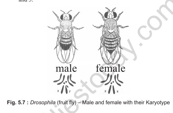
(v) It has 4 pairs of chromosomes, all different in size and shape and easily distinguishable. Three pairs (II, III and IV) are autosomes and the fourth pair (I) is the sex chromosome.
(vi) Females have XX chromosomes and males have XY chromosomes. The Y chromosome is characteristically J-shaped.
(vii) It has many variations that can be seen with a low-power microscope.
Morgan studied eye colour in Drosophila. Normal eye colour is red, and the mutant is white-eyed.
Linkage is the phenomenon in which certain genes stay together during inheritance through generations without any change or separation, because they are located on the same chromosome. Linked genes are an exception to Mendel's law of independent assortment. Mendel's third law is therefore not universally applicable. It applies only to genes on different chromosomes.
Bateson and Punnett (1906) reported that two pairs of alleles in sweet pea (Lathyrus) do not assort independently. They crossed a blue-flowered, long-pollen variety (BBLL) with a red-flowered, round-pollen variety (bbll). The F1 hybrids were test-crossed with the recessive parent. The expected ratio of 1 : 1 : 1 : 1 was not obtained. The observed ratio was 7 : 1 : 1 : 7, with parental combinations in excess (blue with long pollen, and red with round pollen).
On this basis, Bateson and Punnett gave the coupling and repulsion hypothesis. Coupling is when two dominant characters come from the same parent and tend to stay together. Repulsion is when two dominant characters come from different parents and tend to stay separate. T.H. Morgan reported the same phenomenon in Drosophila and named it linkage. He stated that coupling and repulsion are the two aspects of linkage.
Another example of linkage was reported in maize (Zea mays). Crossing a coloured, fully turgid endosperm variety with a colourless, shrunken endosperm variety, and then test-crossing the offspring, gave excess parental combinations.
Key points about linkage
- Linkage is identified by a test cross.
- Linkage is inversely proportional to the distance between genes on a chromosome. The closer the genes, the stronger the linkage and the lower the recombination.
- Sturtevant (1913) proposed the theory of linear arrangement of genes on chromosomes.
- All genes linked together on one chromosome, which do not show independent assortment, form a linkage group.
- The number of linkage groups in an organism equals its haploid number of chromosomes. In Drosophila, n = 4, so there are 4 linkage groups. In garden pea, n = 7, so there are 7 linkage groups. In maize, n = 10, so there are 10 linkage groups.
- Prokaryotes (bacteria, cyanobacteria or blue-green algae and Mycoplasma) have one linkage group.
- Recombination is the rearrangement of linked genes due to crossing over. It also occurs by chance, through the independent separation of chromosomes during gametogenesis and their random union at fertilisation.
- Sturtevant used the frequency of recombination between gene pairs as a measure of distance between genes and mapped their positions on chromosomes. Genetic maps are used as a starting point in sequencing a whole genome, such as the Human Genome Project.
Types of linkage
- Complete linkage: only parental combinations are produced, e.g. in male Drosophila.
- Incomplete linkage: some recombinations are produced along with parental combinations.
Recombinations are produced by crossing over.
CROSSING OVER
Crossing over is the process of forming new recombinations of genes due to the exchange of corresponding segments between non-sister chromatids of homologous chromosomes. It starts in the pachytene substage of prophase I of meiosis and ends in the diplotene substage. It occurs at a point on the chromosome called the chiasma (plural chiasmata). Crossing over occurs at single, double or multiple positions, depending on the length of the chromosome.
Crossing over is directly proportional to the distance between linked genes. The farther apart the genes, the greater the chance of crossing over.
Factors affecting crossing over
(i) Radiations: radiations such as X-rays increase the frequency of crossing over.
(ii) Temperature: any change in temperature increases the frequency of crossing over.
(iii) Sex: crossing over occurs more often in female than in male Drosophila.
(iv) Heterochromatin: the presence of the centromere and heterochromatic regions (such as telomeres) decreases the occurrence of crossing over.
Importance of crossing over
(i) It produces recombinations, or new gene combinations, which form variations. Variations are the raw material for the formation of new species and for evolution.
(ii) Useful recombinations produced by crossing over are used in breeding programmes to produce new varieties of plants and animals.
(iii) It is used in preparing linkage maps.
Linkage or chromosome map is a graphical representation of the linear arrangement of genes and the relative distances between them on a chromosome. It is like a road map that shows locations and the distances between them. The first chromosome maps were prepared by Sturtevant in 1911 for two chromosomes of Drosophila.
Map unit: 1% recombination (or crossing over) between two linked genes equals 1 map unit, or 1 centiMorgan (cM), named after Thomas Hunt Morgan. 100% crossing over is called 1 Morgan (M), and 10% crossing over is called 1 deciMorgan (dM).
Problem 1: Individuals homozygous for genes c and d (cc dd) were crossed with the wild type (++). The F1 dihybrid was test-crossed and gave the following offspring:
++ = 950, cd = 800, +d = 130, +c = 110. What is the distance between genes c and d?
Answer:
Parental combinations = 950 + 800 = 1750.
Recombinant (non-parental) combinations = 130 + 110 = 240.
Total offspring = 1750 + 240 = 1990.
Recombination frequency = (240 / 1990) × 100 = 12%.
The distance between genes c and d is 12 map units (cM).
Problem 2: The crossing over frequencies between genes X and Y, X and Z, and Y and Z are 10%, 19% and 29% respectively. What is the sequence of genes on the chromosome?
Answer: The distance between X and Y is 10 m.u., between X and Z is 19 m.u., and between Y and Z is 29 m.u. Since 10 + 19 = 29, gene X lies between Y and Z. The linear order is Y - X - Z.
Check point 3: Fill in the blanks
(i) ................... is the father of experimental genetics.
Answer: T.H. Morgan.
(ii) Linked genes are located on the same ................... and show low ................... .
Answer: Chromosome; recombination.
(iii) The number of linkage groups in prokaryotes is ................... .
Answer: One.
(iv) All the genes linked together on a single chromosome are known as a ................... .
Answer: Linkage group.
(v) ................... are the factors which induce mutation.
Answer: Mutagens.
SEX DETERMINATION IN HUMAN AND OTHER ANIMALS
History of sex chromosomes
Henking (1896), working on the squash bug (Pyrrhocoris) and other insects, traced a nuclear structure through spermatogenesis and observed that 50% of the sperms received it and 50% did not. He called it the X-body but could not explain its significance. McClung (1901) found that this X-body was a chromosome in grasshoppers, and named it the X-chromosome. Wilson and Stevens (1905), working on Drosophila, found that one pair of chromosomes looked alike in females but differed in males. The odd chromosome in males was named the Y-chromosome.
Methods of sex determination
1. On the basis of sex chromosomes
(a) XY and XX system: found in most fishes, amphibians, reptiles and mammals. The Y chromosome determines maleness, and the absence of Y determines femaleness. The male is heterogametic (produces two types of sperms: X-containing and Y-containing) and the female is homogametic (produces one type of ova, all X-containing). X-containing sperms are called gynosperms and Y-containing sperms are called androsperms. In humans, the genetic make-up of the sperm determines the sex of the child. Women are unfairly blamed for producing daughters, which is a false notion.
(b) XO and XX system: found in bugs, grasshoppers and locusts. The male is heterogametic (produces sperms with or without an X chromosome) and the female is homogametic.
(c) Sex mechanism in Drosophila: the diploid complement is 8 (6 + 2), with 3 pairs of autosomes and 1 pair of sex chromosomes. The sex mechanism is XY and XX, but maleness is not decided by the Y chromosome, which is genetically inactive or inert. According to the genic balance theory of C.B. Bridges, the sex in Drosophila is determined by the ratio of the number of X chromosomes to the number of autosomal sets (ploidy). The ploidy numbers for haploid, diploid and triploid individuals are 1, 2 and 3.
Gynandromorphs are individuals in which some body regions are male and others female. This happens in Drosophila, moths and other insects when one X chromosome is lost in one of the cleaving cells. The cell with XX forms a female region, and the cell with XO forms a male region.
(d) ZZ and ZW system: the sex mechanism is opposite to that of mammals. The X chromosome is represented by Z and the Y by W. The actual mechanism is ZZ (male) and ZW (female). It is found in birds (chicken, fowl), some butterflies and moths. The male is homogametic (produces similar sperms) and the female is heterogametic (produces two types of ova). In birds, sex is decided by the ova of the female and not by the sperms of the male.
(e) Sex determination in honey bees (and wasps, ants): in honey bees the males (drones) are haploid and develop from unfertilised eggs, while females are diploid and develop from fertilised eggs. Male development from unfertilised eggs is called arrhenotoky. Drones do not have a father and cannot have sons, but they have a grandfather (the mother's father) and grandsons (through daughters). In males, the chromosome number is 16 and in females it is 32. Sperm production involves an equational division (not meiosis), since males are already haploid. The queen and the workers are both females, and workers are sterile. The difference develops because of food: embryos fed on royal jelly (salivary secretion of worker bees) throughout development become queens, and those fed on bee bread (honey plus partly digested pollen) become workers. The fertile members of the colony are the drones (haploid males) and the queen (diploid female).
Thelytoky is parthenogenesis in which only females develop. In turkeys and lizards (Lacerta saxicola), the colony consists only of diploid females that develop from diploid eggs. Males are absent.
2. On the basis of ploidy
In some insects, sex chromosomes are not differentiated, and sex is determined by the ploidy of the individual. Haploid individuals are male and diploid individuals are female.
3. On the basis of environmental factors
In some animals, sex chromosomes are not differentiated and there is no difference in ploidy. Sex is then decided by environmental conditions.
- Bonellia viridis (an echiuroid worm): the male (3-4 mm) lives inside the uterus of the female (100 mm). The fertilised egg, when released, becomes a male if it contacts the proboscis of a female, and a female if it settles on a rock or vegetation.
- Alligator and crocodile: at 30 °C or below, hatchlings are female. At about 34 °C, they are male.
- Turtle: below 28 °C produces males, and above 31 °C produces females.
- Snails: even adults can change sex.
According to Ferguson and Joanen (1982), temperature-dependent sex determination may have been responsible for the extinction of dinosaurs. The enzyme aromatase, which converts testosterone into oestrogen, is critical in temperature-dependent sex determination in reptiles.
Sex determination based on the time of fertilisation
(1) Progamic: sex is determined before fertilisation, e.g. males of honey bee.
(2) Syngamic: sex is determined during fertilisation, e.g. humans.
(3) Epigamic: sex is determined after fertilisation, e.g. Bonellia.
Check point 4: Answer the following
(i) What is the number of allosomes and autosomes in a human sperm?
Answer: A human sperm has 23 chromosomes: 1 allosome (X or Y) and 22 autosomes.
(ii) Which sex mechanism is found in honey bees?
Answer: Haplodiploidy (arrhenotoky): males are haploid and females are diploid. Sex is determined by the ploidy of the individual.
(iii) Who discovered the Y-chromosome?
Answer: Wilson and Stevens (1905), in Drosophila.
(iv) What is the sex mechanism in Drosophila?
Answer: XY and XX, where maleness is determined by the ratio of X chromosomes to autosomal sets (genic balance theory), not by the Y chromosome.
(v) Which type of gametes determines the sex in birds?
Answer: The ova (eggs) of the female, because the female is the heterogametic sex (ZW).
(vi) Which sex mechanism is found in birds?
Answer: ZZ (male) and ZW (female).
(vii) What is the diploid complement of chromosomes in Drosophila?
Answer: 8 (6 + 2), i.e. 3 pairs of autosomes and 1 pair of sex chromosomes.
(viii) What is the role of royal jelly in honey bees?
Answer: Embryos fed on royal jelly throughout development develop into queens (fertile diploid females). Those fed on bee bread become workers.
(ix) Write the difference between arrhenotoky and thelytoky.
Answer: Arrhenotoky is parthenogenesis in which only males develop from unfertilised eggs while females develop from fertilised eggs, e.g. honey bees. Thelytoky is parthenogenesis in which only diploid females develop from diploid eggs and males are absent, e.g. turkey birds and the lizard Lacerta saxicola.
(x) What is the sex mechanism in grasshopper?
Answer: XO (male) and XX (female). The male is heterogametic.
(xi) Which sex in Drosophila is heterogametic?
Answer: Male (XY).
MUTATION
Mutation is a phenomenon that alters the DNA sequence, and consequently changes the genotype and phenotype of an organism. The term was first used by Hugo de Vries (1900) for sudden phenotypic changes that are heritable. Darwin called these mutations 'sports'. The first scientific study of mutation was made by T.H. Morgan (1910) in Drosophila. About 500 mutations are now known in Drosophila.
Types of mutation
- Gene mutation (point mutation): involves a change in a single nucleotide. It produces new alleles of a gene. There are two types: induced mutation (produced in response to specific external factors or chemicals) and spontaneous mutation (occurs randomly or naturally due to internal reasons).
- Chromosomal mutation: involves a change in the structure or number of chromosomes.
- Change in structure: deletion or deficiency (a chromosome segment is lost), duplication (addition of a chromosome segment), inversion (a segment is inverted by 180 degrees), and translocation (exchange of segments between non-homologous chromosomes).
- Change in number: polyploidy (change in the number of sets of chromosomes) and aneuploidy (change in the number of a pair or pairs of chromosomes).
Muller (1927) was the first to produce induced mutations in Drosophila by exposing them to X-rays. Most mutation studies have been carried out in microorganisms, such as Neurospora (red mould), E. coli and bacteriophages (viruses that infect bacteria).
Flow Chart 5.1: Types of Mutation
Fig. 5.2 : Types of chromosome aberrations
Mutagens are physical or chemical factors that induce mutations.
(1) Physical mutagens (radiations):
- Ionising radiations, e.g. X-rays, beta rays and gamma rays. They have high penetrating power.
- Non-ionising radiations, e.g. UV rays (first used by Altenburg, 1930). They have low penetrating power.
X-rays were first shown to be mutagenic by H.J. Muller (1927) in Drosophila, and later by L.J. Stadler (1928) in maize and barley.
(2) Chemical mutagens: C. Auerbach first used chemicals to induce mutations during World War II. She used mustard gas, which has a delayed effect after 2 to 3 generations. Other chemical mutagens include ethyl urethane, formaldehyde, phenol, maleic hydrazide, hydroxylamine, hydrazine, nitrous acid, methyl methane sulphonate (MMS) and ethyl methane sulphonate (EMS). EMS is the most extensively used chemical mutagen in crop plants today. Carcinogenic chemicals such as nicotine and nitric oxide (NO) also act as mutagens.
Mutation in crop plants: the Sharbati Sonora variety of wheat was developed by gamma-ray mutation of the Sonora-64 variety (Mexican dwarf wheat). It was developed by M.S. Swaminathan and was used in the Green Revolution in India.
HUMAN CHROMOSOMES
Tjio and Levan (1956) reported the correct number of human chromosomes: 2n = 46, which is 23 pairs (22 pairs of autosomes and 1 pair of sex chromosomes). Sex chromosomes are also called allosomes or heterosomes. Females have XX and males have XY.
The Y chromosome determines maleness. It carries the gene Sry (sex determining region of Y), which codes for TDF (testis determining factor). TDF is required for the development of male sex, and its absence leads to femaleness. Sry is the smallest human gene, with 14 base pairs, and it is located at the tip of the short arm of the Y chromosome. The longest human gene is Dystrophin, the gene for muscular dystrophy, with about 2400 kbp (2.4 Mbp), on the X chromosome.
Karyotype and karyogram
The diploid complement of chromosomes, obtained at mitotic metaphase, is called the karyotype. It is studied from cultured white blood cells taken from bone marrow. When chromosomes are arranged in decreasing order of length and position of the centromere, the arrangement is called a karyogram.
In the standard karyotype, human chromosomes are stained for banding patterns and arranged in seven groups (A to G) on the basis of decreasing length and centromere position. Group A has 3 pairs, B has 2 pairs, C has 7 pairs, D and E have 3 pairs each, and F and G have 2 pairs each. The X chromosome falls in group C and the Y chromosome in group G.
Philadelphia chromosome: the abnormal 22nd chromosome, named after Nowell and Hungerford, who worked in Philadelphia (USA). It has a deletion in the longer arm and is associated with blood cancer (leukaemia).
Comparison with apes: in apes the chromosome number is 2n = 48 (23 pairs of autosomes and 1 pair of sex chromosomes). Chromosomes 3 and 6 of humans are identical to those of apes. The 2nd chromosome of humans has probably been formed by the fusion of two ape chromosomes. Inversions have occurred in chromosomes 1 and 18. More than 95% of DNA is identical between humans and chimpanzees. In humans, 99.9% of DNA is identical across all races.
Check point 5: Answer the following
(i) Number of allosomes and autosomes in a human sperm.
Answer: One allosome (X or Y) and 22 autosomes (see Check point 4, part (i)).
(ii) What is the condition 2n - 1 (of chromosomes) called?
Answer: Monosomy.
(iii) What is the gender of a person with Turner's syndrome?
Answer: Female.
(iv) Who discovered the correct number of human chromosomes?
Answer: Tjio and Levan (1956).
(v) What does the term 'hyperploidy' mean?
Answer: Addition of one or more chromosomes to the normal diploid complement.
(vi) Which chromosome contains the smallest gene of the human genome?
Answer: The Y chromosome (the Sry gene).
(vii) What are the sex chromosomes in Klinefelter's syndrome?
Answer: XXY (or XXXY and similar combinations with more than one X and one Y).
(viii) With which disease is the Philadelphia chromosome associated?
Answer: Blood cancer (leukaemia).
(ix) Which syndrome has XYY sex chromosomes?
Answer: Criminal syndrome (Jacob's syndrome), also called meta male or super male.
(x) Which chromosomes are similar in apes and humans?
Answer: Chromosomes 3 and 6.
GENETIC DISORDERS
All genetic disorders (also called genomic mutations) can be broadly classified into two categories: (I) chromosomal disorders (aberrations), which can be structural or numerical, and (II) Mendelian disorders.
Numerical variations are of two types: euploidy and aneuploidy.
1. Euploidy variations
The chromosome number is a multiple of the haploid set, other than the diploid. This arises due to failure of cytokinesis after telophase. Individuals may be triploid (3n), tetraploid (4n), pentaploid (5n), and so on. These are called polyploid individuals. Polyploidy is common in plants but does not survive in human beings. It does survive in insects such as Drosophila and honey bees.
2. Aneuploidy variations
The diploid chromosome number changes by the addition or loss of one, two or a few chromosomes. Addition of one or more chromosomes to the diploid set is called hyperploidy. Loss of one or more chromosomes is called hypoploidy.
- Monosomy: 2n - 1 = 45 chromosomes
- Trisomy: 2n + 1 = 47 chromosomes
- Tetrasomy: 2n + 2 = 48 chromosomes
- Nullisomy: 2n - 2 = 44 chromosomes
Aneuploidy develops due to non-disjunction (non-separation) of chromosomes, generally during meiosis I.
(I) Aneuploidy of sex chromosomes
(i) Turner's syndrome (XO):
- It is monosomy (2n - 1) of the sex chromosomes. Total chromosome number is 45 (44A + X).
- Individuals are female because the Y chromosome is absent.
- Prominent symptoms are webbed neck, broad shoulders, low-set pinna and short stature.
- Reproductive organs and secondary sexual characters are underdeveloped, and the individuals are sterile. The ovaries are rudimentary. Such individuals do not menstruate or ovulate.
- Frequency: about 1 in 2500 births.
(ii) Klinefelter's syndrome (XXY or XXXY):
- It develops mainly due to trisomy of the sex chromosomes (2n + 1). Total chromosome number is generally 47 (44A + XXY). Conditions such as XXXY or XXXXY are also called Klinefelter's syndrome (nX + XY). In all such cases the number of chromosomes is more than 46.
- The more X chromosomes present, the greater the mental retardation.
- Individuals are male because of the Y chromosome.
- Male reproductive organs and secondary sexual characters are poorly developed. Breast development (gynaecomastia) can sometimes occur.
- Such individuals are tall and sterile.
- Frequency: about 1 in 700 live births.
(iii) Meta female (super female), XXX or XXXX:
- It develops mainly due to trisomy of the sex chromosomes. Sex mechanism is nX + XX, where n is 1 or more. Chromosome number is generally 47 (44A + XXX).
- Mental retardation increases with the number of X chromosomes.
- Individuals are female and may be fertile or sterile.
(iv) Meta male (super male) or criminal syndrome (Jacob's syndrome), XYY:
- It develops generally due to trisomy of the sex chromosomes (44A + XYY). There is more than one Y chromosome. Chromosome number is generally 47. Sex mechanism is nY + XY.
- Individuals are male, with a robust body and a criminal bent of mind. The crime may be non-violent and petty. They may be fertile or sterile.
No other aneuploidy of sex chromosomes has been reported in humans.
(II) Aneuploidy of autosomes: Down's syndrome
First described by Langdon Down (1866). It is the most common congenital disorder, with a frequency of about 1 in 700 live births. It is a case of trisomy of chromosome 21 (G group). The diploid number is 47 (45A + XX or XY). It can occur in males or females, since it is an autosomal disorder.
The frequency of Down's syndrome is higher in the children of older mothers, because of weakening of the spindle fibres of the 21st pair of chromosomes in oocytes that formed during foetal life. The frequency does not change with the father's age, because spermatogenesis is completed in a short time (about 65 days).
Symptoms:
- The life span is generally short (around 18 years).
- The upper eyelid is folded (epicanthic fold), as in the Mongolian race.
- Short stature, a rounded and flat face, an open mouth, a furrowed tongue, and saliva that keeps dribbling.
- Simian crease is prominent in the middle of the palm. Many loops are present on the finger tips.
- Mental and psychomotor development is retarded. IQ is less than 25 (idiocy).
- One of the causes of neuronal degeneration in Alzheimer's disease is the development of extracellular plaques of beta-amyloid protein. The gene for this precursor protein is on chromosome 21. That is why persons with an extra chromosome 21 develop dementia, similar to Alzheimer's disease, at an early age.
Familial Down's syndrome: in some cases a major part of chromosome 21 is translocated to chromosome 14. The individual has 46 chromosomes but shows Down's syndrome. Such Down's syndrome can run in families.
Other autosomal aneuploidies:
(ii) Edward's syndrome (trisomy of chromosome 18, E group): diploid number is 47. The individual may be male or female with kidney, heart and finger deformities. Life span is very short (around 6 months).
(iii) Patau's syndrome (trisomy of chromosome 13, D group): diploid number is 47. The individual may be male or female. Life span is very short (around 3 months). Malformations include hare-lip, cleft palate, cardiovascular disorders, microcephaly, eye defects (blindness), deafness, brain disorders and polydactyly (extra digits).
Check point 6: Answer the following
(i) In Turner's syndrome, what is the total chromosome number?
Answer: 45 (44A + X).
(ii) Which disorder is caused by trisomy of chromosome 21?
Answer: Down's syndrome.
(iii) Why does Down's syndrome frequency depend on the age of the mother and not the father?
Answer: The 21st chromosome pair in oocytes, formed during foetal life, weakens with the mother's age, causing non-disjunction. Spermatogenesis takes only about 65 days, so paternal age has no major effect.
SEX CHROMATIN (BARR BODY)
M.L. Barr and Bertram (1940) observed a darkly stained body inside the nucleus, close to the nuclear membrane, in the nerve cells (neurons) of female cats. Since the presence or absence of this body helps to determine sex, it was called the sex chromatin body or Barr body. It was later found in the interphase nuclei of many mammalian cells.
The number of Barr bodies is always one less than the number of X chromosomes.
- Normal female (XX): 2 - 1 = 1 Barr body.
- Normal male (XY): 1 - 1 = 0 Barr bodies.
- Turner's syndrome (XO) has no Barr body, while Klinefelter's syndrome (XXY) has one.
Barr bodies are seen in the drum-stick shape in the neutrophils (a type of white blood cell) of females. They are absent in RBCs.
Applications of Barr body study
(i) In international sports: a Barr body test on cheek epithelium or hair root cells can be used to identify the sex of athletes in the Olympics and other national or international games.
(ii) In forensic science: the study of Barr bodies can identify the sex of criminals from their blood cells (neutrophils) or hair root cells.
(iii) Prenatal diagnosis: in amniocentesis, the amniotic fluid containing floating foetal cells is taken from the pregnant mother around 6 to 10 weeks of pregnancy. The foetal cells are examined for Barr bodies. The absence of a Barr body indicates a male child. Chorionic villi sampling (CVS) is a better method than amniocentesis for chromosomal and biochemical studies.
Check point 7: Answer the following
(i) The sex chromatin body is called ................... .
Answer: Barr body.
(ii) The number of Barr bodies in a normal female (XX) is ................... .
Answer: 1.
(iii) The frequency of the occurrence of Down's syndrome depends on the age of the ................... .
Answer: Mother.
(iv) In Down's syndrome, the chances of Alzheimer's disease are ................... .
Answer: More.
(v) The sex chromatin body appears in ................... nuclei.
Answer: Interphase.
(vi) Barr bodies are lacking in normal ................... .
Answer: Males.
(vii) The Turner's syndrome is ................... in sex, and Down's syndrome can be male or female.
Answer: Female.
Check point 11: Briefly explain why?
(i) Amniocentesis is done.
Answer: To find the sex of the foetus (absence of Barr body indicates a male) and to detect chromosomal and biochemical disorders from the foetal cells.
(ii) Sex chromatin body is called 'Barr body'.
Answer: It is named after its discoverer, M.L. Barr.
(iii) The frequency of the occurrence of Down's syndrome depends on the age of the mother.
Answer: The spindle fibres of the 21st pair of chromosomes in oocytes, formed during foetal life, weaken with age, so non-disjunction is more likely.
(iv) In Down's syndrome chances of Alzheimer's disease are more.
Answer: The gene for the precursor of beta-amyloid protein is on chromosome 21, so persons with an extra chromosome 21 develop its symptoms early.
(v) The sex chromatin body appears in neutrophils and not in RBCs.
Answer: Mature RBCs have no nucleus, whereas neutrophils have a nucleus in which the Barr body is seen as a drum-stick.
(vi) Down's syndrome is called Mongoloid idiocy.
Answer: The upper eyelid is folded as in the Mongolian race, and the patients are mentally retarded (IQ less than 25).
(vii) Barr bodies are lacking in normal males.
Answer: The number of Barr bodies is one less than the number of X chromosomes, and males (XY) have only one X.
(viii) The Turner's syndrome is female but Down's syndrome can be male or female.
Answer: Turner's syndrome (XO) lacks a Y chromosome, so it is female. Down's syndrome is autosomal, so it can occur in either sex.
MENDELIAN INHERITANCE OF TRAITS IN HUMANS
Mendelian traits are either autosomal or sex-chromosomal.
I. Autosomal traits are of several types.
(a) Polygenic traits are governed by several genes (multiple genes) and show graded variation, e.g. IQ, skin colour, hair colour, height and wheat kernel colour.
IQ (intelligence quotient): it is a measure of intelligence and is known to be governed by 11 sets of genes.
IQ = (mental age / actual age) × 100. Mental age is calculated by psychological tests. A normal IQ is 90 to 109.
Skin colour in humans: Davenport studied the polygenic inheritance of skin colour. Skin colour depends on the amount of melanin pigment, which depends on the number of genes and on sunlight. Skin colour is controlled by 3 to 7 genes on different loci. Curl Stern used at least 3 genes (A, B and C) with cumulative effects on melanin. Each dark-skin allele is incompletely dominant over the light-skin allele. AABBCC has 6 units of darkness (darkest skin), AaBbCc, AABbcc or AAbbCC have 3 units, and aabbcc has no darkness (lightest skin). The results form a bell-shaped curve, called a normal distribution.
In a cross between AaBbCc and AaBbCc, there are 64 combinations. The phenotypic ratio is 1 (6 units) : 6 (5 units) : 15 (4 units) : 20 (3 units) : 15 (2 units) : 6 (1 unit) : 1 (0 units), which corresponds to the counts of 1/64, 6/64, 15/64, 20/64, 15/64, 6/64 and 1/64.
Common autosomal traits in humans:
| Trait | Dominant form | Recessive form |
|---|---|---|
| Shape of hair | Curly hair | Straight hair |
| Ear lobe | Free lobe | Fused lobe |
| Tongue rolling (longitudinal) | Rollers | Non-rollers |
| PTC paper tasting | Taster | Non-taster |
| Sickle cell anaemia | Normal | Affected |
| Tay-Sachs disease (infantile amaurotic idiocy) | Normal | Affected |
| Cystic fibrosis | Normal | Affected |
| Thalassaemia | Normal | Affected |
| Myopia, hypermetropia, astigmatism | Affected | Normal |
| Myotonic dystrophy | Affected | Normal |
(Sickle cell anaemia, Tay-Sachs disease, cystic fibrosis and thalassaemia are autosomal recessive, so the affected form is recessive. Myopia and myotonic dystrophy are autosomal dominant, so the affected form is dominant.)
Sickle cell anaemia is an autosomal recessive disorder of the blood. RBCs become sickle-shaped due to sickle haemoglobin (HbS), caused by a defect in the beta chain. In normal haemoglobin (HbA), the 6th position of the beta chain has glutamic acid, coded by the CTC triplet in DNA, which gives the GAG codon in mRNA. A point mutation changes CTC to CAC in DNA, producing GUG in mRNA. This changes glutamic acid to valine (Val), which changes the shape of haemoglobin, and RBCs become sickle-shaped. The gene is located on chromosome 11. In heterozygous condition (HbA HbS), RBCs remain normal at sea level, but at high altitude, where the partial pressure of oxygen is low, some RBCs become sickle-shaped. In homozygous condition (HbS HbS) the abnormality is lethal. Sickle cell anaemia is a qualitative disorder.
PTC paper is a paper dipped in phenyl thio carbamide (PTC) that tastes bitter (the source says sour). People who can taste it are tasters, and those who cannot are non-tasters. Tasters have the genotype TT or Tt, and non-tasters have tt.
Tay-Sachs disease (infantile amaurotic idiocy) is an autosomal recessive disorder caused by deficiency of the enzyme hexosaminidase. The affected child is mentally retarded, blind, and has a very short life span (around 3 years).
Cystic fibrosis is a common genetic disorder among Caucasians in which thick, salty mucus blocks the respiratory tract. Its gene is on chromosome 7. The homozygous recessive condition produces a defective protein that normally regulates the chloride transport channel. The pancreas and liver are also affected.
Thalassaemia is an autosomal recessive blood disorder. Haemoglobin is abnormal because of non-synthesis or reduced synthesis of one of the globin chains.
- There are two forms: alpha thalassaemia (production of alpha-globin chain affected) and beta thalassaemia (production of beta chain affected).
- In heterozygous condition, it provides protection against malaria, similar to sickle cell anaemia.
- In sickle cell anaemia the beta chain is abnormal in quality, due to a point mutation. In thalassaemia there is a deficiency in quantity of the alpha or beta chain. So sickle cell anaemia is a qualitative disorder, and thalassaemia is a quantitative disorder.
- The alpha chain is coded by two closely linked genes (HBA1 and HBA2) on chromosome 16, and the beta chain by a single gene (HBB) on chromosome 11. The number of alpha-globin molecules depends on mutation in one or more of the 4 alleles, and the number of beta-globin molecules depends on one or both alleles.
- Alpha thalassaemia is common in South Asia (India) and West Africa. Beta thalassaemia is common among people near the Mediterranean Sea, such as Italians, Greeks and some Africans.
- The term thalassaemia is of Greek origin (thalassa = sea, emia = blood).
- Treatment includes blood transfusion or bone marrow transplantation.
Myotonic dystrophy is an autosomal dominant disorder. It is characterised by muscle wasting (muscular dystrophy), along with cataract and endocrine disorders. There is general muscular weakness, beginning in the muscles of the hands, feet, neck and face, and later extending to the heart. It can appear at any time from birth to adulthood.
Check point: Fill in the blanks with suitable words/terms
(i) ............ and ............ are autosomal recessive disorders of blood.
Answer: Sickle cell anaemia and thalassaemia.
(ii) Cystic fibrosis is the disorder of ............ system.
Answer: Respiratory.
(iii) The gene of sickle cell anaemia is associated to ............ chromosome.
Answer: 11th.
(iv) ............ and ............ are the examples of polygenic traits in human.
Answer: IQ and skin colour.
(v) ............ thalassaemia is governed by 4 alleles.
Answer: Alpha.
(vi) ............ chain/s is/are affected in sickle cell anaemia.
Answer: Beta.
(vii) Myotonic dystrophy is an autosomal ............ disorder.
Answer: Dominant.
(viii) ............ ear lobe is dominant to ............ ear lobe in human.
Answer: Free; fused.
Problems based on monogenic traits
Problem 1: What is the probability of the fused ear lobe condition in a child if both parents are heterozygous for the ear lobe trait?
Answer: Ll x Ll gives LL : Ll : Ll : ll in the ratio 1 : 2 : 1, i.e. 3 free lobed to 1 fused lobed. So there is a 25% chance (1/4) that the child will have fused ear lobes.
Problem 2: Predict the genotype of the 'taster' parents for PTC, who have a non-taster child.
Answer: The non-taster child is tt. Each parent must contribute a t allele. Since a parent with TT cannot produce a tt child, both parents must be Tt.
Problem 3: Two parents, one curly haired and one straight haired, have one straight-haired child. What are the chances that the second child will be curly haired?
Answer: A straight-haired child (cc) can be produced only if the curly-haired parent is heterozygous (Cc). This is a test cross (Cc x cc). The chances of curly and straight hair are 50% and 50%, i.e. 1 : 1. So there is a 50% chance that the second child will be curly haired.
II. Sex-limited traits
- They are autosomal traits, and they follow the Mendelian pattern.
- Their expression (phenotype) is limited to one of the two sexes.
- E.g. beard and moustaches in males, milk production in females, and Adam's apple in males.
III. Sex-controlled or sex-influenced traits
- They are autosomal traits, and they follow the Mendelian pattern.
- They are expressed in both sexes but with different frequency.
- E.g. pattern baldness in humans, horns in sheep, and gout (a type of arthritis) in humans.
Pattern baldness is an autosomal dominant trait.
- The homozygous dominant genotype (BB) is rare, but it expresses baldness in both sexes.
- The homozygous recessive genotype (bb) is non-bald in both sexes.
- The heterozygous genotype (Bb) is bald in males and non-bald in females.
In females the dominant allele behaves as recessive. In males, the extra dose of testosterone makes the heterozygous condition express baldness. So baldness is more common in males. Similarly, the heterozygous condition for horns (Hh) produces horns in males but not in females.
IV. Traits due to metabolic disorders
Archibald Garrod, a British physician known as the father of human genetics, reported heritable metabolic disorders in his book 'Inborn Errors of Metabolism'. These disorders arise from mutations in autosomal genes responsible for the production of specific enzymes. They appear in the homozygous recessive condition. Examples include alkaptonuria, phenylketonuria, albinism, cretinism and tyrosinosis.
Phenylketonuria
- Gene is on chromosome 12. Appears in homozygous recessive condition (pp).
- Phenylalanine hydroxylase (E1) is not formed, so phenylalanine (an essential amino acid) accumulates in the liver and forms phenylketone bodies. These pass into the urine, since the kidney absorbs them poorly. This gives the disease its name.
- Children are mentally retarded (IQ less than 25) due to brain damage. The damage can be limited if the child is fed a low-phenylalanine diet.
- Carrier (heterozygous) individuals can be identified by special tests.
Cretinism
- Tyrosine combines with iodine in the presence of enzyme E2 to form thyroxine. In homozygous recessive condition (tt), thyroxine is not formed because enzyme E2 is not available.
- The child becomes physically, mentally and sexually retarded. He or she is a dwarf, sterile, and has a low IQ.
Alkaptonuria
- In homozygous recessive condition (hh), the enzyme homogentisic acid dioxygenase (E8) is absent, so homogentisic acid (alkapton) is not converted into maleylacetoacetic acid.
- Homogentisic acid appears in the urine, which turns black on exposure to air. This is how the disorder got its name.
- Such children are prone to arthritis. Their joints become black.
- Deficiency of the enzyme adenosine deaminase, due to a point mutation, causes severe combined immunodeficiency disease (SCID).
Albinism
- Melanin is synthesised from DOPA, which comes from the metabolism of tyrosine. In homozygous recessive condition (aa), the enzyme (E3 or E4) is absent, so melanin is not synthesised.
- Such children are albinos and are sensitive to skin cancer.
Check point: Fill in the blanks, each with a suitable word / term
(i) The pattern baldness in human is ................... disorder.
Answer: Autosomal (sex-influenced).
(ii) In ................... (disorder) homogentisic acid passes through urine.
Answer: Alkaptonuria.
(iii) In ..................., an inborn metabolic disorder, there is brain damage.
Answer: Phenylketonuria.
(iv) The milk production in females is ................... trait.
Answer: Sex-limited.
(v) ................... wrote the book 'Inborn errors of metabolism'.
Answer: Archibald Garrod.
(vi) In case of ................... the urine turns black on exposure.
Answer: Alkaptonuria.
(vii) The deficiency of enzyme ................... causes SCID.
Answer: Adenosine deaminase.
(viii) The inheritance of sex-influenced traits ................... Mendelian pattern.
Answer: Follows.
V. Multiple allele traits
In these traits, instead of having two alleles (one dominant and one recessive), the gene has three or more alleles. Examples: ABO system and Rh system of blood groups.
ABO blood group system
- This system has four blood groups: A, B, O and AB.
- Landsteiner (1901), known as the father of blood group studies, discovered groups A, B and O. Decastello and Sturli (1902) discovered group AB.
- There are two types of antigens: antigen A and antigen B. The blood group is named after the antigen present on the RBC. Group A has antigen A, group B has antigen B, group AB has both A and B, and group O has no antigen.
- Antibodies are present against the antigen that is missing in the blood. Group A has anti-B antibodies (beta). Group B has anti-A antibodies (alpha). Group AB has no antibodies. Group O has both anti-A and anti-B antibodies (alpha and beta).
- Antigens are glycolipids (on RBCs) or glycoproteins (in other tissues). They are also called isoagglutinogens (represented by I). Some antigens are present in sweat and salivary glands.
- Antibodies (agglutinins) are pure proteins, the gamma globulins, present in the plasma. Antibodies against antigen A and antigen B are called alpha and beta antibodies.
Genetic basis: there are three alleles (IA, IB and IO, or LA, LB and LO) on the long arm of chromosome 9. Alleles A and B are both dominant over O, and they show co-dominance with each other. Allele O is recessive to both. There are 6 genotypes: LALA or LALO (group A), LBLB or LBLO (group B), LOLO (group O), and LALB (group AB). L is used to honour Landsteiner. I can also be used in place of L, so LALB can be written as IAIB, LBLO as IBi, and LOLO as ii.
Summary of ABO system: 2 types of antigens, 2 types of antibodies, 3 types of alleles, 4 types of blood groups and 6 types of genotypes.
Inheritance of blood groups
Problem 1: What blood groups can the children have if the parents are group A and group B?
Answer: Assuming the parents are heterozygous (the usual approach when genotypes are not given), the father is IAIO (group A) and the mother is IBIO (group B). The children can be IAIB (AB), IAIO (A), IBIO (B) or IOIO (O). So the children may have any of the four blood groups.
To find all possible genotypes in children, the parents are taken to be heterozygous unless their genotypes are specified. In the table below, IAIB (or LALB) is written as AB only, and IAIO as AO. The table gives the possible blood groups of children.
| Blood group of mother | Blood group of father | Blood group of children - ? |
|---|---|---|
| A | A | A or O |
| A | B | A or B or O or AB |
| A | O | A or O |
| A | AB | A or B or AB |
| B | A | A or B or O or AB |
| B | B | B or O |
| B | O | B or O |
| B | AB | A or B or AB |
| O | A | A or O |
| O | B | B or O |
| O | O | O |
| O | AB | A or B |
| AB | A | A or B or AB |
| AB | B | A or B or AB |
| AB | O | A or B |
| AB | AB | A or B or AB |
| Gametes | IB | IO |
|---|---|---|
| IA | IAIB (AB) | IAIO (A) |
| IO | IBIO (B) | IOIO (O) |
(Parent IAIO gives gametes IA and IO; parent IBIO gives gametes IB and IO; the offspring genotypes are IAIB, IAIO, IBIO and IOIO.)
Problem 2: If the blood group of the mother is A and the child is B, which blood group can the father have?
Answer: The child (group B, genotype IBIO or IBIB) receives O from the mother only if the mother is IAIO. The child's B allele must come from the father. So the father must have allele B. The father's blood group can be B (genotype IBIO or IBIB) or AB (IAIB).
| Blood group of mother | Blood group of child | Blood group of father - ? |
|---|---|---|
| A | A | A or B or O or AB |
| A | B | B or AB |
| A | O | A or B or O |
| A | AB | B or AB |
| B | A | A or AB |
| B | B | A or B or O or AB |
| B | O | A or O or B |
| B | AB | A or AB |
| O | A | A or AB |
| O | B | B or AB |
| O | O | A or O or B |
| O | AB | *This combination is not possible |
| AB | A | A or B or O or AB |
| AB | B | A or B or O or AB |
| AB | O | *This combination is not possible |
| AB | AB | A or B or AB |
Rules of paternity from the table:
- If one parent is AB, the child cannot be group O (IOIO). The reverse is also true, so if the child is AB, the parents cannot both be group O.
Diagnosis of blood groups
Blood drops are placed on two places on a slide. Anti-A serum (alpha antibody) is added to one drop, and anti-B serum (beta antibody) to the other. Antigen A reacts with alpha antibody, and antigen B reacts with beta antibody, causing agglutination (clumping of RBCs). Agglutination is positive (+ve) and its absence is negative (-ve).
| Anti-A (alpha) serum | Anti-B (beta) serum | Blood group |
|---|---|---|
| +ve | -ve | A |
| -ve | +ve | B |
| -ve | -ve | O |
| +ve | +ve | AB |
Blood group percentages in India: group O (most common) about 38.5%, group B about 32.0%, group A about 23.0%, and group AB (rarest) about 6.5%. The percentages vary from region to region. For example, group O is nearly 100% in some South Americans, and 47% in white Americans and western Europeans.
The ABO system has also been traced in anthropoid apes. The chimpanzee mainly has group A, while the rhesus monkey has only group B.
Check point 11: Fill in the blanks, each with a suitable word or statement
(i) A person of blood group 'B' can donate blood to ...... blood group persons.
Answer: B and AB.
(ii) ............... is a common example of a multiple allele trait.
Answer: ABO blood group.
(iii) The antigens of the ABO system are located in/on ............... .
Answer: The surface of RBCs.
(iv) The blood group ............... is most common in India.
Answer: O.
(v) A person of blood group ............... has ....... antibodies and ....... antigen.
Answer: For example, group O has α and β antibodies and no antigen.
(vi) ............... is known as the father of blood groups.
Answer: Landsteiner.
(vii) The blood group ............... was not discovered by Landsteiner.
Answer: AB (discovered by Decastello and Sturli).
(viii) In the ABO blood group the alleles ...... and ...... are dominant over ...... .
Answer: A and B; O.
(ix) In the ABO system the ............... blood group is the universal donor.
Answer: O.
(x) The parents of a child with blood group 'O' can never have ...... blood group.
Answer: AB.
Blood transfusion
When blood is transferred from donor to recipient, the antigens on the donor's RBCs react with the antibodies in the recipient's plasma. The reverse is not a problem in the same way. A group that has no antigen will not cause agglutination, so it can be transfused to anyone. Group O is the universal donor. Group AB has no antibodies, so it can receive blood from anyone and is the universal recipient.
| Blood group | Can accept blood from | Can donate blood to |
|---|---|---|
| A | A and O | A and AB |
| B | B and O | B and AB |
| O | O only | A, B, O and AB |
| AB | A, B, O and AB | AB only |
Universal donor: blood group O. Universal recipient: blood group AB.
The above table can also be represented as:
Rh system of blood groups
The Rh system was discovered in the rhesus monkey (Macaca mulatta), and so it is called the Rh (Rhesus) system. It was first reported by Landsteiner and Wiener (1940).
- The Rh factor is governed by 3 closely placed genes or alleles (C, D and E), called pseudoalleles. Of these, D is the most antigenic or strong.
- The Rh factor is an antigen on the surface of RBCs. A person with the Rh antigen is Rh positive (+ve), and a person without it is Rh negative (-ve).
- For simplicity, the Rh factor is represented by one gene with two alleles: R (dominant) and r (recessive). RR or Rr is Rh +ve, and rr is Rh -ve.
- There are no natural antibodies against the Rh factor. An Rh -ve person does not have Rh antibodies in the plasma. Antibodies can be induced if Rh +ve blood is transfused into an Rh -ve person. These are acquired antibodies.
- Distribution: among European whites, 85% are Rh +ve and 15% are Rh -ve. Among Indians, about 96.5% are Rh +ve and about 3.5% are Rh -ve. Among Africans, 100% are Rh +ve.
- Examples: A +ve (group A with Rh antigen) and AB -ve (group AB without Rh antigen).
- The most common blood group in India is O +ve. The rarest is AB -ve. The universal donor is O -ve, and the universal recipient is AB +ve.
Erythroblastosis foetalis is also called haemolytic disease of the newborn (HDN) or the 'yellow baby' condition. It develops when the parents are incompatible: an Rh +ve father and an Rh -ve mother.
In the first pregnancy, the child is normal, because the mother has no natural antibodies. During birth, the placenta ruptures and the child's blood mixes with the mother's blood. Rh antigens from the child's RBCs enter the mother's blood and induce the formation of antibodies (IgG type) against the Rh factor. In a second Rh +ve child, these acquired antibodies pass through the placenta into the foetus. They cause agglutination and haemolysis of the foetal RBCs. Haemoglobin is released into the plasma, and its haem breaks down into bilirubin, which causes jaundice and makes the baby yellowish. To compensate for the loss of RBCs, erythroblasts (immature RBCs) are released into the foetal blood. This is why the disease is called erythroblastosis foetalis. The third, fourth or later children are also affected.
Prevention: erythroblastosis foetalis can be prevented by giving the Rh -ve mother an injection of anti-D antiglobulin after the first delivery. This makes the Rh antigens inactive. The antibodies formed in the ABO system are IgM type and cannot cross the placenta, so they do not cause similar effects.
Applications of the study of blood groups
(i) ABO and Rh systems are important for blood transfusion.
(ii) Identification of blood groups is used in forensic science to identify criminals.
(iii) Blood group identification is used in paternity disputes and medico-legal cases. It cannot confirm who the father is, but it can confirm who is not the father.
Check point 8: Answer the following
(i) Which blood group is the universal donor?
Answer: Group O.
(ii) What percentage of Europeans are Rh +ve?
Answer: About 85%.
(iii) Which eye colour in Drosophila is dominant?
Answer: Red eye colour.
(iv) Which is a non-compatible match for erythroblastosis foetalis?
Answer: An Rh +ve father and an Rh -ve mother.
(v) Who discovered the Rh factor?
Answer: Landsteiner and Wiener (1940).
(vi) Who is the father of blood group studies?
Answer: Landsteiner.
(vii) What are two applications of the study of blood groups?
Answer: Blood transfusion, and identification of criminals in forensic science (also paternity disputes).
Check point (PDF, additional questions): Answer the following
(i) In which blood group are there no natural antibodies?
Answer: In the Rh system, there are no natural antibodies against the Rh factor, so an Rh -ve person has none unless Rh +ve blood is received. (In the ABO system, group AB has no antibodies in plasma.)
(v) What is the other name of erythroblastosis foetalis?
Answer: Haemolytic disease of the newborn (HDN), or the 'yellow baby' condition.
(vi) Who discovered sex-linked inheritance in Drosophila?
Answer: T.H. Morgan (1910).
(viii) What are the results of reciprocal crosses in sex-linked inheritance?
Answer: Reciprocal crosses between red-eyed and white-eyed Drosophila give different results. A cross of red-eyed females with white-eyed males gives all red-eyed offspring. A cross of white-eyed females with red-eyed males gives red-eyed daughters and white-eyed sons (1 : 1).
SEX-CHROMOSOMAL TRAITS
These traits are governed by genes present on the X or Y chromosome. They are of two categories.
1. X-linked or sex-linked traits are governed by genes on the part of the X chromosome that has no homologue on the Y chromosome. Sex-linked inheritance was discovered by T.H. Morgan (1910) for eye colour in Drosophila. Red eye (natural, wild and dominant) and white eye (mutant and recessive) are the two eye colours.
Morgan observed that reciprocal crosses between red-eyed and white-eyed parents do not give the same results, so the gene for eye colour is not autosomal.
- Red-eyed female (XRXR) x White-eyed male (XrY): all offspring are red-eyed.
- White-eyed female (XrXr) x Red-eyed male (XRY): daughters are red-eyed (XRXr) and sons are white-eyed (XrY), in the ratio 1 : 1.
Since the Y chromosome passes from father only to sons, the X chromosome can never pass from a father to his sons. So sex-linked characters never pass from a male parent to his male offspring.
Examples of sex-linked traits in humans:
(a) Sex-linked recessive traits: haemophilia, colour blindness, Duchenne's (juvenile) muscular dystrophy, deficiency of glucose-6-phosphate dehydrogenase (G-6PD) in RBCs.
(b) Sex-linked dominant traits: defective tooth enamel, and Xg blood group.
Juvenile muscular dystrophy (Duchenne's muscular dystrophy): its gene Dystrophin is the longest gene in the human genome, with 2400 kbp (2.4 Mbp).
Haemophilia is also called 'bleeder's disease' or 'royal disease', because it first appeared in the descendants of Queen Victoria (she was normal but heterozygous for the trait). The pedigree of haemophilia was studied by Haldane. In females, heterozygous individuals are normal but are carriers. In males, the gene is present only on the X chromosome, so the male is hemizygous (neither homozygous nor heterozygous).
- Haemophilia-A is more common (about 80%). It is caused by the absence of blood clotting factor VIII (antihaemophilic globulin, AHG).
- Haemophilia-B is less common (about 20%). It is caused by the absence of blood clotting factor IX (plasma thromboplastin component, PTC). It is also called Christmas disease, because it was first discovered in a Christian family.
- Haemophilia-C is very rare and is caused by the absence of clotting factor XI (plasma thromboplastin antecedent, PTA). It is autosomal recessive and affects both sexes equally.
- Haemophilia in India occurs at about 1 in 10,000. There are more than 1 lakh haemophiliacs in India, of whom about 50,000 are severe cases.
Genotypes for haemophilia:
- In males: XHY = normal; XhY = haemophilic (diseased).
- In females: XHXH = homozygous normal; XHXh = heterozygous normal (carrier); XhXh = homozygous diseased.
(In problems, XH is often written simply as X.)
Sex-linked recessive traits such as haemophilia appear more often in males than females. In males a single recessive allele causes the disease. In females, two recessive alleles (double dose) are needed, which is less likely. For a female to be haemophilic, the father must be haemophilic (a condition that is often unviable in later life), and the mother must be at least a carrier.
Problem 1: If the father is haemophilic and the mother is homozygous normal, predict the genotypes of the children and grandchildren.
Answer: Father XhY x Mother XHXH. Children: all daughters are XHXh (normal carriers), and all sons are XHY (normal). So all children are normal. Grandchildren: a daughter's children may be XHXh x XHY, which gives 50% haemophilic sons (XhY) and 50% normal sons, while all granddaughters are normal (50% homozygous normal and 50% carriers). Haemophilia passes from the grandfather to his grandsons through his daughters. This is called criss-cross inheritance, or skip-generation inheritance.
Colour blindness is the most common sex-linked disorder. A colour-blind person cannot distinguish between red and green. Such people cannot be appointed in traffic police and cannot be issued a driving licence. It is also called daltonism, because John Dalton, the founder of the atomic theory, had this disorder. Red blindness is called protanopia, and green blindness is called deuteranopia. In Europeans it occurs in 8% of males, and in Indians in about 3.6% of males. It is a recessive trait: XcY is a colour-blind male, and XcXc is a colour-blind female.
Problem 2: Miss Pooja, whose father is colour blind, marries Mr Rakesh, whose mother is colour blind. What is the probability of colour blindness in their child if (a) the child is a son, and (b) the child is a daughter?
Answer: Pooja's father is colour blind, so Pooja is a carrier (XCXc). Rakesh's mother is colour blind, so Rakesh receives Xc from her and is colour blind (XcY). (a) Sons get the X from their mother, which is XC or Xc with equal chance, so 50% of the sons are colour blind. (b) Daughters receive Xc from their father, and the X from their mother is XC or Xc. So 50% of the daughters (XcXc) are colour blind, and the other 50% are carriers.
Deficiency of G-6PD is a sex-linked recessive trait. The enzyme glucose-6-phosphate dehydrogenase is present in RBCs for a minor pathway of glycolysis. Persons deficient in this enzyme cannot have Plasmodium in their RBCs, so they are protected from malaria. But if they are given antimalarial drugs, their fragile RBCs can rupture, causing severe anaemia that can be fatal. The percentage of G-6PD-deficient persons is higher in South Africa, a malaria-prone area.
2. Y-linked or holandric traits are governed by genes on the Y chromosome. They pass from father to all of his sons. E.g. hypertrichosis (excessive hair) on the pinna.
Check point 9: Fill in the blanks with suitable terms or words
(i) In ................... traits, the concerned allele cannot pass from father to his sons.
Answer: X-linked (sex-linked).
(ii) ................... is also known as Christmas disease.
Answer: Haemophilia-B.
(iii) Blindness for green colour is known as ................... .
Answer: Deuteranopia.
(iv) Haemophilia is commonly called ................... .
Answer: Bleeder's disease (or royal disease).
(v) Daltonism is the other name for ................... .
Answer: Colour blindness.
(vi) Haemophilia is ................... in males than in females.
Answer: More common.
(vii) The skip-generation inheritance is also called ................... .
Answer: Criss-cross inheritance.
(viii) ................... traits appear in hemizygous condition in ................... .
Answer: Sex-linked; males.
(ix) Haemophilia first appeared in the descendants of ................... .
Answer: Queen Victoria.
(x) ................... is the most common sex-linked disorder.
Answer: Colour blindness.
PEDIGREE ANALYSIS (GENEALOGY)
Human genetics is difficult to study because controlled crosses cannot be made, the number of offspring is small, the life span is long and sexual maturity is reached after about 15 years, and many phenotypes appear only in later life. To overcome these difficulties, Francis Galton, the father of eugenics, recommended pedigree analysis.
Pedigree analysis is the study of the inheritance of a trait over several generations of a family. The pedigree of haemophilia in the descendants of Queen Victoria, studied by Haldane, is a well-known example. The person from whom the case history begins is called the proband (if male) or proposita (if female). Children of a couple are called siblings and are numbered from left to right.
Standard symbols used in pedigree analysis
| Symbol | Meaning |
|---|---|
| Square | Male (normal) |
| Circle | Female (normal) |
| Diamond | Unspecified sex (normal) |
| Filled (shaded) symbol | Affected individual |
| Symbol with a cross or slash | Deceased (dead) individual |
| Horizontal line between square and circle | Mating (marriage) between unrelated individuals |
| Double horizontal line | Consanguineous mating (marriage) |
| Half-filled square or half-filled circle | Heterozygous condition of an autosomal trait |
| Circle with a dot in the centre | Heterozygous condition of a sex-linked trait (males cannot be heterozygous for such traits) |
| Square or circle with a cross inside | Still birth |
| Two lines from one parent point (fraternal twins), can be of the same sex | Fraternal twins |
| Two lines joined at a single point (identical twins), always the same sex | Identical twins (cannot be of different sexes) |
| Arrow | Proband (or propositus) |
(Symbols for the heterozygous condition, twins and still birth are described in the source by their meaning. The exact shapes are shown in Fig. 5.11 of the source.)
Pedigree problems
Problem 1: What type of trait (autosomal recessive, sex-linked, holandric or autosomal dominant) is shown in the given pedigree? (The heterozygous individuals are not indicated.)
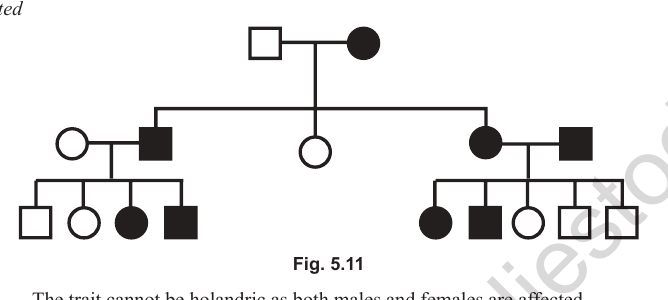
Answer: Autosomal dominant. Reasons: (1) It cannot be holandric, because both males and females are affected. (2) It cannot be sex-linked, because an affected male parent cannot have an affected male offspring. (3) It cannot be autosomal recessive, because both affected (recessive) parents cannot produce normal offspring. In an autosomal dominant trait, affected parents may produce normal or affected offspring.
Problem 2: Which statement regarding the pedigree is correct? (Statements given: (1) the trait is holandric; (2) the parents are homozygous recessive; (3) the parents are heterozygous; (4) the parents are homozygous dominant.)
Answer: Statement (3) is correct. The trait is autosomal recessive, and both parents can be heterozygous. The affected offspring are homozygous recessive. If both parents were homozygous recessive, all offspring would be affected. If the parents were homozygous dominant, all offspring would be normal, and an affected offspring could not arise.
Check point 10: Draw the appropriate symbols used in pedigree analysis and answer the following
(i) Three infants of unspecified sex, against the characters in the pedigree.
Answer: Represented by a diamond symbol.
(ii) Heterozygous condition of a female for a sex-linked trait.
Answer: A circle with a dot in the centre. Males cannot be heterozygous for sex-linked traits.
(iii) Consanguineous marriage.
Answer: A double horizontal line between the square and the circle.
(iv) Affected child of unspecified sex.
Answer: A filled diamond.
(v) Mother from whom the pedigree begins.
Answer: The proposita, shown by a filled circle with an arrow.
(vi) Identical twins.
Answer: Two lines joined at a single point, and they are always of the same sex.
(vii) A normal female.
Answer: An open circle.
NEET NEET Biology Study Material: Principals of Inheritance and Variation
Essential Notes for NEET Biology
Explore essential learning tools for NEET Biology Principals of Inheritance and Variation. This curated collection features in-depth notes and targeted practice questions built around the active 2026 curriculum to streamline your daily revision.
Verified Solutions for NEET Biology
Built using official NCERT guidelines for NEET Biology, these materials provide reliable academic support. Integrating past examination questions and step-by-step solutions helps students understand official NEET grading criteria.
Enhance Accuracy with Online Tests
For peak performance in upcoming evaluations, integrate official Biology sample papers directly into your study schedule. Follow up your revision by attempting online MCQ tests for Principals of Inheritance and Variation to refine calculation speed and precision.
FAQs
Our advanced study package for Principals of Inheritance and Variation includes detailed concepts, diagrams, Mind Maps, and explanation of complex topics to ensure NEET students learn as per syllabus for 2026 exams.
The Mind Maps provided for Principals of Inheritance and Variation act as visual anchors which will help faster recall during high-pressure exams.
Yes, teachers use our NEET Biology resources for lesson planning as they are in simple language and have lot of solved examples.
Yes, You can download the complete, mobile-friendly PDF of the Biology Principals of Inheritance and Variation advanced resources for free.
Yes, our subject matter experts have updated the Principals of Inheritance and Variation material to align with the rationalized NCERT textbooks and have removed deleted topics and added new competency-based questions.