

Official NEET Study Materials for NEET Biology
Access comprehensive study materials and useful resources for Body Fluids and Circulation using the NEET Biology Body Fluids and Circulation Notes. Designed to align with the 2026-27 NEET academic guidelines, these advanced resources help NEET Biology students reinforce core concepts beyond standard textbooks.
Advanced Resources for Biology
View or download the dedicated NEET Biology Body Fluids and Circulation Notes resource below. Engaging with these advanced study guides under focused conditions ensures continuous academic progress and mastery of the 2026-27 curriculum for Body Fluids and Circulation.
Body fluids and circulation explain how blood, tissue fluid and lymph transport materials in the body. This NEET Biology notes page on Body Fluids and Circulation covers open and closed blood vascular systems, single and double circulation, the structure and conducting system of the human heart, cardiac cycle, heart sounds, ECG, heart disorders, blood pressure, arteries, veins and capillaries, the arterial and venous systems, portal systems, the lymphatic system and blood clotting, with check point questions and answers.
While going through the chapter pay special attention to the following -
Body Fluids and Circulation
Terms and definitions -
1. Systemic heart 2. Mitral valve 3. Papillary muscles
4. Myogenic heart 5. Pulse pressure 6. Sphygmomanometer
7. Pace maker 8. Thoracic duct 9. Christmas factor
10. Portal vein
Differences -
1. Open and closed BVS 2. Tachycardia and Bradycardia
3. Single and double circulation 4. 1st and 2nd heart sounds
5. Arteriosclerosis and atherosclerosis 6. Arteries and veins
7. Lymph and serum
Peculiarities -
1. Heart of crocodile 2. BVS of a fish
Disorders -
1. Angina 2. Fibrillation
3. ECG abnormalities 4. Myocardial infarction
Angiology − Study of blood vascular system.
William Harvey (a British scientist) is the ‘Father of Angiology’. He studied blood vascular system (BVS) in a live snake. His monograph on blood circulation was named, ‘Anatomical exercise on the motion of the heart and blood’.
Fabricius (Guru of William Harvey) discovered valves in (longer) veins.
Marcello Malpighii (Founder of histology) discovered blood capillaries.
BVS first time developed in Phylum Annelida. Protozoans to Aschelminthes do not have heart or BVS.
At a Glance
- Blood vascular system
- Open blood vascular system
- Closed blood vascular system
- Type of hearts
- Human heart
- Valves
- Conducting system
- Heart beat
- Control of heart beat
- E.C.G. (E.K.G.)
- Heart disorders
- Blood pressure
- Pulse & pulse pressure
- Arteries, veins and capillaries
- Arterial system in human
- Venous system in human
- Lymphatic system
- Blood clotting
Blood Vascular System is of 2−types.
1. Open blood vascular system. 2. Closed blood vascular system
1. OPEN BLOOD VASCULAR SYSTEM
- In this type of BVS the blood is filled in sinuses or lacunae which together for a large blood filled cavity called Haemocoel.
- Capillaries−network is lacking.
- Such system is present in all the members of Phylum Arthropoda and a few members of Phylum Mollusca (eg. Pila, Unio).
In echinoderms the BVS is very much reduced, and is called Haemal system.
Connecting Concepts
- Cardiology is the study of heart and related functions, while Haematology is the study of blood.
2. CLOSED BLOOD VASCULAR SYSTEM
- In this type of BVS the blood does not come out of the vessels and, hence, haemocoel is lacking.
- The Capillaries−network is present.
- Such system is present in all the members of subphylum Vertebrata, phylumAnnelida* and few members of Phylum Mollusca (eg. Octopus, Squid).
- * The Leech (an annelid), however, has haemocoelomic system, consisting of haemocoelomic channels (not true blood vessels) which are derived from coelomic cavity.
The closed blood vascular system is of 2−types.
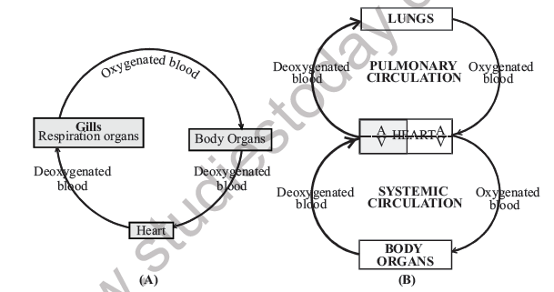
Single Circulation System−
In such system, for one complete circulation through the body, the blood passes only once through the heart . Such animals, therefore, have only deoxygenated blood in the heart. Such heart is known as Venous heart, eg Fishes
[Figure: Fig. 18.1 (A) Single circulation system in fishes - Heart to Gills (respiration organs) carrying deoxygenated blood, from Gills to Body Organs carrying oxygenated blood, and from Body Organs back to the Heart carrying deoxygenated blood. (B) Double circulation system in mammals - the Heart (atrium A and ventricle V on each side) sends deoxygenated blood to the LUNGS and receives oxygenated blood back in the PULMONARY CIRCULATION, and sends oxygenated blood to the BODY ORGANS and receives deoxygenated blood back in the SYSTEMIC CIRCULATION]
Double Circulation System−
In such system, for one complete circulation through the body, the blood passes twice through the heart:
(i) To the lungs and back− It is called Pulmonary circulation or Lesser circulation (due to Lesser distance of the lungs from the heart.
(ii) To the body organs and back− It is called Systemic circulation or Greater circulation (covering greater distance from heart to the body−organs. eg.Amphibians, Reptiles, Birds and Mammals. (Because of single ventricle, the amphibians and reptiles have incomplete double circulation)
Heart
From origin point of view the whole BVS, including heart, is Mesodermal. The heart is one of the first organ to be completed in embryo.
Survey of Hearts in Important Groups
- In earthworm (Phylum Annelida) eight hearts (4−pairs) are present.Two pairs are true hearts and two pairs false.
For details please refer Chapter 7
- In Cockroach (Phylum Arthropoda) the heart is single and dorsal in position. It is 13-chambered (3+10). In Prawn (Palaemon) the heart contains only oxygenated blood because the deoxygenated blood from the body directly goes to the gills. The oxygenated blood from there finally goes to the heart
- InUrochordates(ex.Herdmania)theheartistubularorsac-like.Thishearthas2-‘Pacemakers’ and the flow of blood is reversed periodically. In Amphioxus (Branchiostoma), there is no true heart and blood is collected into Sinus Venosus.
- In fishes the heart is 2−chambered, 1−Atrium(A) and 1−Ventricle(V). As mentioned above, the heart of fishes is Venous heart.
- In Amphibians the heart is 3−chambered (2A+1V). Because of single Ventricle the blood in frog is mixed in this chamber. ( Chapter 7).
- In Reptiles the heart in general, is incompletely 4−chambered. However, in Crocodile and Alligator the heart is completely 4−chambered (2A+2V).
- In Birds and Mammals the heart is always 4−chambered (2A+2V).
HUMAN HEART
- It is 10−12 cm in size, and is present in the Mediastinum (space) of the thoracic-cavity .
- It weighs ~250 g in males and 225 g in females.
- The apex of the heart is ~9 cm to the left of the mid line
- The heart is covered by a transparent serous membrane, the pericardium (the parietal mesoderm/peritoneum). The pericardial fluid between heart and the pericardium keeps the heart moist and protects it from friction and mechanical injury.
- The wall of the heart has 3- coatings, Epicardium (Outer), Myocardium (Middle) and Endocardium (Inner). The myocardium contains cardiac muscles.
- The atria and ventricles are divided by Coronary Sulcus.
- Two Atria are separated by Interatrial septum. In foetus this septum contains Foramen ovale (opening) and the blood from right atrium (oxygenated blood from Placenta) passes to the left atrium. At the time of birth when placenta is detached this passage is closed to form Fossa ovalis. If Foramen ovale is left open after first few weeks of life, it is referred as a ‘hole in the heart’.
- The left side of the heart (Lt. A and Lt. V) contains oxygenated (pure) blood and the right side (Rt. A and Rt. V) has deoxygenated (impure) blood.
- Lt. atrium receives oxygenated blood through 2−pairs of pulmonary veins (2 per lung).
- Rt. atrium receives deoxygenated blood through 1−Precaval or Anterior Vena Cava and 1−Post caval or posterior Vena cava (Rabbit has 2−Precavals).
- Lt. and Rt. ventiricles are separated by interventicular septum.
[Figure: Fig. 18.2 Internal structure of human heart (simplified) - labelled Superior vena cava, Inferior vena cava, Rt. atrium, Eustachian valve, Tricuspid valve, Rt. ventricle, Papillary muscles, Chordae tendinae, Lt. atrium, Bicuspid valve, Lt. ventricle, Systemic aorta, Lt. pulmonary artery, Rt. pulmonary artery, Lt. pulmonary veins]
- Lt.ventricle and Rt. ventricle receive oxygenated and deoxygenated blood from respective atria.
- From Lt.ventricle oxygenated blood is supplied to the body organs through systemic aorta. From Rt. ventricle deoxygenated blood is supplied to lungs through pulmonary aorta
- The wall of the Lt.ventricle, which supplies oxygentated blood to all body organs, is thickest of all the chambers.
- Systemic aorta in Amphibians and reptiles is paired. In birds and mammals it is unpaired (single).In birds it is right and in mammals it is left.
Valves
i. Eustachian valve− A flap of this valve is present at the opening of the post caval and prevents the back flow of the blood. The major portion of this valve is vestigial. ii. Thebasian valve− It is present at the opening of coronary sinus which receives deoxygenated blood from cardiac muscles. iii. Atrio−ventricular valves− In Lt. side there is Bicuspid (2−flaps) valve between Lt. atrium and Lt. ventricle. This is also known as Mitral valve. In between Rt. atrium and Rt. ventricle there is a 3−flapped Tricuspid valve. The flaps of both Bicuspid and Tricuspid valve are attached to Papillary muscles through Chordae Tendinae. These prevent reverting of the valves during contraction of ventricles.
Bicuspid is the only valve in mamalian heart which always remains in contact of the pure (oxygentated) blood. All other type of valves come in contact of deoxygenated blood.
(The base of the anterior papillary muscles is connected to the interventricular septum by a special band of cardiac muscles, called Moderator band.) iv. Semilunar valves− These are present at the base of aorta and each has three pockets which are filled with blood to prevent its back flow to the ventricles.
Conducting System or Nodal Tissue
Like all other vertebrates, in human also, the heart is Myogenic, i.e. the heart beat is generated by muscles and not nerves
- The heart beat is generated through Sino atrial node (SAN) which is also known as Pace maker. It is a bundle of specialized cardiac muscles. (These muscles like other muscles, are not contractile but are specialized for conducting impulses like nerve fibres).
- In human SA node is embedded in the wall of Rt. atrium. In frog’s heart it is present in the wall of Sinus Venosus.
- Atrio−ventricular node (AVN) is present in inter−atrial septum, close to Tricuspid valve. (The AV node is absent in frog’s heart).
- From AV node arises a bundle of specialized muscle fibres, called Bundle of His or Atrio−ventricular bundle. This runs in the anterior part of inter-ventricular septum and then bifurcates. The branches ramify in the walls of ventricles as Purkinje fibres.
[Figure: Fig. 18.3 Conducting system in human heart - S.A. Node in the right atrium, Inter nodal pathway to the A.V. Node, then the Bundle of His in the inter ventricular septum dividing into Purkinje (Purkyne) fibres in the walls of the ventricles; Precaval and Post caval veins, RV, LV and LA are also marked]
Connecting Concepts
- Lt. part of the heart (Lt. atrium and Lt. ventricle), having oxygenated blood, is called Systemic heart.
Connecting Concepts
- In frog’s heart there is a triangular chamber on the dorsal side, called Sinus Venosus. The deoxygenated blood through 2−precavals and 1−postcaval is first filled into this chamber and from here it goes to Rt. atrium. Sinus Venosus is lacking in the heart of birds and mammals. (For detailed structure of Frog’s heart, please refer Chapter 6)
- Amongst non-chordates or invertebrates, Mollusca is the only phylum in which the heart is Myogenic. In Arthropods the heart is Neurogenic ( Heart beat is generated by nerve fibres).
Heart Beat
- The rate of heart beat in a adult human is ~72 per minute.
- The single heart beat is called Cardiac cycle and takes 0.8 sec (60 sec / 72 beats ).
- Each Cardiac cycle consists of 3−parts.
1. Atrial systole (0.1 sec)
2. Ventricular systole (0.3 sec)
3. Diastole (joint) (0.4 sec) (The heart beat in newly born child is ~100 per min while in foetus it is ~140 per min.)
| Chamber | Systole | Diastole |
|---|---|---|
| Atrium | 0.1 sec | 0.7 sec |
| Ventricle | 0.3 sec | 0.5 sec |
- Stroke volume ~70 ml
- It is the amount of blood pumped out from each ventricle per beat.
- Cardiac output ~5 litre
- It is the output of the heart per unit time (70 ml x 72 beats per min.)
- Cardiac index ~ 3.2 litre / m2 It is Cardiac output per square meter of the body surface per min. i.e. (5 lit. / 1.6 m2).
Factors affecting heart beat−
- Heart beat increases during excitement, eating, exercise, fever and pregnancy.
- Heart beat decreases during sleep and shock.
- Heart beat is more in males than in females.
- Heart beat increases with the increase of altitude (Heart beat ∝ altitude).
- The heart beat increases with the decrease of animal size (Heart beat ∝ 1/size). If elephant, human and mouse are compared the heart beat will be highest in mouse and lowest in elephant.
- Heart beat ∝ 1/BP (except in exercises, in which both blood pressure (BP) and the rate of heart beat, increase).
Control of Heart beat−
(a) Chemical Control
(i) Adrenaline− This hormone stimulates heart beat either directly or through sympathetic nervous system.
(ii) Thyroxine− This increases metabolic rate and hence heart beat.
(b) Nervous Control Medulla oblongata has two regulatory centre −
(i) Accelerator centre− It functions through sympathetic nervous system (SNS) and increases heart beat by the secretion of epinephrine or adrenaline.
(ii) Depressor centre− It functions through parasympathetic nervous system (PSNS) by the secretion of acetylcholine. It decreases heart beat.
Heart Sound
In each cardiac cycle, though the heart produces 4−sounds but only 1st and 2 st and 2 st nd sound can nd sound can nd be heard by Stethoscope. These sounds are described below:
1. 1st Sound −
- It sounds like LUBB.
- It is of longer duration.
- It is of lower frequency (low pitch).
- It is louder (volume ↑).
- When ?− At the beginning of ventricular systole and coincides with the ‘R’ wave of ECG.
Why ?− Mainly because of closing of Bicuspid and Tricuspid valves.
Connecting Concepts
- PSNSfibrestotheheartpassthrough Vagus (10th cranial nerve). If Vagus is cut the secretion of acetylcholine is also cut and the heart beat increases due to continuous supply of adrenaline. If, on the other hand, Vagus is stimulated the secretion of acetylcholine through PSNS fibres increases and heart beat decreases.
- The increase of Heart beat is called Tachycardia, while decrease of heart beat is called Bradycardia.
2. 2nd Sound −
- It sounds like DUP.
- It is of shorter duration.
- It is sharper (high frequency).
- It is softer (low volume) When ?− At the end of ventricular systole and coincides with the ‘T’ wave of ECG.
Why ?− Because of closing of the semilunar valves of systemic aorta and pulmonary aorta.
E.C.G. (E.K.G.)
The instrument which records electrical activity of the heart muscles is called Electrocardiograph. The sketch obtained on the graph paper is called electrocardiogram (or electrokardiogram).
- The standard symbols used for ECG are PQRST, where P* represents atrial depolarisation; QRS* complex represents ventricular depolarisation and T* represents ventricular repolarisation. (P, R & T are deflection waves).
[Figure: Fig. 18.4 An Electrocardiogram (ECG) - a trace over 0 to 0.8 sec showing the P wave, the QRS complex and the T wave, with PR interval (0.18 s), QRS interval (0.08 s), ST interval (0.32 s), atrial systole (0.1 s), ventricular systole (0.3 s) and joint diastole (0.4 s)]
Intervals in Normal ECG
| Interval | Average duration | Event |
|---|---|---|
| PR (or PQ) | 0.18 sec (180 mili sec) | Atrial depolarisation and conduction through AV node |
| QRS | 0.08 sec (80 ms) | Ventricular depolarisation and atrial repolarisation |
| ST | 0.32 sec (320 ms) | Ventricular repolarisation |
Abnormality of ECG
1. In rheumatic heart disease (valves damaged) and arteriosclerotic heart desease (due to formation of plaque or calcification) there is inflammation of atria and AV node, which results in the lengthening of PR interval.
2. When heart muscles receive insufficient oxygen (lesser blood supply− heart ischaemia), the ST segment is depressed and T wave is flatenned.
3. In acute myocardial infarction the ST segment is elevated and Q & R waves are enlarged.
4. In case of hyperkalemia (K+↑↑) T wave is very tall and slender. * ‘P’ - wave occurs slightly before the onset of Atrial contraction (systole). * QRS - waves (complex) begin slightly before the onset of ventricular systole. * ‘T’-wave occurs slightly before the end of ventricular systole.
Connecting Concepts
- Resting potential in cardiac muscle fibres is − 90 mV and not − 70 mV as in an average sized neuron. Cardiac muscles do not show tetanus also.
[Figure: Fig. 18.5 Abnormalities in ECG - (1) Rheumatic fever: lengthening of PR interval; (2) Heart ischaemia: ST segment depressed; (3) Myocardial infarction: ST segment elevated with enlarged Q and R waves; (4) Hyperkalemia: T wave taller. The normal trace with P, Q, R, S, T and the PQ (PR) and ST segments is shown for comparison]
Heart Disorders
1. Heart Ischaemia − The blood supply to the heart muscles (myocardium) is reduced
(a) Angina pectoris− Blood supply to the heart muscles is reduced because of narrowing of the coronary artery. In such a case there is no problem in routine work but pain develops during exercise or exertion.
(b) Myocardial infarction− In this case the blood supply is totally blocked and the part of heart tissue becomes dead. The death results mainly from fibrillation.
2. Heart Block− In atrio−ventricular block there is delay between atrial and ventricular systole. PR > 0.2 sec. In such a case AV node is either not stimulated or stimulation is delayed. In bundle branch-block QRS > 0.1 sec.
(If atrial impulse suddenly fails to be transmitted to the ventricles, it is called Ventricular escape or Adam’s syndrome).
3. Rheumatic Heart Disease− It develops due to recurrence of rheumatic fever. The heart valves are damaged due to formation of antibodies. It is an autoimmune disorder.
4. Persistent ductus arteriosus − In foetus pulmonary aorta and systematic aorta are connected by ductus arteriosus. At birth it degenerates. Rarely it persists and the blood supply to systemic aorta is reduced which causes congestion in lungs.
5. Fibrillation - A rapid, irregular, abnormal, asynchronous and ineffective contraction of cardiac muscles is called fibrillation. Atrial fibrillation may occur due to Rheumatic heart disease and myocardial infarction. The ventricular fibrillation may be produced by electrocution. If ventricular fibrillation lasts for more than a few minutes, it can be fatal. It can be stopped by electric shocks or by electronic defibrillators. The latter can be implanted in the persons of high risks.
Atherosclerosis− It is the disorder of arteries (blood vessels) in which cholesterol (plaque) is deposited.
Arteriosclerosis− It is the hardening of arteries due to calcification in old age. The elasticity of the vessles is reduced. This may also result from atherosclerosis.
Thrombosis− It is the clotting of blood inside the blood vessels.
BLOOD PRESSURE
The pressure exerted by the blood on the walls of blood vessels is called blood pressure (BP). The blood pressure in all the arteries is relatively very high, but drops with distance away from the heart.
- BP decreases from arteries to veins (arteries > capillaries > veins)
- BP is measured by Sphygmomanometer on Brachial artery of upper arm (generally of left side).
- In adults the normal BP is 120/80.
- 120 mm of Hg is systolic pressure during contraction of ventricles, and 80 mm of Hg is diastolic pressure during relaxation of ventricles.
- In children the BP is low as compared to adults.
- In old age the average BP is 150/90, as the arteries become less elastic.
- The chemicals which are vasodialator decrease the blood pressure while vasoconstrictors increase the blood pressure. The Histamine is a vasodialator and adrenaline is a vasoconstrictor.
PULSE
Pulse is a wave of distension, i.e., a rhythmic expansion and relaxation (not contraction) of arteries.
- Pulse is felt at radial artery, of wrist.
- With the help of pulse the heart beat can be measured.
Pulse Pressure
- It is the difference between systolic and diastolic pressure.
- In normal adult the value of pulse pressure is 40 mm of Hg (120 mm−80 mm).
ARTERIES, VEINS AND CAPILLARIES
Each artery has three coats, viz (i) Tunica externa (adventitia), (ii) T. media and (iii) T. interna (intima).
(1) Tunica externa mainly consist of white fibrous connective tissue (collagen fibres).
(2) Tunica media contains elastic fibres and smooth muscles. The arteries near the heart have lot of elastic fibres, and those farther from the heart have proportionately more smooth muscles (muscular tissue).
(3) Tunica interna has endothelial layer (simple squamous epithelium) or intima layer with elastic fibres.
(In atherosclerosis the cholesterol or other fatty materials are deposited in sub−intimal layer).
| Arteries | Veins |
|---|---|
| 1. More elastic | 1. Less elastic |
| 2. Tunica media very thick | 2. Tunica media thin |
| 3. Lumen narrow | 3. Lumen broader |
| 4. Valves absent | 4. Valves (semilunar) present in (longer) veins |
| 5. Lie deeper in the body | 5. Generally superficial |
| 6. All, except* pulmonary artery, carry oxygenated blood | 6. All, except pulmonary vein, carry deoxygenated blood. |
| 7. Blood flow is jerky or discontinuous | 7. Blood flow is continuous |
| 8. These vessels carry blood away from the heart. | 8. These carry blood towards heart. |
| 9. They contain ~13% of the blood at a time | 9. They contain ~65% of the blood at a time |
[Figure: Fig. 18.6 Histology of blood vessels - (a) Artery with Tunica externa (collagen fibres), Tunica media (smooth muscles and elastic fibres) and Tunica interna or Endothelium; (b) Vein with the same three layers but a thin media; (c) Capillary with only Endothelium]
* Like pulmonary artery, the Umbilical artery of foetus also carries deoxygenated blood.
Connecting Concepts
- In arterioles, the smallest branches of arterial system, the tunica media does not have elastic fibres and consists of smooth muscles only. The arterioles, therefore, can regulate the amount of blood flowing to the tissues.
Capillaries
- Tunica externa and T. media are absent. There are no collagen fibres, no elastin fibres and no smooth muscles.
- Only endothelial lining with basement membrane is present. (The capillary network supplying outer layer of larger blood vessels is called Vasovasorum)
ARTERIAL SYSTEM IN HUMAN
2− major aorta − 1. Pulmonary aorta, 2. Systemic aorta Pulmonary aorta arises from Rt. ventricle and supplies deoxygenated blood to the lungs.
Systemic aorta arises from Lt. ventricle and gives out following important arteries.
(a) Coronary artery− It supplies blood to the heart muscles.
(b) Innominate artery− (Rt. Brachio- cephalic artery) − It bifurcates into Rt. subclavian and Rt. common carotid artery.
(c) Lt. common carotid artery− It divides into external and internal carotid arteries.
(d) Lt. subclavian − it arises independently from the aorta.
[Figure: Fig. 18.7 Arterial system in human - labelled External carotid, Internal carotid, Vertebral artery, Rt. carotid artery, Lt. common carotid artery, Rt. subclavian artery, Lt. Subclavian artery, Brachio-cephalic or Innominate artery, Systemic aorta, Coronary artery, LV, Mammillary artery, Brachial artery, Bronchial artery, Thoracic aorta, Dorsal aorta, Oesophageal artery, Intercostal arteries (9 pairs), Diaphragm, Phrenic artery, Abdominal aorta, Coeliac artery, Anterior mesenteric artery, Renal artery, Posterior mesenteric artery, Lt. common iliac artery, Internal iliac artery and External iliac artery]
Blood supply−
1. Internal carotid supplies oxygenated blood to the brain while external carotid supplies blood to the other parts of the head.
2. Brachial arises from subclavian artery and supplies blood to the arm.
3. Mammary artery also arises from subclavian artery and supplies blood to the pericardium.
Systemic aorta now takes a turn and comes to the dorsal side of the heart. This dorsal aorta is differentiated into Thoracic aorta (in thoracic region) and Abdominal aorta (in abdominal region).
Connecting Concepts
- In mammals there is Lt. systemic aorta. In birds it is Rt. systemic aorta. In both it is unpaired. In amphibians (Frog) and reptiles the systemic aorta is paired.
Important arteries arising from thoracic aorta−
1. Oesophageal artery− supplies blood to oesophagus.
2. Intercostal artery− supplies blood to intercostal muscles in between the ribs.
3. Bronchial artery− supplies oxygenated blood to the lungs.
Important Arteries arising from abdominal aorta−
(a) Paired arteries− e.g. Phrenic artery, Renal artery and Gonadial artery.
Phrenic artery− supplies blood to diaphragm.
Renal artery− supplies blood to kidney.
Gonadial artery− supplies blood to gonads.
(b) Unpaired arteries− e.g. Coeliac artery, Anterior (superior) mesenteric and Posterior (inferior) mesenteric arteries.
Coeliac artery− supplies blood to stomach, pancreas, spleen and liver etc.
Anterior mesentric artery− supplies blood to small intestine.
Posterior mesentric artery− supplies blood mainly to the large intestine.
In the posterior most region the abdominal aorta bifurcates into Rt and Lt. common iliac arteries. Each iliac artery then divides into external and internal iliacs. External iliac enters the leg and outer branch of it is called Femoral artery.
VENOUS SYSTEM IN HUMAN
Portal Vein − It is a vein that arises as capillaries from one organ and terminates as capillaries in the other organ. Portal vein is different from other veins as it does not carry blood directly to the heart.
The system formed by portal veins is called Portal system.
Portal system is of 2-types
Minor portal system−
- The portal vein of this system is shorter.
- Such system is present between hypothalamus (the floor of diencephalon)and anterior pituitary.
- This system is meant for transferring the releasing factors (Hormones) from Hypothalamus to the Anterior pituitary.
- This portal system is also known as Hypophyseal portal system as it terminates into Hypophysis -cerebri (pituitary).
Major portal system−
- The portal veins in this system are longer.
- There are two such portal systems in vertebrates.
(a) Hepatic portal system−
- It is present in all vertebrates.
- It starts from alimentary canal and terminates into liver.
- It brings digestive end products to the liver before releasing them (in modified form) into the blood.
(b) Renal portal system−
- It is present in lower vertebrates (fishes and amphibians) only. In reptiles it is reduced.
- It starts from fins in fishes and from hind limbs in amphibians (frog).
- It terminates into kidney. (For details of this system, in ‘Frog’, please refer chapter 6
For details of this system, in ‘Frog’, please refer chapter 6
Connecting Concepts
- The name of the portal system is given after the name of the organ into which the portal vein terminates, e.g. Hepatic portal system : the vein terminates into liver and Renal portal system : the vein terminates into kidney.
[Figure: Fig. 18.8 Venous system in human - labelled Precaval (Rt. brachiocephalic vein), Internal jugular vein, External jugular vein, Subclavian vein, Diaphragm, Hepatic veins, Renal vein, Gonadial vein, Inferior vena cava or post caval, Rt. common iliac vein, Internal iliac vein, Ext. iliac vein, Femoral vein and Saphenous vein]
The Venous system Venous system V in human includes 1−Precaval and 1−Postcaval.
- Precaval or superior venacava is formed by the combination of 2−Brachiocephalic veins.
- Each brachiocephalic vein is formed by the union of internal jugular vein and subclavian vein. External jugular runs down to join subclavian vein.
- The venous blood from brain is collected by internal jugular vein.
- The subclavian vein (below clavicle bone) collects deoxygented blood from arms.
- Thoracic blood is collected by Azygos, Hemizygos, Oesophageal, Intercostal and Bronchial veins.
- Postcaval or inferior venacava is formed by the joining of Rt. and Lt. common iliac vein.
- Hepatic vein, renal vein and gonadial veins join postcaval.
- From hind limbs the venous blood is collected through Femoral and Saphenous veins. The Femoral vein ascends in to the abdominal region and becomes external iliac vein.
- The renal portal system is absent in mammals.
LYMPHATIC SYSTEM
- It is the characteristic feature of all vertebrates.
- Lymphatic vessels are comparable to blood capillaries and veins. There is no counter part of arteries in lymphatic system. (All systems, except CNS, have lymphatic circulation .)
- As in longer veins, the valves are present in the lymphatic vessels also.
- In lower vertebrates (fishes and amphibians) the lymphatic hearts are also present ( In frog there are 4−such hearts, 2-Thoracic and 2 - abdominal).
- In mammals lymphatic hearts are absent but lymph nodes are present.
Connecting Concepts
- The Saphenous vein is the longest vein in human body and is also used in coronary artery bypass grafting (CABG).
- The lymph capillaries in human form lymph vessels, which join to form lymphatic ducts.
- The lymphatic duct of the left side is called Thoracic duct. In Rt. side there is Rt. lymphatic duct.
- Both Lt. thoracic duct and Rt. lymphatic duct pour the lymph into subclavian vein of their side.
Lymph −
- It is mostly described as Blood minus RBCs. It can also be represented as plasma plus WBCs. In lymph the number of WBCs is lesser but the number of lymphocytes is greater than blood.
- In comparison to blood plasma, the proteins in lymph are fewer.
- Like plasma the lymph also contains fibrinogen and hence can clot.
- The lymph outside the lymph vessels is known as tissue fluid or interstitial fluid or tissue fluid or interstitial fluid or tissue fluid extra cellular fluid (ECF). cellular fluid
BLOOD CLOTTING (HAEMOSTASIS)
The clot or coagulam is a dark -reddish brown ‘scum’ formed mainly by a network of threads in which dead or damaged blood elements are trapped.
- It is the property of plasma.
- Normal blood clotting time is 3−10 min.
- The clot inside the blood vessels is called thrombus. A moving thrombus is called embolus.
- In haemophilia (a sex linked disease) the blood clotting is delayed.
- According to Macferlane hypothesis there are 13−factors responsible for blood clotting (or coagulation). The 4−factors are primary and 9−factors are accessory for this process.
Primary factors
(I) Fibrinogen (II) Prothrombin (III) Thromboplastin (Thrombokinase)
(IV) Calcium ions
- The first 3−factors are proteinaceous in nature.
- The Prothrombin (II factor) is synthesized in liver in the presence of vitamin K.
- The Thromboplastin is released from injured platelets or injured tissue. This converts prothrombin into thrombin in the presence of calcium ions. The thrombin converts fibrinogen into fibrin, the latter forms the clot. The blood cells are entangled in the network of fibrin. A straw -coloured fluid, separated from clot, is called Serum. As the Serum does not have fibrinogen, it can not clot.
[Figure: Fig. 18.9 Role of primary factors (I to IV) in blood clotting - Thromboplastin or Thrombokinase (III), an enzyme complex from injured platelets or tissues, with calcium ions (IV) converts Prothrombin (II) into Thrombin; Thrombin converts Fibrinogen (I) into Fibrin]
Connecting Concepts
- Heparin (a polysaccharide) is a natural anticoagulant in the blood and preventstheconversionofprothrombin into thrombin in circulating blood. It is secreted from liver (mainly) and mast cells.
Accessory factors
(V) Labile factor (Proaccelerin) − It helps in complete conversion of prothrombin into thrombin.
(VI) No separate entity, hence No specific name (existence doubtful). existence doubtful). existence doubtful (VII) Stable factor (Proconvertin) − It accelerates the formation of active thromboplastin.
(VIII) Anti−haemophilic globulin (AHG)−The absence of this factor delays blood clotting causing Haemophilia−A. This type of haemophilia is most common (80%).
(IX) Plasma thromboplastin co−factor (PTC)− The deficiency of this factor causes Haemophilia−B. Approximately 20% of haemophilic patients have this type of haemophilia. The IX factor is also known as Christmas factor, and Haemophilia B is known as ‘Christmas disease’.
(X) Stuart Prower factor− It helps in the conversion of prothrombin into thrombin.
(XI) Plasma thromboplastin antecedent (PTA)− It activates the inactive christmas factor. The deficiency of this causes a rare type of bleeder-disease, called Haemophilia -C.
(XII) Hageman’s factor or Glass factor − It converts inactive PTA into active form. It also dilates blood vessels for increasing their permeability.
(XIII) Fibrin stabilizing factor− It causes polymerization of soluble fibrin into insoluble fibrin and also inhibits depolymerization.
Factors slowing blood clotting−
(a) Inside body− Heparin is a natural anticoagulant present in the blood. It prevents conversion of prothrombin into thrombin and does not allow blood coagulation inside blood vessels.
(b) Outside body− sodium citrate, sodium oxalate, double oxalate and EDTA (ethylene diamine tetra acetic acid) and the other chelating agents bind with free calcium and prevent the conversion of prothrombin into thrombin. Dicumarol and Warfarin inhibit the action of vitamin K (anti- prothrombin function) and thus prevent blood clotting.
Factors hastening blood clotting−
(a) Inside body− Vitamin K, which is essential for the synthesis of prothrombin in the liver, if injected into the body, during haemorrhage, stimulates blood clotting.
(b) Outside body− The blood oozing out from injured part can be coagulated by applying pressure or providing rough surface (for injuring more platelets) or by increasing the temperature (to activate blood clotting− enzymes).
CHECK POINT QUESTIONS
1. Fill in the blanks with suitable word/statement :
(i) ........................ is called father of Angiology.
Answer: William Harvey
Teacher's Note:
a) He studied the blood vascular system in a live snake.
(ii) ....................... valve is present between Lt. atrium and Lt. ventricle.
Answer: Bicuspid (mitral) valve
Teacher's Note:
a) The tricuspid valve lies on the right side.
(iii) In fish heart there is ..................... circulation system.
Answer: single
Teacher's Note:
a) Fish have a venous heart with only deoxygenated blood.
(iv) All arthropods have .................... blood vascular system
Answer: open
Teacher's Note:
a) The blood fills sinuses forming the haemocoel.
(v) ....................... is called pace maker of human heart.
Answer: Sino-atrial node (SA node)
Teacher's Note:
a) It lies in the wall of the right atrium.
(vi) 'Lesser circulation' is between ...................... and .......................
Answer: the heart and the lungs (pulmonary circulation)
Teacher's Note:
a) It covers a lesser distance than the systemic circulation.
(vii) Bicuspid and tricuspid valves are .......................during joint diastole.
Answer: open
Teacher's Note:
a) Blood flows from the atria into the ventricles in this phase.
(viii) In mammals the systemic aorta lies in ..............side of the heart.
Answer: left
Teacher's Note:
a) In birds it is on the right side.
1. Differentiate between :
(i) Open and closed blood vascular system.
Answer:
| Open blood vascular system | Closed blood vascular system |
|---|---|
| Blood is filled in sinuses or lacunae forming a haemocoel | Blood does not come out of the vessels; haemocoel is lacking |
| Capillary network is lacking | Capillary network is present |
| Present in Arthropoda and a few Mollusca (Pila, Unio) | Present in Vertebrata, Annelida and a few Mollusca (Octopus, Squid) |
Teacher's Note:
a) Use a table with at least three points.
(ii) Pulmonary and systemic circulation
Answer:
| Pulmonary (lesser) circulation | Systemic (greater) circulation |
|---|---|
| Blood goes from the heart to the lungs and back | Blood goes from the heart to the body organs and back |
| Covers the lesser distance | Covers the greater distance |
| Starts at the right ventricle (pulmonary aorta) and ends in the left atrium | Starts at the left ventricle (systemic aorta) and ends in the right atrium |
Teacher's Note:
a) The pulmonary circulation carries deoxygenated blood in the artery.
(iii) Myogenic and Neurogenic heart.
Answer:
| Myogenic heart | Neurogenic heart |
|---|---|
| Heart beat is generated by the cardiac muscles themselves | Heart beat is generated by nerve fibres |
| Found in vertebrates (including humans) and Mollusca | Found in Arthropods |
Teacher's Note:
a) The SA node is the pace maker of a myogenic heart.
(iv) Tachycardia and Bradycardia.
Answer:
| Tachycardia | Bradycardia |
|---|---|
| Increase in the heart beat rate | Decrease in the heart beat rate |
| Occurs in excitement, exercise, fever | Occurs in sleep and shock |
Teacher's Note:
a) Tachy means fast and brady means slow.
(v) Cardiac output and cardiac index.
Answer:
| Cardiac output | Cardiac index |
|---|---|
| Output of the heart per unit time = 70 ml x 72 beats per minute, about 5 litre | Cardiac output per square metre of the body surface per minute |
| About 5 litre per minute | About 3.2 litre per m2 (5 litre ÷ 1.6 m2) |
Teacher's Note:
a) Cardiac index = cardiac output ÷ body surface area.
(vi) SA node and AV node
Answer:
| SA node (Sino-atrial node) | AV node (Atrio-ventricular node) |
|---|---|
| Called the pace maker of the heart | Passes the impulse on to the Bundle of His |
| Embedded in the wall of the right atrium (in frog, in the sinus venosus) | Present in the inter-atrial septum close to the tricuspid valve (absent in frog) |
| Generates the heart beat | Gives rise to the Bundle of His and Purkinje fibres |
Teacher's Note:
a) The AV node is absent in the frog heart.
(vii) Foramen ovale and fossa ovalis
Answer:
| Foramen ovale | Fossa ovalis |
|---|---|
| An opening in the inter-atrial septum of the foetus | A depression formed in the septum after birth when the opening closes |
| Lets blood pass from right atrium to left atrium in the foetus | Has no passage; if the foramen is left open it is called a hole in the heart |
Teacher's Note:
a) Foramen ovale closes when the placenta is detached at birth.
1. Name the followings :
(i) Instrument used for measuring Blood pressure.
Answer: Sphygmomanometer
Teacher's Note:
a) It is used on the brachial artery of the upper arm.
(ii) Standard symbols used for ECG.
Answer: P, Q, R, S and T (PQRST)
Teacher's Note:
a) P is atrial depolarisation, QRS is ventricular depolarisation and T is ventricular repolarisation.
(iii) The blood vessel at which pulse is felt.
Answer: Radial artery of the wrist
Teacher's Note:
a) Pulse is a rhythmic expansion of arteries.
(iv) The artery that carries deoxygenated blood.
Answer: Pulmonary artery (pulmonary aorta); the umbilical artery of the foetus also carries it
Teacher's Note:
a) Veins carry deoxygenated blood except the pulmonary vein.
(v) Disorder of arteries due to cholesterol deposition.
Answer: Atherosclerosis
Teacher's Note:
a) Arteriosclerosis is hardening by calcification.
(vi) Difference between systolic and diastolic pressure
Answer: Pulse pressure (normally 40 mm of Hg)
Teacher's Note:
a) 120 - 80 = 40 mm of Hg.
(vii) When heart muscles are suddenly damaged by inadequate blood supply
Answer: Myocardial infarction (heart attack)
Teacher's Note:
a) The blood supply is totally blocked and the tissue dies.
(viii) Acute chest pain when enough oxygen is not reaching heart muscles.
Answer: Angina pectoris
Teacher's Note:
a) Pain develops on exertion due to narrowing of the coronary artery.
1. What are the target organs of the following blood vessels?
(i) Phrenic artery
Answer: Diaphragm
Teacher's Note:
a) It arises from the abdominal aorta.
(ii) Coeliac artery
Answer: Stomach, pancreas, spleen and liver
Teacher's Note:
a) It is an unpaired artery.
(iii) Internal carotid artery
Answer: Brain
Teacher's Note:
a) The external carotid supplies the other parts of the head.
(iv) Bronchial artery
Answer: Lungs (supplies oxygenated blood to the lungs)
Teacher's Note:
a) It arises from the thoracic aorta.
(v) External iliac artery
Answer: Leg (its outer branch is the femoral artery)
Teacher's Note:
a) It arises from the common iliac artery.
1. Answer the followings :
(i) From which organ does the Hepatic portal system collect the blood?
Answer: From the alimentary canal (digestive tract); it brings digestive end products to the liver
Teacher's Note:
a) It starts in the alimentary canal and ends in the liver.
(ii) Which blood vessel collects the blood from brain?
Answer: Internal jugular vein
Teacher's Note:
a) It joins the subclavian vein to form the brachiocephalic vein.
(iii) From which portal system does the anterior pituitary receive releasing hormones.
Answer: Hypophyseal portal system (the minor portal system from the hypothalamus)
Teacher's Note:
a) The portal vein is short in this system.
(iv) Which is the longest vein in human body?
Answer: Saphenous vein
Teacher's Note:
a) It is also used in coronary artery bypass grafting.
(v) At what stage of cardiac cycle does the Ist heart sound appear?
Answer: At the beginning of ventricular systole, due to closing of the bicuspid and tricuspid valves
Teacher's Note:
a) It coincides with the R wave of ECG and sounds like LUBB.
1. Answer the followings :
(i) According to Macferlane hypothesis how many factors are responsible for blood clotting?
Answer: 13 factors (4 primary and 9 accessory)
Teacher's Note:
a) Learn the four primary factors first.
(ii) Which Haemophilia is also known as christmas disease?
Answer: Haemophilia B (deficiency of factor IX, the Christmas factor)
Teacher's Note:
a) About 20 percent of haemophilic patients have this type.
(iii) Name the natural anticoagulant of the blood?
Answer: Heparin
Teacher's Note:
a) It is secreted mainly by the liver and mast cells.
(iv) Which vitamin is responsible for the synthesis of Prothrombin?
Answer: Vitamin K
Teacher's Note:
a) Prothrombin is made in the liver.
(v) What are the primary factors for blood clotting?
Answer:
1. Fibrinogen (I)
2. Prothrombin (II)
3. Thromboplastin or Thrombokinase (III)
4. Calcium ions (IV)
Teacher's Note:
a) The first three are proteins.
(vi) The absence of which factor causes classical haemophilia?
Answer: Factor VIII, the anti-haemophilic globulin (AHG), causing Haemophilia A
Teacher's Note:
a) Haemophilia A is the most common type (80 percent).
(vii) What is the name of left lymphatic duct
Answer: Thoracic duct
Teacher's Note:
a) It pours lymph into the left subclavian vein.
(viii) Which blood clotting factor is inorganic in nature?
Answer: Calcium ions (factor IV)
Teacher's Note:
a) The first three factors are proteinaceous.
1. Differentiate between :
(i) Thrombus and Embolus
Answer:
| Thrombus | Embolus |
|---|---|
| The clot formed inside the blood vessel | A moving thrombus |
Teacher's Note:
a) An embolus can block a vessel far from where it formed.
(ii) Lymph and ECF
Answer:
| Lymph | ECF (tissue fluid) |
|---|---|
| The fluid present inside the lymph vessels | The lymph outside the lymph vessels, also called interstitial fluid |
| Flows through the lymphatic system | Fills the spaces between the cells |
Teacher's Note:
a) Both are mostly blood minus RBCs.
(iii) The role of Dicumarol and Sodium oxalate in blood clotting
Answer:
| Dicumarol | Sodium oxalate |
|---|---|
| Inhibits the action of vitamin K (anti-prothrombin function), so prothrombin is not made | Binds with free calcium ions and prevents conversion of prothrombin into thrombin |
| Prevents clotting inside the body | Prevents clotting of blood outside the body |
Teacher's Note:
a) Warfarin acts like dicumarol.
(iv) Thrombin and Thrombokinase
Answer:
| Thrombin | Thrombokinase (Thromboplastin) |
|---|---|
| Active enzyme formed from prothrombin | Enzyme complex released from injured platelets or tissues |
| Converts fibrinogen into fibrin | Converts prothrombin into thrombin in the presence of calcium ions |
Teacher's Note:
a) Thromboplastin is factor III.
(v) Serum and Plasma
Answer:
| Serum | Plasma |
|---|---|
| Straw-coloured fluid separated from the clot | Fluid part of the blood before clotting |
| Does not contain fibrinogen, so it cannot clot | Contains fibrinogen, so it can clot |
Teacher's Note:
a) Serum = plasma minus clotting factors.
NEET NEET Biology Study Material: Body Fluids and Circulation
Core Study Kit: NEET Biology Body Fluids and Circulation
Explore essential learning tools for NEET Biology Body Fluids and Circulation. This curated collection features in-depth notes and targeted practice questions built around the active 2026 curriculum to streamline your daily revision.
NCERT-Aligned Solutions for Body Fluids and Circulation
Designed around the official curriculum, these study guides guarantee standard compliance. Reviewing step-by-step solutions clarifies complex sub-topics within Body Fluids and Circulation and demystifies standard marking schemes for Biology evaluations.
Next Steps in Your Exam Preparation
Wrap up your chapter revision by testing your knowledge against standard objective question formats. Explore our full library of free, up-to-date sample papers and online assessments to secure top grades in your NEET examinations.
FAQs
Our advanced study package for Body Fluids and Circulation includes detailed concepts, diagrams, Mind Maps, and explanation of complex topics to ensure NEET students learn as per syllabus for 2026 exams.
The Mind Maps provided for Body Fluids and Circulation act as visual anchors which will help faster recall during high-pressure exams.
Yes, teachers use our NEET Biology resources for lesson planning as they are in simple language and have lot of solved examples.
Yes, You can download the complete, mobile-friendly PDF of the Biology Body Fluids and Circulation advanced resources for free.
Yes, our subject matter experts have updated the Body Fluids and Circulation material to align with the rationalized NCERT textbooks and have removed deleted topics and added new competency-based questions.