

Advanced Study Material for NEET Biology: Biotechnology Principles and Processes
Review targeted academic resources with the NEET Biology Biotechnology Principles and Processes Notes. Built according to official NEET standards for the 2026-27 term, these downloadable NEET Biology study materials support effective daily practice and deeper conceptual understanding for Biotechnology Principles and Processes.
Practice NEET Biology Resources: Biotechnology Principles and Processes
Navigate directly to the advanced Biology study materials using the digital viewer below. Each resource pack includes detailed conceptual summaries and solved practice questions, allowing students to instantly reinforce their learning.
NEET Biology: Biotechnology - Principles and Processes Notes
At a glance
- Recombinant DNA technology
- Tools of r-DNA technology: enzymes, cloning vectors, competent hosts
- Process of rDNA technology
- Polymerase chain reaction (PCR)
- Bioreactors
Terms and definitions
- Recombinant DNA technology
- Genetically modified organisms (GMO)
- In vitro fertilization (IVF)
- Cosmid
- Phasmid
- Marker sequences
- Taq polymerase
Biotechnology is the application of techniques using live organisms to get desired products for human welfare. It includes recombinant DNA, gene cloning and gene therapy.
“Fermentation” by some microorganisms is the oldest biotechnological process. By biotechnology a wide range of products is obtained, such as yoghurt (curd), cheese and butter from milk, vinegar from molasses and antibiotics like penicillin from certain fungi. Organic acids (acetic, lactic and citric acids), food items (bread, dosa, idli) and alcoholic beverages (beer, wine, whisky) are also obtained. These are included in old biotechnology, which is based on the natural capabilities of microorganisms.
Modern biotechnology using genetically modified organisms was made possible only when man learnt to alter the chemistry of DNA and construct recombinant DNA. This key process is called recombinant DNA technology.
Biotechnology is an integrated branch of biology which involves biochemistry, microbiology, genetics, cell and molecular biology, immunology, environmental sciences and engineering sciences.
Examples of biotechnological applications are GMO (genetically modified organisms), IVF (in vitro fertilization), test tube babies, DNA vaccines and gene therapy.
Two main techniques used in biotechnology are:
- Genetic engineering.
- Biochemical engineering processes which help in the growth of the desired microbe/eukaryote cell in large quantities in a sterile medium by using tissue culture techniques. They produce products in bulk, such as antibiotics, vaccines and enzymes.
Connecting concept: In 1972 genetic engineering was started by Paul Berg. Berg (1972) was able to introduce a gene of the SV-40 virus into a bacterium with the help of lambda phage. Berg is often considered the “father of genetic engineering”.
Genetic engineering is the manipulation of genes by man in vitro. It refers to artificial synthesis, isolation, modification, combination, addition and repair of the genetic material (DNA) to alter the phenotype of the host organism to suit human needs.
Three techniques of genetic engineering are:
- rDNA (recombinant DNA technology)
- Gene cloning/gene amplification
- Gene therapy
RECOMBINANT DNA TECHNOLOGY
rDNA technology is hybridization of DNA from different sources to achieve the desired genotype and phenotype in an organism. It is based on two important discoveries in bacteria:
- Presence of plasmids in bacteria, which can replicate along with, and independent of, chromosomal DNA.
- Restriction endonucleases, which can break DNA at specific sites. They are appropriately called molecular scissors or biological scissors.
Tools of rDNA technology
- Enzymes
- Cloning vector
- Competent host
1. Enzymes
Different kinds of enzymes are used in genetic engineering (recombinant DNA technology).
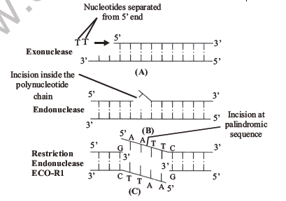
Cleaving enzymes: These enzymes are used to break DNA molecules. They are of three types: exonucleases, endonucleases and restriction endonucleases.
Connecting concept: Eukaryotes do not have restriction enzymes.
- Exonucleases: They remove nucleotides from the terminal ends (either 5′ or 3′) of DNA in one strand of double stranded DNA.
- Endonucleases: They make cuts at specific positions within the DNA. These enzymes cut only one strand of the double stranded DNA.
- Restriction endonucleases: Restriction endonucleases are called “molecular scissors or biological scissors”. They recognise and cut double stranded DNA at specific points called palindromic sequences. Palindromic sequences are ones which read the same on both strands in the 5′ → 3′ direction. The same is true about the 3′ → 5′ direction.
5′ —— G↓A A T T C —— 3′
3′ —— C T T A A↑G —— 5′
- Restriction endonuclease was discovered by Arber in 1963 in bacteria. The first one was EcoRI, found in the colon bacterium Escherichia coli.
Nomenclature of restriction endonuclease
- 1st letter: generic name
- 2nd letter: species name
- 3rd letter: strain name
- 4th letter: order in which the enzyme was isolated from that strain of bacteria [Roman numeral]
e.g., EcoRI: E = Escherichia, co = coli, R = strain RY13, I = first enzyme isolated from that strain.
Some examples of restriction enzymes are given below, showing the names of the restriction enzymes (endonucleases), the source (organism from which they have been isolated), their recognition sequence and site of cleavage.
Table 11.1: Some common examples of type II restriction enzymes, their source, recognition sequence and site of cleavage.
| No. | Restriction enzyme | Microbial source | Palindromic sequence and site of cleavage |
|---|---|---|---|
| 1 | Alu I | Arthrobacter luteus | 5′-A-G↓C-T-3′ 3′-T-C↑G-A-5′ |
| 2 | Bam H I | Bacillus amyloliquefaciens H | 5′-G↓G-A-T-C-C-3′ 3′-C-C-T-A-G↑G-5′ |
| 3 | Eco R II | Escherichia coli R245 | 5′-↓C-C-T-G-G-3′ 3′-G-G-A-C-C↑-5′ |
| 4 | Hae III | Haemophilus aegyptius | 5′-G-G↓C-C-3′ 3′-C-C↑G-G-5′ |
| 5 | Hin d III | Haemophilus influenzae Rd | 5′-A↓A-G-C-T-T-3′ 3′-T-T-C-G-A↑A-5′ |
| 6 | Sma I | Serratia marcescens | 5′-C-C-C↓G-G-G-3′ 3′-G-G-G↑C-C-C-5′ |
- Sticky ends are overhangs (stretches of single stranded portion) generated as a result of a cut by a restriction endonuclease into 2 fragments. They are named so because they form hydrogen bonds with their complementary counterparts. The stickiness of the ends facilitates the action of the enzyme DNA ligase. All the fragments cut by a restriction endonuclease will have the same sticky ends. The sticky ends have complementary bases, so they can rejoin.
- DNA ligases (joining or sealing enzymes): In 1969 Har Govind Khorana discovered DNA ligase in T4 bacteriophage.
- They join two individual fragments of double stranded DNA by forming phosphodiester bonds between them. Thus they help in sealing gaps in DNA fragments. Therefore, they act as molecular glue. The enzyme used most often in rDNA technology is T4 DNA ligase.
- Cutting of DNA by restriction endonuclease results in a mixture of DNA fragments of varied length. This is called restriction digestion.
- DNA fragments of various lengths are separated on a gel plate under an electric field by a technique called gel electrophoresis.
- The main dye used with agarose gel electrophoresis is ethidium bromide.
- The restriction digest is subjected to electrophoresis to separate DNA on the basis of length.
- DNA is negatively charged and moves towards the positively charged anode in a gel with definite pore size. Smaller DNA moves faster and farthest.
- Elution is the extraction of DNA bands out of the agarose gel. The DNA is visualised by staining with ethidium bromide followed by exposure to UV radiation. It is visible as a range of coloured bands.
2. Cloning vector
- Vectors are cloning vehicles required to transfer DNA of interest from one organism to another.
Desirable properties of a cloning vector are:
- High copy number
- Presence of an origin of replication (Ori)
- Presence of a selectable marker
- Presence of a unique recognition site or cloning site
- Ability to sustain in a bacterial cell
- Ori is the sequence where replication starts, and any piece of DNA linked here will be replicated. Ori also controls the copy number of the vector.
- Selectable marker allows us to select those host cells that contain the vector from among those which do not. Selectable markers help in identifying and eliminating non-transformants and selectively permitting the growth of transformants. Usually the genes encoding resistance to antibiotics such as ampicillin, chloramphenicol, tetracycline or kanamycin are considered useful selectable markers for E. coli.
- In the cloning site, the vector should have a single recognition site for the restriction enzyme. Presence of more than one recognition site will cut the vector into many fragments.
- Vectors are grouped into bacterial plasmids, bacteriophages, cosmids and phasmids, yeast artificial chromosome (YAC) and bacterial artificial chromosome (BAC).
- Plasmids are extra-chromosomal, circular, double stranded, autonomously replicating DNA sequences in a bacterial cell. A plasmid is a molecular vector analogous to a mosquito, which acts as an insect vector to deliver the malarial parasite into the human body. Plasmids are characterised by marker sequences.
- Cohen et al. (1972) for the first time reported the cloning of DNA by using a plasmid as vector.
- Bacteriophage is a virus which infects bacteria. Bacteriophages have a special sticky “cos” site to accept foreign DNA. As bacteriophages multiply faster, the transformation efficiency is higher in bacteriophage.
Several bacteriophages are being used as cloning vectors, but the most commonly used are lambda (λ) phage and M13 phage. Bacteriophage vectors (i) can be used for large DNA fragments and (ii) can easily be detected at the time of cloning experiments.
- Cosmid can be defined as a hybrid vector derived from plasmids which contains the cos site of lambda phage. Cosmids can be used to clone DNA fragments up to 45 kb in length.
- A phage genome containing an att site and more than one plasmid molecule is called a phasmid.
- Bacterial artificial chromosome (BAC) vectors: These are vectors based on the natural, extrachromosomal plasmid (the fertility or F-plasmid) of E. coli. They contain genes for replication and maintenance of the F-factor, a selectable marker and a cloning site. These vectors can accommodate up to 300–350 kb of foreign DNA and are also being used in genome sequencing projects.
- Yeast artificial chromosome (YAC) vectors: These are used to clone DNA fragments of more than 1 Mb (megabase pairs, 106 bp) in size. Therefore, they have been used in mapping large genomes, e.g., in the Human Genome Project. These vectors contain the telomeric sequence, the centromere and the autonomously replicating sequence from yeast chromosomes. They also contain restriction enzyme sites and selectable marker genes of yeast.
- The first artificial plasmid was synthesised by combining a marker gene to it, e.g., pUC 11 (plasmid developed at the University of California) and pBR 322.
- Cloning sites (recognition sites): Plasmid pBR322 has a variety of unique recognition sites for restriction endonucleases. Two unique sites, Pst I and Pvu I, are located within the ampR gene, and Bam HI, Sal I etc. are within the tetR gene. Some other unique restriction sites are Eco RI, Cla I, Hind III and Pvu II. rop codes for the proteins involved in the replication of the plasmid.
ampR: gene for ampicillin resistance; tetR: gene for tetracycline resistance; ori: origin of replication; rop: gene for a protein involved in replication of the plasmid.
- Agrobacterium tumefaciens is a bacterium containing the Ti (tumour-inducing) plasmid. Infection by this bacterium transforms normal plant cells into tumours and directs them to form a mass of cells called galls, which start secreting chemicals required by the bacteria.
- As the gene transfer occurs naturally, the bacterium is called a natural genetic engineer.
- Retrovirus is a transforming cloning vector for animal cells, which has the ability to transform an animal cell into a cancerous cell.
- Transformation efficiency of a vector is the percentage of competent bacterial host cells receiving the desired DNA at specific recognition sequences, which are palindromic.
Connecting concepts: Transposons are DNA stretches which can move from one DNA molecule to another. They are also called transposable elements, mobile genes or jumping genes. Transposons were first observed by McClintock (1951) in maize plants. They are also used as vectors.
Shuttle vectors can exist in both the eukaryotic cell and E. coli. These vectors contain two types of origin of replication and selectable marker genes: one type functions in the eukaryotic cell and another in E. coli. In the case of plants, a naturally occurring plasmid of the bacterium Agrobacterium tumefaciens, called the Ti plasmid, has been suitably modified to function as a vector. Most of the eukaryotic vectors are shuttle vectors.
3. Competent host
- A host is the organism in which the desired DNA is introduced with the help of a vector. A host should have a specific DNA sequence called the origin of replication [Ori C], which is responsible for initiating replication. E.g., yeast, E. coli, plant and animal cells.
- Various kinds of host cells, including E. coli, yeast, animal and plant cells, are used in genetic engineering. The selection of host cells depends on the purpose of the cloning experiment. For the expression of some eukaryotic proteins, eukaryotic cells such as yeast cells are mostly used.
- Yeasts are the simplest eukaryotic organisms and, like bacteria, are single-celled, genetically well-characterised, and easy to grow and manipulate. They can be grown readily in both small culture vessels and large scale bioreactors. Plant and animal cells may also be used as hosts in gene manipulation experiments and for protein expression, either in tissue culture or as cells in the whole organism, to create genetically modified plants and animals.
- Transfer of DNA into a eukaryotic cell is called transfection.
- Vector mediated gene transfer: For vector mediated gene transfer a competent host is essential. As DNA is a hydrophilic molecule, it cannot pass through membranes. A specific concentration of a divalent cation such as calcium is required. It increases the efficiency with which DNA enters the cell through pores in its cell wall.
- Recombinant DNA (rDNA) can then be forced into such cells by incubating the cells with recombinant DNA on ice, followed by placing them briefly at 42°C (heat shock) and then putting them back on ice. This enables the cell to take up the recombinant DNA.
- Direct or vector-less gene transfer: It is the process of gene transfer into the host cell without using a vector. This is possible by the following methods.
Bacteria can be transformed in the following ways:
- By microinjection, where DNA can be injected directly into the nucleus of the cell to be transformed by using a micropipette. This technique was originally used in animal cells but has subsequently been applied to plant cells.
- Gene gun/biolistic gun: Microscopic particles of gold or tungsten are coated with the DNA of interest and bombarded onto cells. This technique is known as biolistics.
Connecting concept: The science of recombinant technology took birth when Cohen and Boyer (1972) were able to introduce a piece of an antibiotic resistance gene into a plasmid of Salmonella typhimurium. This modified plasmid was then inserted into E. coli to get clones of recombinant DNA. Thus Cohen and Boyer discovered recombinant technology. Recombinant DNA (rDNA) = vector (carrier of DNA) + insert (DNA fragment of interest).
PROCESS OF RECOMBINANT DNA TECHNOLOGY
Steps of recombinant DNA technology
- Isolation of genetic material: The removal of plasmid or genomic DNA from cells is termed isolation. DNA is obtained by treating bacterial cells/plant or animal cells with enzymes such as lysozyme (bacteria), cellulase (plant cells) and chitinase (fungal cells). The purified DNA ultimately precipitates out after the addition of chilled ethanol.
- Purification of DNA: Isolated DNA is associated with RNA and histone proteins, which are removed with the help of the enzymes ribonuclease and protease respectively.
- Fragmentation of DNA with restriction enzymes: Restriction enzyme digestions are achieved by incubating purified DNA molecules with the restriction enzyme under optimal conditions for that specific enzyme.
- Selection of the desirable gene by gel electrophoresis: Electrophoresis is a technique of separation of molecules such as DNA, RNA or protein under the influence of an electrical field, so that they migrate towards the electrode bearing the opposite charge, i.e., positively charged molecules move towards the cathode (−ve electrode) and negatively charged molecules travel towards the anode (+ve electrode) through a medium/matrix. The most commonly used matrix is agarose, which is a polysaccharide extracted from seaweeds. DNA fragments separate according to size through the pores of the agarose gel. Smaller fragments move a longer distance in comparison to larger fragments. Agarose dissolves in hot water; when this solution is cooled, double helices form and become arranged laterally and produce thick filaments. These filaments become cross-linked to form the gel. Pore size depends on agarose concentration. The process is repeated with the vector DNA also.
- Ligation of the gene with the vector in the presence of DNA ligase to make rDNA. The rDNA is then inserted into the recipient host cell after making the cells competent. Selection or screening of transformed cells from non-transformed ones is done with the help of a selectable marker and its insertional inactivation.
- Culturing of the transformed cells on a suitable culture medium to get multiple copies of the desired gene.
Screening: If we ligate a foreign gene in the Bam H1 site of the tetR gene, then bacteria/transformants containing the recombinant plasmid will not grow on tetracycline (as the tetR gene has been inactivated), but will grow on ampicillin.
- If a transformant has a non-recombinant plasmid (without foreign gene), it will grow on both culture media, containing tetracycline and ampicillin. In this case one antibiotic resistance gene helps in selecting the transformants, whereas the other antibiotic resistance gene, which gets inactivated due to insertion of alien DNA, helps in the selection of recombinants.
- The second type of screening is by inserting foreign DNA into the coding sequence of the enzyme β-galactosidase. This enzyme gives blue colour in the presence of a chromogenic substrate in the culture medium. When a foreign gene is inserted into its sequence, the enzyme is not formed and a coloured colony will not be formed. These are identified as recombinant colonies.
- This procedure of selecting a transformant with a recombinant plasmid is called insertional inactivation.
In vitro, multiple copies of the DNA of interest can be synthesised by PCR (polymerase chain reaction).
- This technique was developed by Kary Mullis in 1985.
- It is based on the principle of DNA denaturation (i.e., a DNA molecule, when subjected to high temperature, splits into two strands) and DNA renaturation (joining of single stranded DNA to form double stranded DNA).
- These single stranded molecules synthesise new strands in the presence of the enzyme DNA polymerase.
- Thus a double stranded DNA molecule is duplicated, and multiple copies of the original DNA sequence can be generated by repeating the process several times in a very short time.
The basic requirements of PCR are:
- DNA template: a DNA segment or gene of interest that has to be amplified.
- Two nucleotide primers: usually 10–18 nucleotides long and complementary to the sequences present at the 3′ ends of the DNA segment.
- Enzyme: thermostable DNA polymerase (usually Taq polymerase) is required for the synthesis of new DNA molecules.
Steps of PCR
- The DNA segment to be amplified is heated to a high temperature (94°C). Heating breaks the hydrogen bonds of the DNA strands, forming 2 single strands (denaturation of DNA). Each of the two strands now acts as a template for synthesis of a new DNA strand.
- Denaturation is followed by annealing (joining). During this step, two oligonucleotide primers hybridise to each of the single stranded template DNA. Annealing is carried out at a lower temperature (40–60°C).
- The third and final step is extension. During this step, the enzyme DNA polymerase synthesises the DNA segment between the primers. Usually Taq DNA polymerase, isolated from the thermophilic bacterium Thermus aquaticus, is used. The two primers extend towards each other to copy the DNA segment lying between them. It occurs in the presence of deoxynucleoside triphosphates (dNTPs) and Mg2+ and takes place at 72°C.
- The above three steps complete the first cycle of PCR. The second cycle begins with denaturation of the product of the first cycle. If these cycles are repeated many times, a huge number of copies of the DNA/gene of interest can be obtained.
- Once the gene of interest is amplified, we need to isolate its product. Cells having the gene of interest express its protein, known as recombinant protein, on a small scale in the laboratory.
- The product from rDNA can be obtained on a large scale in bioreactors.
- Bioreactors are large vessels (having a volume of 100 to 1000 litres) in which raw materials are biologically converted into specific products by microbes, plant and animal cells or their enzymes.
- A bioreactor provides the optimal conditions for obtaining the desired product by providing optimum growth conditions such as temperature, pH, substrate, vitamins, oxygen and salts.
- It has an inlet from which a large volume of culture medium is fed and an outlet from which used medium is removed.
- The most commonly used bioreactors are of the stirring type.
Parts of a bioreactor
- Agitator system
- Oxygen delivery system
- Foam control system
- Temperature control system
- pH control
- Sampling port to withdraw a small amount of culture periodically
Downstream processing is the separation and purification of products obtained from bioreactors. The product is converted into a formulation for use, tested for human use, undergoes quality control and is then marketed.
USES OF RECOMBINANT PROTEINS
Some proteins, which form enzymes or hormones, play an important role in the metabolic activity of organisms. Lack or deficiency of these causes diseases. By rDNA technology these proteins are formed in bulk and in much less time in comparison to conventional methods. These proteins are called recombinant proteins and are used for many therapeutic purposes.
Table 11.2: Some recombinant proteins and their therapeutic uses.
| No. | Recombinant protein | Therapeutic use |
|---|---|---|
| 1 | Human insulin | Diabetes mellitus |
| 2 | Human growth hormone | Dwarfism cure |
| 3 | Blood clotting factor VIII/IX | Haemophilia |
| 4 | TPA (tissue plasminogen activator) | Heart attack/strokes |
| 5 | PDGF (platelet derived growth factor) | Stimulates wound healing |
| 6 | Interferon | Treatment of viral infection |
| 7 | Interleukin | Enhances immune reaction |
| 8 | Hepatitis B vaccine | Prevention of infectious disease |
| 9 | Herpes vaccine | Prevention of infectious disease |
| 10 | DNase I | Treatment of cystic fibrosis |
Biology NEET Exam Resources: Biotechnology Principles and Processes
Core Study Kit: NEET Biology Biotechnology Principles and Processes
Access comprehensive study material for Biotechnology Principles and Processes, including revision notes, concept maps, and high-probability questions. These resources are designed in alignment with the latest 2026 NEET syllabus for NEET Biology to support effective exam preparation.
NCERT-Aligned Solutions for Biotechnology Principles and Processes
Built using official NCERT guidelines for NEET Biology, these materials provide reliable academic support. Integrating past examination questions and step-by-step solutions helps students understand official NEET grading criteria.
Next Steps in Your Exam Preparation
Maximize your grade potential by utilizing NEET Biology practice papers in tandem with these structured notes. All printable assignments and interactive mock tests on our platform are updated for the 2026 session and available free of charge.
FAQs
Our advanced study package for Biotechnology Principles and Processes includes detailed concepts, diagrams, Mind Maps, and explanation of complex topics to ensure NEET students learn as per syllabus for 2026 exams.
The Mind Maps provided for Biotechnology Principles and Processes act as visual anchors which will help faster recall during high-pressure exams.
Yes, teachers use our NEET Biology resources for lesson planning as they are in simple language and have lot of solved examples.
Yes, You can download the complete, mobile-friendly PDF of the Biology Biotechnology Principles and Processes advanced resources for free.
Yes, our subject matter experts have updated the Biotechnology Principles and Processes material to align with the rationalized NCERT textbooks and have removed deleted topics and added new competency-based questions.