

Download NEET NEET Biology Useful Resources
Review targeted academic resources with the NEET Biology Neural Control and Coordination Notes. Built according to official NEET standards for the 2026-27 term, these downloadable NEET Biology study materials support effective daily practice and deeper conceptual understanding for Neural Control and Coordination.
Access NEET Study Guides and Summaries
Navigate directly to the advanced Biology study materials using the digital viewer below. Each resource pack includes detailed conceptual summaries and solved practice questions, allowing students to instantly reinforce their learning.
This NEET Biology notes page covers Chapter 21, Neural Control and Coordination, in both parts. It explains the conduction of nerve impulses through axons and synapses, the central, peripheral and autonomic nervous systems, the human brain and spinal cord, reflex action, sense organs including the human eye, ear, tongue and nose, other sensory structures, and disorders of the nervous system and the eye, with the key differences, structures and terms for NEET exam revision.
Topics Covered
1. Conduction of nerve impulse (through axon and synapse)
2. Central nervous system (human brain, spinal cord)
3. Peripheral nervous system (cranial nerves, spinal nerves)
4. Reflex action
5. Autonomic nervous system (sympathetic and parasympathetic)
6. Disorders of the nervous system
7. Receptors (human eye, human ear, tongue, nose, other sensory structures)
8. Disorders of the eye
Important Terms, Differences and Structures to Remember
Terms and definitions: Impulse, Refractory Period, Iter, Reflex, Synapse, Grey matter, Amygdala, Otoconia, Echolocation, Membranous Labyrinth, Algesi-receptors, Uvea, Johnston's organs, Fovea centralis, Helicotrema and Bowman's gland.
Differences: Depolarization and repolarization; Synaptic fatigue and synaptic delay; Excitatory and inhibitory neurotransmitters; Corpora bigemina and corpora quadrigemina; Broca's centre and Brodmann's area; Electrical and chemical synapses; Conditioned and unconditioned reflexes; Dorsal root and ventral root of spinal nerves; Aqueous humour and vitreous humour; Photopic and scotopic vision; Night blindness and colour blindness; Thermoreceptors and caloreceptors; Fenestra ovalis and fenestra rotundus; Basilar membrane and Reissner's membrane.
Structures: Corpus callosum, Choroid plexus, Foramen of Monro, Limbic system, Colliculi, Pons, Ciliary body, Tapetum lucidum, Schlemm canal, Iris, Columella auris, Lateral line organ and Cochlea.
Disorders: Parkinson's disease, Meningitis, Alzheimer's disease, Presbyopia, Glaucoma, Strabismus, Hypermetropia, Cataract and Conjunctivitis.
Nerves: Hypoglossal, Trochlear (Pathetic), Trigeminal (Dentist's nerve), Sensory nerve, Coccygeal and Cholinergic nerve.
GENERAL POINTS ABOUT THE NERVOUS SYSTEM
1. Nervous tissue is ectodermal in origin.
2. Protozoans and porifers do not have nervous tissue.
3. Nervous tissue first evolved in the members of phylum Coelenterata (Cnidaria). The nerve cells in this group are non-polarized, which means the dendrites and axons are not differentiated.
4. In non-chordates the nerve cord is ventral and solid.
5. In chordates the nerve cord (spinal cord) is dorsal and tubular (hollow).
6. Neuron (nerve cell) is the structural and functional unit of the nervous system.
7. A multipolar neuron has at least two dendrites.
CONDUCTION OF NERVE IMPULSE
An impulse is a wave of depolarization. The conduction of a nerve impulse is an electrochemical phenomenon. It can be studied in two parts: (1) conduction through the axon and (2) conduction through the synapse.
Conduction through axon
1. The axon membrane, called axolemma, contains voltage-gated channels for sodium (Na+) and potassium (K+) ions. In myelinated fibres these voltage gates are present only at the nodes, while in non-myelinated fibres they are distributed all along the axon membrane.
2. The sodium-potassium pump is also distributed in the axolemma. For the expense of 1 ATP, 3 Na+ move out into the extracellular fluid (ECF) and 2 K+ move into the intracellular fluid (ICF).
3. Na+ is maximum in the ECF (about 150 mmol/L, or 10 times the Na+ in ICF), while K+ is maximum in the ICF (about 150 mmol/L, or 30 times the K+ in ECF).
4. The axolemma is slightly permeable to K+ but almost impermeable to Na+ at rest.
(a) Resting stage
1. The potential difference in an average-sized neuron at rest is -70 mV.
2. The outer surface of the axolemma, facing the ECF, has a positive charge, while the inner surface, facing the ICF, has a negative charge. The sign used for potential difference is the charge inside the membrane, or in the axoplasm.
3. The potential difference is maintained with the help of the Na+-K+ pump.
4. Resting potential values: in RBCs about -10 mV; in cardiac muscles about -90 mV; in skeletal muscles about -100 mV.
5. The Na+ and K+ voltage gates remain closed during this stage.
(b) Action potential
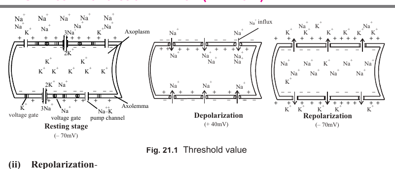
There are two phases of the action potential: depolarization and repolarization.
(i) Depolarization
1. The threshold value for generating a nerve impulse is -50 mV to -55 mV. A stimulus of lesser intensity does not generate an impulse.
2. All neurons follow the all or none law. Either the impulse is not generated at all, or it is generated to its full magnitude. There are no weak impulses.
3. When the stimulus brings the potential difference to the threshold value, Na+ gates open and sodium enters the axoplasm (Na+ influx) down its concentration gradient.
4. Because Na+ moves into the axoplasm, the polarity reverses: the outer surface becomes negatively charged and the inner surface becomes positively charged.
5. The potential difference rises from -55 mV to 0 and then reaches up to +40 mV. This is called the spike potential.
6. During depolarization the K+ gates remain closed.
[Figure: Threshold value and depolarization. A graph of potential in millivolts against time shows the resting stage at -70 mV, the threshold at -50 mV to -55 mV, depolarization up to +40 mV through Na+ influx, repolarization back to -70 mV through K+ efflux and hyperpolarization to -80 mV. Labels: Na+ gates open, K+ gates open, Resting stage, Depolarization, Repolarization and Hyperpolarization.]
(ii) Repolarization
Repolarization is completed in two phases.
First phase
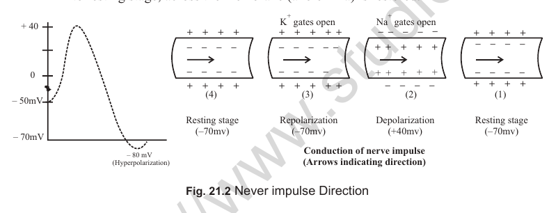
1. The Na+ gates close and the K+ voltage gates open.
2. K+ moves from the axoplasm (ICF) into the ECF down its concentration gradient (K+ efflux).
3. The outer surface facing the ECF develops a positive charge and the inner surface develops a negative charge. The charges are similar to the resting potential, but the positive charge is due to more K+ rather than Na+.
Second phase
1. The Na+-K+ pump becomes active again and the ionic distribution across the axolemma is restored to the resting stage.
[Figure: Nerve impulse conduction in four stages (1 to 4) across the axolemma, showing the Na+-K+ pump channel, Na+ voltage gate and K+ voltage gate, with the axoplasm inside and the axolemma as the membrane. The stages are Resting stage (-70 mV), Depolarization (+40 mV) with Na+ gates open, Repolarization with K+ gates open, and Resting stage again (-70 mV). Arrows show the direction of impulse conduction.]
[Figure: Direction of nerve impulse. The impulse travels in both directions from a stimulus given in the middle of an axon.]
Refractory period
1. The refractory period is the minimum time between two consecutive stimuli that can generate impulses. It can also be defined as the time required for repolarization, or recovery of the resting potential.
2. In an average neuron the refractory period is about 1/1000 second, or 1.0 ms (millisecond).
3. The first phase of repolarization is called the absolute refractory period. In this period a second impulse cannot be generated, whatever its magnitude.
4. The second phase is called the relative refractory period. In this period an impulse can be generated only if the stimulus is of greater magnitude.
Conduction of impulse through synapse
The junction between two neurons is called a synapse. It includes the presynaptic membrane of the terminal button of the previous axon, a synaptic cleft and the postsynaptic membrane of the dendrite. There are two types of synapses in humans.
1. Electrical synapses: the synaptic cleft is about 0.2 nm and conduction through such a synapse is similar to that in an axon. If the stimulus is given in the middle of an axon, the impulse travels in both directions, but through a synapse the conduction is one way only.
2. Chemical synapses: the synaptic cleft is about 20 nm wide. Transmission requires a chemical neurotransmitter, the most common of which is acetylcholine. The conduction through a chemical synapse is one way only.
Process at a chemical synapse
1. The terminal buttons, or synaptic knobs, contain synaptic vesicles filled with a neurotransmitter. The advantage of chemical synapses is that the neurotransmitter can differ between synapses.
2. In place of voltage gates, the postsynaptic membrane has ligand-gated channels. Its receptors bind the neurotransmitter, which opens the channels.
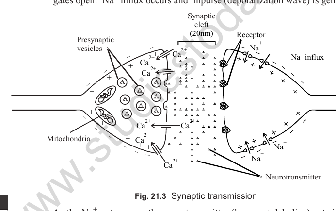
3. When the impulse reaches the terminal button, Ca++ gates open and calcium influx occurs through the presynaptic membrane.
4. The synaptic vesicles become active and rupture on contact with the presynaptic membrane. The neurotransmitter is released by exocytosis into the synaptic cleft.
5. The neurotransmitter binds to the receptors on the postsynaptic membrane and Na+ gates open. Na+ influx occurs and a depolarization wave is generated.
6. The amount of neurotransmitter released is proportional to the amount of Ca++ influx.
7. As the Na+ gates open, acetylcholine is immediately destroyed by the enzyme acetylcholinesterase. For the other neurotransmitter, noradrenaline, the enzyme is mono-amine oxidase.
[Figure: Synaptic transmission. Labels: Presynaptic membrane, Synaptic vesicles, Neurotransmitter, Mitochondria, Synaptic cleft (20 nm), Receptor, Postsynaptic membrane, Ca2+ influx and Na+ influx.]
Synaptic delay and synaptic fatigue
1. Synaptic delay is the time taken for Ca++ influx, release of the neurotransmitter and binding of the neurotransmitter with the receptors.
2. Synaptic fatigue develops when acetylcholine is completely exhausted. During this period the impulse cannot be transmitted.
3. Neurons never undergo such fatigue. Fatigue develops only at the junction of two neurons, that is, at synapses.
4. The neurotransmitter is formed by Nissl's granules in the cell body of the neuron and then reaches the synaptic vesicles of the terminal buttons.
Velocity of nerve impulse
1. In frogs the velocity is about 25 m/s in myelinated fibres and about 0.5 m/s in non-myelinated fibres.
2. In humans the velocity is about 100 to 120 m/s in myelinated fibres and about 2 m/s in non-myelinated fibres.
3. The velocity depends on three factors:
(i) Presence or absence of a myelin sheath. Conduction in myelinated fibres is called saltatory conduction because the impulse jumps (saltare means to jump) from node to node. It is faster and more economical because depolarization occurs in a smaller area, only at the nodes.
(ii) Number of synapses. Every synapse causes a delay, so the velocity falls as the number of synapses increases. Velocity is inversely proportional to the number of synapses.
(iii) Diameter of the fibre. A larger cross-section area has more voltage gates, so depolarization is faster and the velocity is greater. Velocity is directly proportional to diameter.
Neurotransmitters
1. Excitatory neurotransmitters allow the impulse to travel from one neuron to the next without diminishing. Examples: acetylcholine, adrenaline, noradrenaline and serotonin.
2. Inhibitory neurotransmitters check the transmission of the impulse. They increase the permeability of the axolemma to chloride ions instead of Na+, or they open K+ voltage gates. Hyperpolarization therefore develops in place of depolarization and the impulse is not conducted. Examples: glycine (an amino acid) and GABA (gamma-amino butyric acid).
DIVISIONS OF THE NERVOUS SYSTEM
The nervous system has three parts:
(i) Central nervous system (CNS): brain and spinal cord.
(ii) Peripheral nervous system (PNS): cranial nerves and spinal nerves.
(iii) Autonomic nervous system (ANS): sympathetic nervous system (SNS) and parasympathetic nervous system (PSNS).
The autonomic nervous system is also called the vegetative or visceral nervous system. Some texts include cranial nerves, spinal nerves, SNS and PSNS together in the peripheral nervous system.
1. CENTRAL NERVOUS SYSTEM (CNS)
Brain and its coverings
1. In all vertebrates the brain is enclosed in a brain box called the cranium.
2. The brain is covered by two coatings in frogs and three coatings in mammals. These coatings are called meninges (singular: meninx).
3. Duramater: the outer meninx, close to the cranium, made of dense fibrous connective tissue.
4. Arachnoid: the middle coating, made of delicate serous membrane. It is absent in the frog's brain.
5. Piamater: the innermost membrane, made of fine connective tissue. It contains many blood vessels that nourish the brain.
6. The human brain weighs about 1.5 kg (about 2% of body weight).
7. The brain cavities are filled with cerebrospinal fluid (CSF), a lymph-like fluid secreted by the choroid plexuses. CSF flows into the subarachnoid and subdural spaces through the Foramen of Magendie (dorsal) and the Foramina of Luschka (lateral) of the medulla oblongata. CSF nourishes the brain, removes metabolic wastes and acts as a shock absorber for the brain and spinal cord.
Parts of a typical vertebrate brain
1. Fore brain (prosencephalon): telencephalon (cerebrum and olfactory lobes) and diencephalon.
2. Mid brain (mesencephalon): optic lobes.
3. Hind brain (rhombencephalon): metencephalon (cerebellum) and myelencephalon (medulla oblongata).
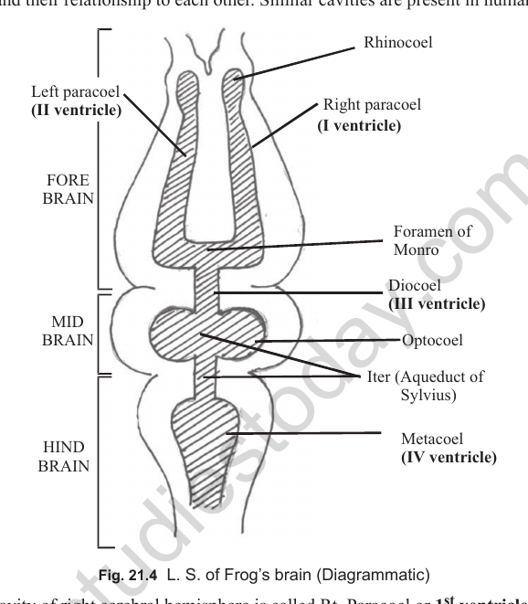
[Figure: Longitudinal section of the frog's brain (diagrammatic). Labels: Rhinocoel, Right paracoel (I ventricle), Left paracoel (II ventricle), Foramen of Monro, Diocoel (III ventricle), Optocoel, Iter (Aqueduct of Sylvius), Metacoel (IV ventricle). Divisions shown are Fore brain, Mid brain and Hind brain.]
Brain cavities (ventricles)
1. The cavity of the right cerebral hemisphere is the right paracoel, or 1st ventricle. The left paracoel is the 2nd ventricle.
2. The cavity of the diencephalon is the diocoel, or 3rd ventricle.
3. The two paracoels are connected to the diocoel through the Foramen of Monro.
4. The cavity of the mid brain is the Iter, or Aqueduct of Sylvius. It lies between the diocoel and the metacoel.
5. The cavity of the optic lobes is the optocoel. In mammals the optic lobes are solid and the optocoel is absent.
6. The cavity of the hind brain is the metacoel, or 4th ventricle.
7. Thus there are four ventricles in the brains of frogs and humans. Each ventricle has an inner lining of ependymal epithelium.
8. The number of ventricles in the heart of frogs and humans is only 1 and 2 respectively.
HUMAN BRAIN
Three prominent parts of the human brain are visible: (i) cerebrum, (ii) brain stem, made of the mid brain, pons and medulla oblongata, and (iii) cerebellum.
Cerebrum
1. The cerebrum consists of two cerebral hemispheres.
2. Each hemisphere is divided into four lobes:
(i) Frontal lobe (FL): towards the forehead.
(ii) Parietal lobe (PL): towards the middle.
(iii) Temporal lobe (TL): close to the ear.
(iv) Occipital lobe (OL): in the posterior-most region.
3. The lobes are named after the cranial bones over them. The boundaries of the lobes are marked by deep fissures or sulci (singular: sulcus).
4. Gyri (ridges) and sulci (grooves) in each lobe increase the surface area for accommodating more neurons.
5. The outer 2 to 4 mm of the cerebrum is grey matter. It is called neocortex in mammals and cortex in frogs.
6. In the subcortical part of the cerebrum there are groups of cell bodies (cytons) and axons, called nuclei and tracts respectively.
7. The two cerebral hemispheres are connected internally by a transverse band of myelinated nerve fibres called the corpus callosum. This band is present in all eutherian mammals, including humans. Its anterior part is the genu and its posterior part is the splenium.
8. Inside the cerebrum there are other nerve tracts, such as association fibres and projection fibres. The internal capsule is an example of projection fibres.
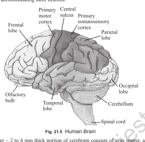
[Figure: Human brain, lateral view. Labels: Frontal lobe, Primary motor cortex, Central sulcus, Primary somatosensory cortex, Parietal lobe, Occipital lobe, Temporal lobe, Olfactory bulb, Cerebellum and Spinal cord.]
Important nuclei (ganglia) of the forebrain
There are two important nuclei in the forebrain: (i) basal ganglia and (ii) limbic system.
Basal ganglia
1. The basal ganglia consist of two main structures: the corpus striatum (the biggest nucleus of the basal ganglia) and the red nucleus.
2. The corpus striatum is further divided into the lenticular nucleus and the caudate nucleus.
3. The lenticular nucleus has two parts: the pallidum and the putamen.
4. The caudate nucleus is divided into a head and a tail. It regulates motor activities and initiates locomotion.
Limbic system
1. The limbic system is a fork-like neural link between the cerebrum and the brain stem. The term limbic system is preferred over rhinencephalon (nose brain), which is now obsolete.
2. It includes the limbic lobes, the hippocampal lobe, a part of the thalamus, the hypothalamus and the amygdala.
3. The amygdala is an almond-shaped structure at the end of the tail of the caudate nucleus. It controls mood, especially anger, and emotional behaviour such as fear and aggression.
4. The hippocampal lobe converts short-term memory into long-term memory. It also deals with the sense of smell.
[Figure: Basal ganglia and limbic system of the human brain. Labels: Head of caudate nucleus, Tail of caudate nucleus, Caudate nucleus, Thalamus, Post. column of fornix, Hippocampus, Lenticulate nucleus (Pl = Pallidum; Pt = Putamen), Amygdala (nucleus), Parahippocampus, Limbic lobe, Frontal lobe (FL), Temporal lobe (TL), Parietal lobe (PL) and Occipital lobe (OL).]
[Figure: Limbic system and cerebral areas. Labels: Frontal lobe, Motor area, Premotor area, Prefrontal area, Broca's area, Lateral sulcus, Auditory area, Wernicke's area, Temporal lobe, Parietal lobe, Central sulcus and Parieto-occipital sulcus.]
Functions of the cerebrum
(I) Frontal lobe: contains the prefrontal area, premotor area and motor area.
1. Broca's centre is the motor centre for speech. It is dominant in the left hemisphere of right-handed persons, and vice versa. It starts the movement of the lips, tongue and larynx to produce speech.
2. The frontal lobe is the centre of knowledge, wisdom, reasoning, judgement, logic, intelligence, creativity, ideas and dreams. It also governs will power and personality.
(II) Parietal lobe: contains somatosensory areas.
1. Brodmann's area for speech is a sensory area present in this lobe.
2. It is the centre for perceiving pain, pressure, taste, touch and temperature.
(III) Temporal lobe: contains the olfactory area for smell and the auditory area for hearing. It also contains Wernicke's area for understanding speech.
(IV) Occipital lobe: the area for vision and visual interpretation.
Diencephalon and hypothalamus
1. The dorsal wall of the thalamus (the roof of the diocoel) is the epithalamus. The ventral wall (the floor of the diocoel) is the hypothalamus. The lateral walls are the optic thalami.
2. The hypothalamus of a typical brain has the following functions: (i) osmoregulation, (ii) perspiration, (iii) thermoregulation, (iv) urge for hunger, (v) urge for thirst, (vi) pleasure, (vii) love and hate, (viii) sexual behaviour, (ix) satiety (satisfaction) centre, (x) carbohydrate metabolism and (xi) sleep.
3. The hypothalamus is the highest centre of the autonomic nervous system (SNS and PSNS). It also governs the secretion of the anterior pituitary through releasing hormones.
4. The thalamus is the relay centre of the body. It selects the information to be sent to the cerebrum.
Mid brain
1. In humans, as in other mammals, there are four optic lobes, which are solid and have no optocoel. This condition is called corpora quadrigemina.
2. The two anterior lobes, the superior colliculi, perceive stimulation from the head and eye movements (visual reflex).
3. The two posterior lobes, the inferior colliculi, perceive stimulation from head and ear movements (auditory reflex).
Hind brain
Cerebellum
1. Like the cerebrum, the cerebellum has gyri and sulci.
2. It is divided into three peduncles (connecting stalks): the superior peduncle connects the cerebellum to the mid brain; the middle peduncle connects the cerebellum to the pons; and the inferior peduncle connects the cerebellum to the medulla oblongata.
3. The median peduncle contains Purkinje cells (the Purkinje cells contain thousands of dendrites).
4. The cerebellum has grey matter on the surface and branched white matter (arbor vitae) in its deeper parts.
5. It coordinates faster muscular movements and maintains the speed of locomotion. It maintains the balance of the body, which is an unconscious activity, so it is also called the gyroscope of the body.
Pons
1. The pons varolii, present only in mammals, controls breathing.
2. It has a pneumotaxic centre, which acts as the switch on and off point for breathing.
Medulla oblongata
1. It controls involuntary activities: (a) breathing, (b) heartbeat, (c) vasomotor activity and (d) peristalsis.
2. It has dorsal and ventral groups of neurons that regulate normal and faster breathing respectively.
3. The accelerator and depressor centres of the medulla regulate heartbeat. The vasomotor centre controls blood pressure.
4. It controls reflexes such as coughing, sneezing, vomiting and hiccupping.
5. Unlike other parts of the brain, the medulla oblongata has outer white matter and inner grey matter.
6. If the medulla oblongata is destroyed, involuntary functions such as heartbeat and breathing stop, resulting in death.
2. PERIPHERAL NERVOUS SYSTEM (PNS)
The peripheral nervous system includes the cranial and spinal nerves.
Cranial nerves
1. The cranial nerves can be sensory, motor or mixed.
2. Sensory nerves carry impulses from receptors to the CNS. Their axons face the CNS.
3. Motor nerves carry impulses from the CNS to effector organs such as muscles and glands. Their axons face the effector organs.
4. Mixed nerves carry impulses both to and from the CNS. Some axons face the CNS and others face the effector organs.
5. Cranial nerves arise from the brain, not from the cranium.
6. Fishes and amphibians have 10 pairs of cranial nerves.
7. Reptiles, birds and mammals (group Amniota) have 12 pairs of cranial nerves.
8. The largest (thickest) cranial nerve is the 5th, the trigeminal nerve, also known as the dentist's nerve.
9. The longest cranial nerve in humans is the 10th (vagus), and the shortest is the 4th (trochlear).
| No. | Name of Cranial Nerve | Origin | Nature | Distribution / Function |
|---|---|---|---|---|
| 1st | Olfactory | Fore brain | Sensory | Nasal epithelium: smell |
| 2nd | Optic | Fore brain | Sensory | Retina: vision |
| 3rd | Oculomotor | Mid brain | Motor | Extra-ocular muscles of the eyeball (3 rectus and 1 oblique muscle): movement of eyeball |
| 4th | Trochlear (Pathetic) | Mid brain | Motor | Extra-ocular muscle (superior oblique): movement of eyeball |
| 5th | Trigeminal | Pons (Hind brain) | Mixed | Three branches: (i) Ophthalmic (sensory): eyelids, conjunctiva and forehead; (ii) Maxillary (sensory): upper jaw, gums, lips and cheek; (iii) Mandibular (mixed): lower jaw, mainly mastication |
| 6th | Abducens | Pons | Motor | Extra-ocular muscle (external rectus): movement of eyeball |
| 7th | Facial | Pons | Mixed | Sensory part: taste buds of the anterior two-thirds of the tongue. Motor part: movement of facial muscles for expression. In rabbits it has three branches (palatine, hyomandibular and chorda tympani); chorda tympani is absent in frogs. |
| 8th | Auditory (Statoacoustic) | Pons | Sensory | Two branches: (i) Vestibular: balancing the body; (ii) Cochlear: hearing |
| 9th | Glossopharyngeal | Medulla oblongata | Mixed | Sensory part: taste buds of the posterior one-third of the tongue. Motor part: movement of the pharynx and saliva secretion (parotid gland). |
| 10th | Vagus (Pneumogastric) | Medulla oblongata | Mixed | In fishes three branches: lateralis (lateral line organ), visceralis (visceral organs) and branchialis (gills). In mammals it supplies the lungs, larynx, oesophagus, heart, stomach, kidney and intestine, mainly through the laryngeal and depressor nerves. |
| 11th | Spinal accessory | Medulla oblongata | Motor | Neck and shoulder muscles: movement |
| 12th | Hypoglossal | Medulla oblongata | Motor | Tongue muscles: movement |
Note: each eyeball has six extra-ocular muscles: four rectus (superior, inferior, external and internal) and two oblique (superior and inferior).
Cranial nerves in frogs and mammals
1. Frog. Sensory: 1st, 2nd and 8th. Motor: 3rd, 4th and 6th. Mixed: 5th, 7th, 9th and 10th.
2. Mammals. Sensory: 1st, 2nd and 8th. Motor: 3rd, 4th, 6th, 11th and 12th. Mixed: 5th, 7th, 9th and 10th.
3. The maximum number of cranial nerves (4 pairs) are mixed in nature.
4. The maximum number of cranial nerves (5 pairs) are motor in nature in mammals.
[Figure: Cranial nerves of the frog and the mammal, showing the ganglia on the way of the cranial nerves.]
Spinal cord
1. In humans the spinal cord is 40 to 50 cm long and about 2 cm in diameter.
2. It lies inside the neural canal of the vertebral column.
3. It ends at the 1st lumbar vertebra of the abdominal region.
4. The non-nervous part of the spinal cord, containing the meninges, is called the filum terminale.
5. The bundle of spinal nerves in the posterior-most part of the spinal cord looks like a horse's tail and is called the cauda equina.
6. Development of the spinal cord is completed by the age of 5 or 6 years.
7. The spinal cord is hollow or tubular and has a neurocoel, or central canal, which contains CSF.
8. The outer region of the spinal cord is white matter, made of myelinated fibres. The inner region is grey matter, made of non-myelinated fibres and cell bodies (cytons).
9. In mammals the grey matter is generally butterfly-shaped and forms dorsal (posterior) and ventral (anterior) horns. In the thoracic region lateral horns are also present.
10. The white matter forms dorsal, ventral and lateral funiculi, which contain ascending and descending tracts of myelinated fibres.
11. The meninges around the brain continue over the spinal cord, and the subarachnoid space also contains CSF.
[Figure: Longitudinal section of the spinal cord. Labels: Neurocoel, Spinal cord, Pia mater, Filum terminale, Sacrum and Coccyx.]
[Figure: Transverse section of the spinal cord showing the dorsal and ventral roots of a spinal nerve. Labels: Dorsal funiculus, Dorsal septum, Meninges, Interneuron, Dorsal or posterior root, Dorsal root ganglion, Cyton, Sensory (afferent) neuron, Spinal nerve, Ramus communicans, Motor (efferent) neuron, Ventral or anterior root, Ventral funiculus, Ventral fissure, Ventral horn of grey matter, Lateral horn, Lateral funiculus, Dorsal horn of grey matter and Central canal or neurocoel.]
Spinal nerves
1. All spinal nerves are mixed in nature.
2. Each spinal nerve is formed by the union of two roots:
(i) Dorsal (posterior) root: the sensory root, made of sensory (afferent) fibres. It contains the dorsal root ganglion of pseudo-unipolar neurons.
(ii) Ventral (anterior) root: the motor root, made of motor (efferent) fibres.
3. The smallest branch of a spinal nerve is the ramus communicans. The preganglionic fibres of the SNS pass through this branch.
4. The first spinal nerve is called the hypoglossal nerve (the name of the last cranial nerve is also the same). The last spinal nerve is called the coccygeal nerve.
Number of spinal nerves
1. Frog (other than Rana tigrina): 10 pairs.
2. Rana tigrina: 9 pairs.
3. Human: 31 pairs.
4. Rabbit: 37 pairs.
| Region | Name of Nerves | Number of Pairs (Human) |
|---|---|---|
| Neck | Cervical nerves | 8 |
| Chest | Thoracic nerves | 12 |
| Abdomen | Lumbar nerves | 5 |
| Hip | Sacral nerves | 5 |
| Tail | Caudal (coccygeal) nerve | 1 |
Reflex action
1. A reflex action is an involuntary and automatic response to a stimulus.
2. The pathway of a reflex action is called the reflex arc.
3. Components of the reflex arc: (1) sensory structure or receptor, for example the skin; (2) afferent (sensory) neuron; (3) interneuron (association neuron); (4) efferent (motor) neuron; (5) effector organ, for example a muscle or gland.
4. In monosynaptic reflexes there are no interneurons. The minimum number of neurons taking part is two, as in the knee jerk reflex.
[Figure: Reflex arc. Labels: Skin, Receptor, Sensory or afferent neuron, Cell body of sensory neuron, Dorsal root ganglion, First synapse, Association neuron, Spinal cord, Second synapse, Motor or efferent neuron and Effector (muscle).]
Types of reflexes
1. Simple or unconditioned reflexes
1. These reflexes are regulated through the spinal cord and do not need the brain.
2. If the brain is disconnected from the spinal cord by pithing in a frog, these reflexes do not disappear.
3. Examples: blinking reflex, pupillary reflex, withdrawal reflex and knee jerk reflex.
4. An increase in heartbeat after hearing a loud sound is also a simple reflex.
5. Sneezing, coughing, vomiting and swallowing are also simple reflexes.
6. Simple reflexes can be shown by a newborn child, who can sneeze, vomit and blink.
2. Conditioned reflexes
1. These reflexes need the participation of the brain.
2. They require prior learning or training.
3. Their pathway passes through the cerebrum.
4. These reflexes may disappear and reappear.
5. Ivan Pavlov is known as the father of conditioned reflexes. He discovered conditioned reflexes for food in dogs.
6. Examples: typing, swimming, dancing, knitting, driving and cycling.
7. Salivation after tasting food is a simple reflex. Salivation caused by the sound of cutlery, sight or smell is a conditioned reflex.
AUTONOMIC NERVOUS SYSTEM (ANS)
1. The autonomic nervous system is also called the vegetative or visceral nervous system.
2. All its nerves are purely motor in nature.
3. It includes the sympathetic nervous system (SNS) and the parasympathetic nervous system (PSNS).
Sympathetic Nervous System (SNS)
1. The SNS is catabolic in action.
2. It consists of a pair of chains of sympathetic ganglia, one on each side of the vertebral column.
3. The number of ganglia in each chain is about 10 in frogs, about 18 in rabbits and about 22 in humans.
4. The SNS contains two types of fibres: preganglionic and postganglionic.
(A) Preganglionic fibres
1. These fibres arise from the grey matter of the CNS and end in the chain of sympathetic ganglia.
2. They pass through the spinal nerves of the thoracic and lumbar regions. The SNS is therefore also called the thoraco-lumbar outflow.
3. They leave the spinal nerves through the ramus communicans.
4. They are myelinated, and therefore white.
5. They are shorter than postganglionic fibres, because the chain of ganglia lies close to the spinal cord.
(B) Postganglionic fibres
1. These fibres are longer than preganglionic fibres.
2. They are non-myelinated, and therefore grey.
3. They arise from the chain of sympathetic ganglia and reach the visceral organs. At their terminal ends they secrete noradrenaline. The SNS is therefore also called the adrenergic nervous system.
4. Exception: the postganglionic fibres that supply the sweat glands and the blood vessels of skeletal muscles secrete acetylcholine, not noradrenaline.
[Figure: Pre- and postganglionic fibres of the SNS (diagrammatic). Labels: Spinal cord, Ventral root, Spinal nerve, Ramus communicans, Pre-ganglionic fibre, Chain of sympathetic ganglia and Post-ganglionic fibre.]
Parasympathetic Nervous System (PSNS)
1. The PSNS is anabolic in action. It restores body functions for comfort and relaxation.
2. There is no chain of parasympathetic ganglia. The ganglia are either inside the visceral organs or close to them.
(A) Preganglionic fibres
1. They arise from the CNS and pass through either cranial nerves or the spinal nerves of the sacral region. The PSNS is therefore also called the craniosacral outflow.
2. In frogs only the 10th cranial nerve carries these fibres. In mammals the 3rd, 7th, 9th and 10th cranial nerves carry them.
3. The fibres supplying the eyes pass through the 3rd cranial nerve. The fibres supplying the heart pass through the 10th cranial nerve.
4. They are myelinated, and therefore white.
(B) Postganglionic fibres
1. They are shorter than preganglionic fibres.
2. They are non-myelinated, and therefore grey.
3. They secrete acetylcholine at their terminal ends. The PSNS is therefore also called the cholinergic nervous system.
Functions of SNS and PSNS
| Organ or activity | Sympathetic Nervous System (SNS) | Parasympathetic Nervous System (PSNS) |
|---|---|---|
| Peristalsis, salivation and digestive juices | Inhibited | Stimulated |
| Digestion and absorption | Delayed | Faster |
| Urinary bladder | Relaxes, inhibiting micturition | Contracts, causing urination |
| Bronchioles and breathing rate | Bronchioles dilate and breathing rate increases | Bronchioles constrict and breathing rate is normal |
| Heart and blood supply | Coronary artery dilates, heartbeat increases; blood supply to heart and skeletal muscles increases but decreases in the digestive system | Heartbeat normal; blood supply to heart, skeletal muscles and digestive system normal |
| Spleen | Contracts (blood bank), releasing more blood | No such effect |
| Blood clotting time | Decreased | No such effect |
| Pupil | Dilates (mydriasis) in dim light due to contraction of radial muscles | Constricts (miosis) in bright light due to contraction of circular muscles |
| Skin | Sweat glands stimulated for more heat loss; arrector pili contraction produces goose flesh | No parasympathetic nerve supply to skin |
| Reproduction | Stimulates ejaculation of sperms | Stimulates erection of penis by vasodilation |
Disorders of the Nervous System
(1) Hydrocephalus
1. The volume of CSF is abnormally high and the head may be enlarged.
2. It develops due to malabsorption of CSF or an obstruction in its flow.
(2) Meningitis
1. It is inflammation of the meninges.
2. It develops mainly because of bacterial infection (Meningococcus and Streptococcus bacteria).
3. It produces fever, severe headache and stiffness in the neck. The CSF becomes turbid.
(3) Parkinson's disease (paralysis agitans, or shaking palsy)
1. It develops due to deficiency of the neurotransmitter dopamine, which is not produced because specific neurons gradually degenerate. This happens in a part of the basal ganglia.
2. It produces muscle tremors. There is lack of control and coordination of movements, expressionless features and a stooping posture, which gradually cause physical disability.
(4) Multiple sclerosis
1. It is due to demyelination of the white matter in the brain and spinal cord.
2. It is an autoimmune disorder and susceptibility can be genetic.
3. It can cause abnormal sensation, muscular weakness and lack of coordination.
(5) Huntington's chorea
1. It is a genetic disorder (dominant autosomal) that begins after the age of 30 years.
2. There is a deficiency of the neurotransmitter GABA, which causes rapid, involuntary and uncoordinated dancing movements. It is followed by progressive dementia and death.
(6) Alzheimer's disease
1. It was earlier thought to be a prion disease. It is now known to be caused by the deposition of abnormal amyloid (Tau) protein in the brain, which leads to degeneration of neurons.
2. About 5 to 10% of people above 60 years of age are affected.
3. It produces memory loss, dementia, confusion, inability to judge and abnormal behaviour. Death occurs within about 10 years of the first onset.
4. It is mimicked by severe depression due to damage in cholinergic pathways in the brain.
5. Because it involves chromosome 21, it is common among people with Down syndrome. Genes for the disease produce proneness or susceptibility.
RECEPTORS (SENSE ORGANS)
1. The important sensory organs of the human body are the eyes, ears, tongue, nasal epithelium and skin.
2. Receptors can be exteroreceptors or interoreceptors. Exteroreceptors are on the surface of the body and perceive distant stimuli. They are also called teloreceptors, for example the eye and ear. Interoreceptors are present in the visceral organs.
3. Proprioceptors are present in ligaments, tendons and the skeletal muscles of limbs. They detect changes in the position of the body. These receptors are sensory for the detection of earthquake.
| Stimulus | Receptor | Stimulus | Receptor |
|---|---|---|---|
| Light | Photoreceptor | Temperature | Thermoreceptor |
| Pressure | Baroreceptor | Smell | Olfactory receptor |
| Sound | Auditory receptor | Touch | Thigmo / tango receptor |
| Taste | Gustatoreceptor | Water current | Rheoreceptor |
| Cold | Fridgidoreceptor | Warm or hot | Caloreceptor |
| Gravitational force | Georeceptor | Electric current | Galvanoreceptor |
| Pain | Algesireceptor | Balance | Statoreceptor |
HUMAN EYE
1. The human eye is a photoreceptor organ. The photoreceptor part is the retina.
2. It is ecto-mesodermal in origin. The retina, lens, conjunctiva and iris muscles are purely ectodermal.
Coats of the eyeball
The eyeball has three coats:
1. Scleroid (sclerotic) coat: the outermost coat, made of mesoderm. It consists of white fibrous connective tissue, so it looks white. The anterior one-sixth, which is visible and transparent, is the cornea. The remaining five-sixths is white. Six extra-ocular muscles (4 rectus and 2 oblique) are attached to it and move the eyeball.
2. Choroid (uvea): the middle coat, mesodermal in origin. It is pigmented and highly vascular. It is incomplete in the anterior region, where it forms the ciliary body and the iris.
3. Retina: the innermost coat, ectodermal in origin.
Choroid (uvea) in detail
1. The pupil, an opening for the entry of light, is in the centre of the iris. The colour of the eye is the colour of the iris.
2. The iris has two sets of muscles:
(i) Circular muscles: contract in bright light, constricting the pupil (miosis) and reducing the light reaching the retina.
(ii) Radial muscles: contract in dim light, dilating the pupil (mydriasis) and increasing the light reaching the retina. Ophthalmologists use atropine to dilate the pupil.
3. The iris is comparable to the diaphragm of a camera. Radial muscles are regulated by the SNS and circular muscles by the PSNS.
4. The ciliary body secretes aqueous humour, which nourishes the lens and cornea because they have no blood supply of their own. There are two chambers containing aqueous humour: the anterior chamber (between cornea and iris) and the posterior chamber (between iris and lens).
5. Aqueous humour is reabsorbed through the Schlemm canal, a venous channel at the junction of the iris and cornea. If the Schlemm canal is blocked, the pressure of aqueous humour rises and damages the retina. This disorder is glaucoma.
[Figure: Vertical section of the mammalian eye. Labels: Vitreous humour, Suspensory ligaments, Ciliary body, Lens, Blind spot, Fovea, Sclera, Choroid, Retina, Optic nerve, Muscle, Iris, Pupil, Cornea, Conjunctiva and Aqueous humour.]
Lens
1. The human eye lens is biconvex, circular, living, multicellular and non-vascularized. It is purely ectodermal in origin.
2. Its position is fixed, but its focal length can be adjusted by thinning or thickening the lens. This is called the power of accommodation.
3. Ciliary muscles, arising from the ciliary body, adjust the focal length and help in accommodation.
4. The focal length of the human eye lens is 1.5 cm at rest, with a refractive power of 66.7 dioptres.
5. Suspensory ligaments (zonules of Zinn) keep the lens suspended in the centre.
6. In frogs the lens is spherical and its focal length is fixed. To change focus, the lens moves to and fro.
Retina
1. The retina is the sensory coat of the eye.
2. In the human eye the retina is inverted, meaning the sensory cells lie opposite to the entry of light. The retina of vertebrates develops from nervous epithelium, as an outgrowth of the diencephalon. In invertebrates the retina develops from skin epithelium and is not inverted.
3. From outer to inner, the retina has four prominent layers: (i) pigmented epithelium, (ii) sensory layer, (iii) bipolar neuron layer and (iv) ganglionic layer with optic nerve fibres.
4. The pigmented epithelium absorbs oblique (scattered) light rays to prevent internal reflection.
5. Amacrine cells and horizontal cells lie at the inner and outer borders of the bipolar neurons respectively. They transfer information sideways to sharpen contrast.
6. The sensory layer has two types of receptor cells: rods and cones.
7. Each human eye has about 120 million rods, which is 20 times the number of cones.
8. The yellow spot on the visual axis is the macula lutea. It contains only cones and no rods. The sharpest vision is at the central concave point of the yellow spot, called the fovea centralis.
9. The blind spot is where the optic nerve leaves the eye. Rods and cones are absent there, so no vision occurs at this point.
10. Rods are sensitive to dim light and give black and white vision. Night vision, or vision in dim light, is called scotopic vision. The pigment in rods is rhodopsin (visual purple).
11. Cones are sensitive to bright light and give colour vision, called photopic vision. The pigment in cones is photopsin (visual violet).
12. According to the trichromatic theory, there are three types of cones in humans:
(i) Red-sensitive cones, pigment erythropsin.
(ii) Green-sensitive cones, pigment chloropsin.
(iii) Blue-sensitive cones, pigment cyanopsin.
13. Humans see clearly in the visible spectrum, from about 380 nm to 760 nm. Best vision is at 5050 Å (505 nm, yellow light).
14. Insects, bony fishes, birds and mammals (except domestic animals) have colour vision. Honey bees can see ultraviolet light.
15. In nocturnal animals such as the owl, only rods are present (cones absent).
16. In animals with glowing eyes, such as cattle and cats, the pigmented layer is replaced by a shining layer called tapetum lucidum. It contains cysteine and guanine crystals and reflects light back to the sensory layer, so the eyes glow in the dark.
17. Night blindness (nyctalopia) occurs when rhodopsin is not synthesized due to vitamin A (retinol) deficiency.
[Figure: Histology of the retina (H = horizontal cell; A = amacrine cell). Layers from the outer side: (I) Pigmented epithelium, containing pigmented (melanin) cells, (II) Sensory layer, containing rods and cones, (III) Bipolar neuron layer, and (IV) Ganglionic cell layer (nerve fibre layer). Arrows show the direction of light and the direction of impulses.]
Between the retina and lens
Vitreous humour is a jelly-like mucous connective tissue between the lens and the retina. It is a permanent refractive medium and maintains the shape of the eyeball. The image formed on the retina is inverted (upside down).
Glands of the eye
1. Lacrimal glands (tear glands): one in each upper eyelid. They produce tears that clean and moisten the eyeball. Tears also contain germicidal enzymes. These are modified sweat glands and are absent in aquatic mammals.
2. Harderian glands: present in amphibians, reptiles, birds and a few mammals such as whales and rats. They produce an oily secretion that lubricates the nictitating membrane.
3. Meibomian (tarsal) glands: present on the inner side of the upper eyelids. They produce an oily secretion that prevents tears from overflowing onto the cheeks. They are modified sebaceous glands. Infection of these glands can produce styes.
4. Glands of Zeis: present at the base of the eyelashes. They keep the eyelashes soft, supple and smooth.
Disorders of the eye
(i) Myopia (short sightedness)
1. It is mainly congenital. The eyeball is elliptical, so the visual axis is increased.
2. The image is formed in front of the retina.
3. A biconcave (diverging) lens is used for correction.
(ii) Hypermetropia (long sightedness)
1. It is also congenital. The visual axis is shortened.
2. A biconvex (converging) lens is used for correction.
[Figure: Myopia and hypermetropia, with image formation without a lens (I) and after using a lens (II).]
(iii) Astigmatism
1. The surface of the cornea or lens, or both, is uneven, so some parts of the visual field are not in proper focus.
2. A cylindrical lens is used for correction.
(iv) Cataract
1. The transparency of the cornea or lens, or both, is reduced. The lens becomes cloudy or opaque.
2. For correction, either the cornea is grafted or an intra-ocular lens (IOL) is surgically implanted.
(v) Glaucoma
1. The Schlemm canal, which absorbs aqueous humour, is blocked.
2. The pressure of the aqueous humour increases. Normal intraocular pressure is 10 to 20 mm of Hg. This can damage the retina and cause blindness.
(vi) Presbyopia
1. It is an old-age defect caused by weakening of the ciliary muscles. The lens cannot thicken properly to view nearby objects, which is a case of poor accommodation.
2. A biconvex lens is used for correction.
(vii) Nyctalopia (night blindness)
1. It is caused by deficiency of vitamin A (retinol), so rhodopsin is not synthesized.
2. The affected person cannot see clearly in dim light.
(viii) Daltonism (colour blindness)
1. It is a sex-linked (genetic) disorder and the most common sex-linked disorder.
2. The affected person cannot distinguish between red and green. It is tested with the Ishihara chart.
3. Types: anopia (complete colour blindness), protanopia (red colour blindness), deuteranopia (green colour blindness) and tritanopia (blue colour blindness).
4. The defect lies in the red- and green-sensitive cones.
(ix) Conjunctivitis (eye flu)
1. It is an allergic, bacterial or viral infection of the conjunctiva, the covering over the eyeball and the inner side of the eyelids.
(x) Strabismus (heterotropia, squint)
1. It is an abnormal alignment, generally horizontal, of the two eyes.
Visual acuity
Visual acuity is measured with the Snellen letter chart, viewed from a distance of 6 m (20 ft). If the subject reads the chart from a distance of x metres, visual acuity is written as 6/x. Normal vision is 6/6.
HUMAN EAR
1. The ear is a statoacoustic organ, meaning it serves both balance and hearing. In fishes, its primary function is balance.
2. The human ear has three parts: (i) external ear (pinna and auditory meatus), (ii) middle ear (tympanic cavity and ear ossicles) and (iii) internal ear (vestibular apparatus and cochlea).
3. The ear drum (tympanum) lies between the external and middle ear. Between the middle and internal ear are two membrane-bound windows: the oval window (fenestra ovalis) and the round window (fenestra rotundus).
(i) External ear
1. The pinna (auricle), a characteristic of mammals, directs sound vibrations towards the auditory meatus. It is made of elastic cartilage.
2. The auricular muscles of the pinna are vestigial in humans.
3. Ceruminous glands in the lining of the auditory meatus secrete ear wax (cerumen). Ear wax is sticky, traps dust particles and prevents fungal and bacterial infection.
4. The pinna is absent in egg-laying mammals (prototherians) and in some eutherians such as whales and dolphins.
(ii) Middle ear
1. The cavity of the middle ear is called the tympanic cavity. It develops from a pharyngeal outgrowth and is therefore endodermal in origin.
2. The Eustachian tube connects the middle ear to the pharynx. It prevents the rupturing of the tympanum during louder sound by equalizing the pressure on the back of the membrane. It opens during yawning, swallowing and chewing.
3. The tympanic cavity contains three ear ossicles, which run from the tympanum to the oval window in this order: (i) malleus (hammer-shaped), (ii) incus (anvil-shaped) and (iii) stapes (stirrup-shaped). This order can be remembered as MIS.
4. The ossicles conduct and also amplify sound vibrations.
5. Between the malleus and incus is a hinge joint. Between the incus and stapes is a ball-and-socket joint.
6. The stapes is the smallest bone in the mammalian body. It fits on the oval window.
[Figure: Mammalian ear. Labels: Semicircular canals (part of the vestibular apparatus), Pharyngotympanic (auditory) tube or Eustachian canal, Auricle (pinna), Malleus, Incus, Temporal bone, Auditory nerve, Cochlea, Tympanic membrane, Lobule, Helix, Stapes, External acoustic (auditory) meatus and Vestibular apparatus.]
(iii) Internal ear
1. The oval window is the inlet for sound vibrations. Because the oval window is smaller than the tympanum, the pressure of sound on it is amplified about 20 times.
2. The sensory part of the internal ear is the membranous labyrinth, filled with endolymph.
3. The membranous labyrinth is surrounded by the bony labyrinth, formed by the temporal bone. The fluid between the membranous and bony labyrinths is perilymph.
4. The internal ear has two parts: the vestibular apparatus (for balance) and the cochlea (for hearing).
1. Vestibular apparatus
1. It is the sensory part for balance. It is well developed in fishes too.
2. It consists of three semicircular canals, one utricle and one saccule.
3. Two of the semicircular canals are vertical and one is horizontal. Each canal is surrounded by a bony canal, with perilymph between them.
4. At one end of each canal is an ampulla. The ampulla has sensory hair cells bathed in endolymph. Their processes, or cilia, form a bundle called the cupula, which is embedded in a jelly-like mass called the crista, forming an acoustic ridge.
5. The hair cells of the ampullae (semicircular canals) detect rotational acceleration. When a person moves to the right, the cupula also moves to the right and stimulates the hair cells. The vestibular branch of the 8th cranial nerve carries these impulses to the brain.
6. Vertigo is the sensation of rotation in the absence of actual rotation.
7. The utricle has a group of hair cells on its floor called the macula utriculi, bathed in endolymph. These hair cells are covered by an otolithic membrane in which calcium carbonate crystals (otoconia, or otoliths) are embedded. They detect linear horizontal acceleration.
8. The saccule has a group of hair cells in its walls in a semi-vertical position, called the macula sacculi. They are also covered by an otolithic membrane with otoconia. They detect linear acceleration in the vertical direction.
9. Acceleration in any direction displaces the otoliths in the opposite direction. This distorts the hair cell processes and generates an impulse.
[Figure: Vestibular apparatus and cochlea (membranous labyrinth). Labels: Semicircular canal, Ampulla, Crista (hair cells), Utricle, Saccule, Macula, Endolymphatic duct, Receptor neurones (to auditory nerve), Cochlea, Vestibular canal, Tympanic canal, Median canal, Tectorial membrane, Reissner's membrane, Basilar membrane, Hair cells, Helicotrema, Perilymph and Endolymph.]
2. Cochlea
1. The cochlea is the sensory structure for hearing. It is a coiled structure with 2 and 3/4 coils in humans.
2. In cross-section the cochlea shows three canals:
(i) Vestibular canal (scala vestibuli)
(ii) Cochlear, or median, canal (scala media)
(iii) Tympanic canal (scala tympani)
3. The vestibular and tympanic canals are filled with perilymph. The cochlear canal is filled with endolymph and is the actual part of the membranous labyrinth.
4. The roof of the scala media, separating it from the scala vestibuli, is Reissner's membrane. The floor of the scala media, separating it from the scala tympani, is the basilar membrane.
5. The hair cells for hearing lie on the basilar membrane and form the organ of Corti, the ultimate sensory part for hearing. The hair cells are supported by Deiters' cells. They are covered by a secretory (non-cellular) tectorial membrane, into which the hair cell processes are embedded. The tectorial membrane helps stimulate the hair cells to generate impulses.
6. The vestibular canal and tympanic canal are connected at the far end by a passage called the helicotrema.
7. The cochlear branch of the 8th cranial nerve carries impulses to the brain.
Route of sound vibrations
Auditory meatus → Tympanum → Ear ossicles → Oval window → Scala vestibuli → Helicotrema → Scala tympani → Basilar membrane (organ of Corti) → Cochlear nerve → Brain.
Vibrations that move through the round window are absorbed in the middle ear.
Hearing range and frequency
1. The audible range in humans is 20 to 20,000 Hz.
2. The outer coil of the cochlea detects the highest audible frequencies (10,000 to 20,000 Hz), the middle coil detects medium frequencies (about 5,000 Hz) and the innermost coil detects the lowest frequencies (about 100 Hz).
3. A newborn child can hear the sound of higher frequency (about 35,000 to 40,000 Hz).
4. Dogs can hear ultrasonic sound above 20,000 Hz.
5. Bats can produce and hear ultrasonic sound of about 100,000 Hz. They locate objects by echolocation, which is also called acoustic vision.
6. Human hearing intensity ranges from 0 to 120 dB (decibel, named after Alexander Graham Bell). Whispering is about 10 to 20 dB, conversation is about 40 to 80 dB and jet planes are about 120 dB.
Frog's ear
1. The pinna and external auditory meatus are absent in frogs, and the ear starts at the tympanum.
2. The single ear ossicle is the columella auris. It is homologous to the stapes of mammals.
3. The round window (fenestra rotundus) is absent. Only the fenestra ovalis is present.
4. The bony labyrinth is formed by the prootic bone. In humans it is formed by the temporal bone.
5. The cochlea is absent, so the organ of Corti is also absent. In its place are the pars basilaris and the lagena for hearing.
6. Dogs cannot produce ultrasonic sound, so echolocation is absent in dogs. In bats, the cry produces the ultrasonic sound.
TONGUE
1. The lining of the tongue is stratified squamous epithelium.
2. Papillae containing taste buds are mainly found on the tongue. They project from the surface and make it rough.
3. A typical mammal has four types of papillae:
(i) Filiform papillae: pointed and without taste buds.
(ii) Fungiform papillae: umbrella-shaped and carry taste buds.
(iii) Circumvallate (vallate) papillae: circular and the largest, carrying taste buds.
(iv) Foliate papillae: leaf-shaped and carry taste buds.
4. In humans only three types of papillae are present (the foliate papillae are absent). The circumvallate papillae are few in number and are arranged at the back of the tongue in an inverted V shape.
5. Taste buds for sweet and salty taste are at the tip of the tongue. Taste buds for sour taste are on the lateral sides and for bitter taste on the posterior-most part of the tongue.
6. Inorganic and organic acids (H+) taste sour. Glycerol, chloroform and lead and beryllium salts taste sweet. Quinine, nicotine, caffeine, urea, morphine and salts of Ca++ and Mg++ taste bitter.
7. The facial (7th) and glossopharyngeal (9th) cranial nerves supply the taste buds.
8. In frogs there are only two types of papillae, filiform and fungiform. Taste buds are present only in the fungiform papillae, and are also distributed in other parts of the buccal cavity.
[Figure: Tongue. Labels: Vallate papillae, Fungiform papillae and Filiform papillae. Taste tones: Bitter, Sour, Salty and Sweet. All papillae have taste buds, except the filiform papillae.]
NOSE
1. The nasal or olfactory lining is called the Schneiderian membrane and is sensory for smell. It has three types of cells:
(1) Receptor cells: derivatives of bipolar neurons that form olfactory hairs, or sensory cilia.
(2) Supporting cells: columnar epithelial cells.
(3) Glandular cells of Bowman's gland: these secrete mucus.
2. The strength of smell depends on the number of receptors stimulated.
3. Dogs and dogfish have a keen sense of smell.
[Figure: Olfactory epithelium. Labels: Nasal cavity, Supporting cell, Mucus, Bowman's gland, Axons, Receptor cells and Olfactory hair (sensory cilia).]
OTHER SENSORY STRUCTURES
1. Lateral line organ: present in fishes and the tadpole larva of frogs. It contains neuromast cells, which are sensory for water vibrations. Such receptors are called rheoreceptors.
2. Jacobson's organ: occurs in snakes and lizards. It is an olfactory structure near the internal nostrils (choanae). It is also present in frogs, rabbits and humans but is best developed in snakes and lizards.
3. Johnston's organ: present in insects. It is both an auditory and a proprioceptor organ. It is best developed in mosquitoes, where it lies in the second segment (pedicel) of the antenna.
4. Loreal pit: present in pit vipers. It is a thermoreceptor organ located between the eye and the ear of the snake.
5. Whiskers (barbules): present in rats, cats and catfishes. They are thigmo or tango receptors, sensory for touch.
6. Ampullae of Lorenzini: present in the snout region of cartilaginous fishes such as the dogfish. They are thermoreceptors.
7. Skin receptors:
(i) Meissner's corpuscles and Merkel's discs: sensory for touch (thigmoreceptors).
(ii) Krause's end bulbs: sensory for cold (fridgidoreceptors).
(iii) Ruffini's end bulbs: sensory for heat (caloreceptors).
(iv) Pacinian corpuscles: sensory for pressure (baroreceptors).
(v) Free nerve endings: sensory for pain (algesireceptors).
Connecting Concepts
1. In myasthenia gravis, an autoimmune disease of skeletal muscles, antibodies are formed against the acetylcholine receptors of neuromuscular junctions. Muscle degeneration starts after the age of 30 years. Besides blockage of muscle receptors, there is also a deficiency of GABA in the caudate nucleus.
2. Parkinson's disease (shaking palsy) develops due to deficiency of dopamine in a part of the basal ganglia.
3. If the vagus nerve is cut in a frog, the heartbeat increases, because the PSNS fibres passing through it are cut and acetylcholine, which reduces heartbeat, is not secreted.
4. If the vagus nerve is stimulated, the heartbeat decreases because more acetylcholine is secreted at the SA node.
5. Simple reflexes can be shown by a newborn child. This is the basis for distinguishing simple and conditioned reflexes.
Check Point
Differentiate between
1. Nerve cord of chordates and non-chordates.
2. Synaptic fatigue and synaptic delay.
3. Resting potential and action potential.
4. Electrical synapses and chemical synapses.
5. Excitatory and inhibitory neurotransmitters.
6. Depolarization and repolarization.
7. Stimulus and impulse.
8. Presynaptic membrane and postsynaptic membrane.
9. Medullated and non-medullated nerve fibres.
10. Dorsal and ventral roots of the spinal cord.
11. Grey matter and white matter.
12. Simple reflex and conditioned reflex.
13. Preganglionic and postganglionic fibres of the SNS.
14. Adrenergic and cholinergic nerves.
15. Thoraco-lumbar outflow and craniosacral outflow.
16. The functions of adrenaline and acetylcholine.
17. White and grey fibres of the SNS.
Which of the following are not functions of the SNS?
(i) Contraction of urinary bladder. (ii) Peristalsis. (iii) Salivation. (iv) Pupil dilation. (v) Sweating. (vi) Ejaculation of semen. (vii) Contraction of arrector pili muscles. (viii) Contraction of spleen. (ix) Glycogenolysis. (x) Anabolic activity.
Mention the precise location of the following in the human brain
1. Broca's centre. 2. Corpus callosum. 3. Amygdala lobe. 4. III ventricle. 5. Aqueduct of Sylvius. 6. Thalamus. 7. Limbic lobe. 8. Pons. 9. Pia mater. 10. Hippocampal lobe. 11. Occipital lobe. 12. Optic lobe. 13. Foramen of Monro. 14. Medulla oblongata.
Fill in the gaps (Nervous system)
(i) The posterior, non-nervous part of the spinal cord is called ..........
(ii) The facial (cranial) nerve in humans passes through ........... ganglion.
(iii) The ........... is the largest or thickest cranial nerve.
(iv) The number of spinal nerves in Rana tigrina is ........... pairs.
(v) ........... is the name of the last cranial nerve in humans.
(vi) The number of sensory cranial nerves in humans is ........... .
(vii) Funiculus is the name for ........... matter of the spinal cord.
(viii) In humans the number of spinal nerves in the neck region is ........... pairs.
(ix) The number of cranial nerve pairs present in amniotes only is ........... .
(x) ........... is the smallest branch of a spinal nerve.
Fill in the gaps (Eye and ear)
(i) ........... is the pigmented layer in the eye.
(ii) In the human eye, ........... is comparable to the diaphragm of the camera.
(iii) Lens and cornea are nourished by ........... .
(iv) Adjustment of focal length of the eye lens is called ........... power.
(v) The three basic layers of the retina are ........... .
(vi) ........... lens is used for correcting hypermetropia.
(vii) ........... is used for testing colour blindness.
(viii) Cylindrical lens is used for correcting ........... .
(ix) The eye develops from ........... (germ layer or layers). The sensory cells in the blind spot are ........... .
(x) ........... is an old-age defect of the eye due to weakening of the ciliary muscles.
(xi) Short sightedness due to change in the shape of the eyeball is called ........... .
(xii) Vision in dim light is known as ........... .
(xiii) ........... is a jelly-like mucous connective tissue between the lens and retina.
(xiv) Modified sweat glands that produce tears are ........... .
Fill in the gaps (Ear, tongue and nose)
(i) ........... canal connects the pharynx to the middle ear.
(ii) Ear wax is secreted by ........... glands.
(iii) The fluid filling the membranous labyrinth is ........... .
(iv) ........... papillae do not have taste buds.
(v) The audible range in humans is ........... to ........... Hz.
(vi) Sensory structures (receptors) for cold are ........... .
(vii) ........... are the largest papillae on the human tongue.
(viii) Nasal glands that secrete mucus are ........... .
(ix) ........... is the balancing part of the internal ear.
(x) The passage connecting the vestibular and tympanic canals is called ........... .
Check Point: Fill in the gaps (1-5) with suitable words to complete the following statements
The sensory structure for hearing in the cochlea is ..........(1).......... which is covered by a non-secretory covering, ..........(2).........., and is dipped in ..........(3).......... fluid. The sound vibrations enter into the internal ear through a membrane-bound window, ..........(4).......... . The other window between the middle and internal ear is called ..........(5)..........
Match the following
Column A: 1. Loreal pit (organ), 2. Meissner's corpuscle, 3. Johnston's organ, 4. Pacinian corpuscle.
Column B: A. Auditory receptor, B. Thigmoreceptor, C. Thermoreceptor, D. Algesireceptor, E. Baroreceptor, F. Proprioceptor, G. Olfactory receptor.
Check Point (Ear, eye and sense organs, other differentiations)
1. Retina of invertebrate and vertebrate eyes.
2. Glaucoma and cataract.
3. The role of circular muscles and radial muscles of the iris.
4. Ciliary body and ciliary muscles.
5. The eye lens of frog and rabbit.
6. Mention the location and one function of each of the following: proprioceptors, tapetum lucidum, Schlemm canal, amacrine cells and fovea centralis.
Useful Resources and Notes for NEET Biology Neural Control and Coordination
Comprehensive Study Resources for Neural Control and Coordination
Access comprehensive study material for Neural Control and Coordination, including revision notes, concept maps, and high-probability questions. These resources are designed in alignment with the latest 2026 NEET syllabus for NEET Biology to support effective exam preparation.
Understanding Marking Schemes
Designed around the official curriculum, these study guides guarantee standard compliance. Reviewing step-by-step solutions clarifies complex sub-topics within Neural Control and Coordination and demystifies standard marking schemes for Biology evaluations.
Complete Revision for Biology
Wrap up your chapter revision by testing your knowledge against standard objective question formats. Explore our full library of free, up-to-date sample papers and online assessments to secure top grades in your NEET examinations.
FAQs
Our advanced study package for Neural Control and Coordination includes detailed concepts, diagrams, Mind Maps, and explanation of complex topics to ensure NEET students learn as per syllabus for 2026 exams.
The Mind Maps provided for Neural Control and Coordination act as visual anchors which will help faster recall during high-pressure exams.
Yes, teachers use our NEET Biology resources for lesson planning as they are in simple language and have lot of solved examples.
Yes, You can download the complete, mobile-friendly PDF of the Biology Neural Control and Coordination advanced resources for free.
Yes, our subject matter experts have updated the Neural Control and Coordination material to align with the rationalized NCERT textbooks and have removed deleted topics and added new competency-based questions.