

Official NEET Study Materials for NEET Biology
Review targeted academic resources with the NEET Biology Chemical Coordination and Integration Notes. Built according to official NEET standards for the 2026-27 term, these downloadable NEET Biology study materials support effective daily practice and deeper conceptual understanding for Chemical Coordination and Integration.
Advanced Resources for Biology
View or download the dedicated NEET Biology Chemical Coordination and Integration Notes resource below. Engaging with these advanced study guides under focused conditions ensures continuous academic progress and mastery of the 2026-27 curriculum for Chemical Coordination and Integration.
This NEET Biology notes page on Chemical Coordination and Integration covers hormonal and nervous coordination, parahormones, pheromones, every endocrine gland with its hormones, their functions and disorders (pituitary, pineal, thymus, gonads, placenta, gastro-intestinal tract, pancreas, thyroid, parathyroid, adrenal, kidney and heart), the three modes of hormonal action, and the second messenger mechanisms of peptide, catecholamine, steroid and thyroid hormones, along with the chapter's check point questions.
CHEMICAL COORDINATION AND INTEGRATION
Important points to pay special attention to while going through the chapter
Terms and definitions: 1. Pheromones, 2. Prostaglandins, 3. Hypophysis, 4. Somatomedins, 5. Antidiuresis, 6. Isthmus, 7. Brain sand, 8. Adenohypophysis, 9. Islets of Langerhans, 10. Eunuchoidism, 11. Neuro hormones.
Differences: 1. Diabetes mellitus and Diabetes insipidus; 2. Cushing's syndrome and Conn's syndrome; 3. Pseudo-pregnancy and Pseudo-precocity; 4. Hypoparathyroidism and Hyperparathyroidism; 5. Corticotropins and Corticoids; 6. Hormones and Parahormones; 7. Hypoglycemia and Hyperglycemia.
Biologists: 1. F Sanger; 2. Bayliss and Starling; 3. Thomas Addison; 4. Banting and Best; 5. Von Euler; 6. Karlson.
Hormones and chemicals: 1. Catecholamines; 2. Insulin; 3. hCG; 4. Aldosterone; 5. Vasopressin; 6. Progesterone; 7. Parathormone; 8. Melatonin; 9. Secretin; 10. Erythropoietin; 11. Enterogastrone.
Diseases and disorders: 1. Cretinism; 2. Tetany; 3. Acromegaly; 4. Myxoedema; 5. Glycosuria; 6. Addison's disease; 7. Gynaecomastia; 8. Osteoporosis; 9. Simmond's disease.
At a Glance:
- Parahormones / local hormones
- Pheromones or ecto hormones / semiochemicals
- Endocrine glands
- Pituitary gland
- Pineal gland
- Thymus gland
- Gonads
- Placenta
- Gastro-intestinal tract (git)
- Pancreas
- Thyroid gland
- Adrenal gland
- Kidney
- Heart
- Hormonal action
TYPES OF COORDINATION
There are two types of coordinating systems in the body: (1) Hormonal (chemical) coordination and (2) Nervous (neural) coordination.
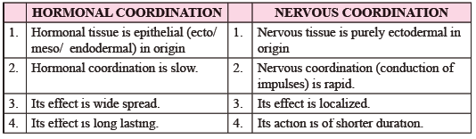
| Hormonal coordination | Nervous coordination |
|---|---|
| 1. Hormonal tissue is epithelial (ecto/meso/endodermal) in origin. | 1. Nervous tissue is purely ectodermal in origin. |
| 2. Hormonal coordination is slow. | 2. Nervous coordination (conduction of impulses) is rapid. |
| 3. Its effect is widespread. | 3. Its effect is localized. |
| 4. Its effect is long lasting. | 4. Its action is of shorter duration. |
HORMONAL ACTION OVERVIEW
Endocrine glands are ductless glands. They pour their secretions directly into the blood or body fluid, and such secretions are not carried through ducts. Exocrine glands, in contrast, have ducts. The study of endocrine glands is called endocrinology. The father of endocrinology is Thomas Addison. The secretions of endocrine glands are called hormones, meaning 'to excite or stimulate'. Secretin was the first hormone, discovered by Bayliss and Starling (1902). The term 'hormone' was coined by Starling (1905).
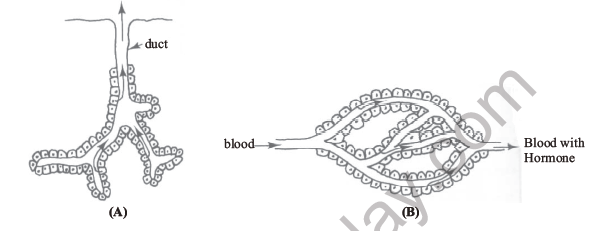
[Figure: Fig. 22.1 (A) Exocrine gland with a duct, (B) Endocrine gland with blood supply, showing blood entering and blood with hormone leaving.]
PARAHORMONES / LOCAL HORMONES
Parahormones are short-lived chemicals which are produced from almost all body cells (except mammalian RBCs). These chemicals affect the functioning of either the neighbouring cells (paracrine hormones) or the same cells from which they are secreted (autocrine hormones). Most of the local hormones are paracrine type and include prostaglandins, neurotransmitters and interferons.
(a) Prostaglandins
They were discovered by Von Euler (1935) in the semen of humans, and were thought to be the secretion of the prostate gland, hence the name.
They are derivatives of polyunsaturated fatty acids (e.g. arachidonic acid).
They regulate BP by contraction and relaxation of smooth muscles of blood vessels.
They also regulate peristalsis and secretion of the digestive juices.
They cause pain at the injury site and induce fever. They also promote inflammation. (The pain killers inhibit the secretion of prostaglandins.)
Prostaglandins also cause contraction of uterine muscles, vas deferens and oviduct etc.
(b) Neurotransmitters / neuro-regulators
These chemicals are peptides and are synthesized in the nerve cells. They help in transmitting nerve impulses to muscles or glands, or across the synapses.
Neuro-regulators are of two types:
1. Excitatory: e.g. acetylcholine, nor-epinephrine, serotonin and dopamine etc.
2. Inhibitory: e.g. glycine, gamma amino butyric acid (GABA), endorphins and nitric oxide (NO) etc.
(c) Interferons
They are low molecular weight proteins secreted by virus-infected cells, and they provide resistance to the neighbouring cells. (For details, refer to 'Immune system'.)
PHEROMONES OR ECTO HORMONES / SEMIOCHEMICALS
The term 'pheromone' was given by Karlson. Pheromones are volatile and odorous. They travel through air and alter the functioning of the other members of the same species. The 'confusion technique' is based upon the use of pheromones. The important examples of pheromones are bombycol, geradiol and muskone. Bombycol is secreted by Bombyx mori (silkmoth) as a sex attractant for mating. This was the first pheromone discovered. Geradiol is secreted by honey bees for aggregation, and muskone is secreted from musk deer.
[Figure: Fig. 22.2 Human endocrine glands. Labelled structures are hypothalamus, pituitary gland (hypophysis), pineal gland, parathyroid gland, thyroid gland, thymus, adrenal gland, pancreas, ovary (inset) and testis.]
ENDOCRINE GLANDS
Chemical nature of hormones
1. Amino acid derivatives: e.g. thyroxine, adrenaline (both tyrosine derivatives) and melatonin (tryptophan derivative).
2. Steroids (lipids): these are cholesterol derivatives. e.g. estrogen and progesterone (from ovary and placenta), testosterone (from testis) and hormones of adrenal cortex (corticoids).
3. Peptides/proteins: the maximum number of hormones are peptides or proteins. Some are glycoproteins (FSH, LH, TSH and hCG), while others are pure proteins, e.g. hormones of pancreas, parathyroid, pituitary and thymus.
(1) PITUITARY GLAND
It is called the master gland (master of the endocrine orchestra), as it regulates the functioning of other endocrine glands like the thyroid, adrenal cortex, testes and ovaries. However, the secretions of the pancreas, thymus and pineal body are not regulated by the hormones of the pituitary.
The pituitary is a single or unpaired gland weighing about 0.5 g. As it is present below the diencephalon of the fore-brain, it is also known as hypophysis cerebri. In mammals it is ectodermal in origin. It is present in the 'sella tursica' cavity of the basisphenoid bone, and is attached to the floor of the diencephalon (hypothalamus) of the fore-brain through a stalk called the infundibulum.
[Figure: Fig. 22.3 Pituitary gland showing the third ventricle (diocoel), infundibulum, pars tuberalis, pars nervosa (posterior lobe), pars intermedia (middle lobe), pars distalis (anterior lobe), sella tursica cavity and sphenoid bone.]
The pituitary consists of two parts: (1) Adenohypophysis and (2) Neurohypophysis.
(1) Adenohypophysis develops from Rathke's pouch, a diverticulum of the buccal cavity, and includes (i) the anterior lobe (pars distalis) and (ii) the middle lobe (pars intermedia).
(2) Neurohypophysis is directly connected to the hypothalamus (diencephalon) part of the fore-brain, and includes the posterior lobe (pars nervosa).
(A) Anterior lobe: 6 hormones
(i) Growth hormone or somatotrophic hormone (STH)
(ii) Prolactin (PRL), also called mammotropin or LTH (luteotropic hormone)
(iii) Adrenocorticotropic hormone (ACTH)
(iv) Thyroid stimulating hormone (TSH)
(v) Follicle stimulating hormone (FSH)
(vi) Luteinizing hormone (LH)
STH and PRL are secreted from acidophilic (40%) cells. ACTH is secreted from chromophobic cells, while the other three, TSH, FSH and LH, are secreted from basophilic (10%) cells.
TSH, FSH and LH are glycoproteins, while the first three are peptides or proteins.
Except prolactin, all other hormones of the anterior pituitary are tropic hormones, because they stimulate other endocrine glands. Prolactin stimulates mammary glands, which are not endocrine but exocrine in nature.
Growth hormone (STH)
It is an anabolic hormone and stimulates protein synthesis. The catabolism of proteins and amino acids is decreased.
It stimulates lipolysis, i.e. the breakdown of lipids for energy production.
It decreases the use of glucose for energy production and conserves carbohydrates as glycogen.
It reserves calcium and phosphate, and causes the elongation of bones.
Hyposecretion of STH: in children, dwarfism (midgets with stunted growth); in adults, Simmond's disease (protein synthesis is decreased and lactation in females is reduced).
Hypersecretion of STH: in children, gigantism (overgrowth of the body); in adults, acromegaly, i.e. gorilla appearance (free ends or extremities like fingers, toes and chin are elongated). The affected person develops osteoarthritis, gynecomastia and hirsutism (coarse and pigmented hair on the chest, back etc.).
Connecting concept: Growth hormone is considered a tropic hormone, though it does not affect the endocrine organs directly. It stimulates the liver for the secretion of a hormone-like chemical, somatomedin. Somatomedin then stimulates the growth of bones, muscles or body organs.
Prolactin (PRL)
It stimulates mammary glands for the synthesis of milk. It is a non-tropic hormone.
Adrenocorticotropic hormone (ACTH)
It stimulates the secretion of corticoids from the adrenal cortex. It also regulates melanin pigment in human skin.
Thyroid stimulating hormone (TSH)
It stimulates the growth of thyroid follicles. It also promotes the utilization of iodine by the gland for the synthesis of thyroxine.
Follicle stimulating hormone (FSH)
It is commonly called the gametogenic hormone, as it regulates spermatogenesis in males and oogenesis in females. In females it also stimulates ovarian follicles for the secretion of estrogen.
Luteinizing hormone (LH)
It is responsible for ovulation, i.e. the release of the female gamete from the ovary. It also helps in the formation of the corpus luteum, and stimulates the corpus luteum for the synthesis of mainly progesterone.
In males, this hormone stimulates the interstitial cells or Leydig cells for the synthesis of testosterone, and is therefore called the interstitial cell stimulating hormone (ICSH).
Hypothalamic control over the anterior pituitary
The chemical factors released from the hypothalamus affect the secretions of the anterior pituitary through the hypophyseal portal system. There are six releasing factors or hormones:
(1) Growth hormone releasing factor (GRH): stimulates the secretion of growth hormone from the anterior pituitary.
(2) Growth hormone release inhibiting hormone (GIH): also known as somatostatin, it inhibits the secretion of growth hormone.
(3) Thyrotropin releasing hormone (TRH): stimulates the secretion of TSH (thyrotropin).
(4) Corticotropin releasing hormone (CRH): stimulates the secretion of ACTH.
(5) Gonadotropin releasing hormone (GnRH): stimulates the anterior pituitary for the secretion of FSH and LH.
(6) Prolactin inhibitory hormone (PIH): [The production of prolactin releasing hormone (PRH) is doubtful in humans.]
(B) Middle lobe
(i) Intermedin: secreted in fishes, amphibians and reptiles. It causes darkening of the skin by dispersing pigmented cells (chromatophores).
(ii) Melanocyte stimulating hormone (MSH): secreted in birds and mammals. It also causes darkening of the skin by synthesizing more melanin and by causing dispersal of melanocytes.
In humans the middle lobe is rudimentary, and MSH is either vestigial or not secreted in adults. The skin pigmentation in humans is regulated by ACTH.
(C) Posterior lobe
The hormones of the posterior lobe are actually produced from the axons of neurons of the hypothalamus. These hormones are therefore called neurohormones. Two neurohormones are secreted (released) from the posterior pituitary.
Oxytocin (Pitocin): a small peptide of 9 amino acids. It causes contraction of smooth muscles, mainly of the mammary glands and uterus. The contraction of mammary glands causes the release of milk, so the hormone is called the 'milk-let-down factor'. The contraction of uterine smooth muscles during mature pregnancy causes the expulsion of the child (parturition), so it is also known as the 'birth hormone'.
Vasopressin (Pitressin): causes constriction of blood vessels and raises blood pressure. It also helps in the absorption of water from the glomerular filtrate in the uriniferous tubules of the kidney, thus reducing the amount of water in urine and concentrating it. It is commonly called the anti-diuretic hormone (ADH).
Hyposecretion of ADH: water absorption is reduced and the amount of water in urine is increased. This condition of diluted urine (diuresis), without sugar or glucose, is called diabetes insipidus. (It is different from diabetes mellitus, in which sugar or glucose passes with urine.)
Check point (Fill in the blanks)
(i) ... of pituitary develops from Rathke's pouch.
(ii) Luteinizing hormone in males is called ... .
(iii) ... is also known as somatostatin.
(iv) The hormones of anterior pituitary are chemically ... .
(v) ... coined the term 'hormone'.
(vi) ... was the first hormone to be discovered.
(vii) Prostaglandins and interferons are included in the category of ... .
(viii) Posterior pituitary is under the direct control of ... .
(ix) Sella tursica, the cavity of basisphenoid, lodges the ... gland.
(x) Deficiency of ... causes diabetes insipidus.
Answers: (i) Adenohypophysis; (ii) ICSH; (iii) GIH (growth hormone release inhibiting hormone); (iv) proteins (peptides or glycoproteins); (v) Starling; (vi) Secretin; (vii) local hormones (parahormones); (viii) the hypothalamus; (ix) pituitary (hypophysis); (x) ADH (vasopressin).
(2) PINEAL GLAND
It is ectodermal in origin. It is unpaired (single) and is attached to the epithalamus of the diencephalon. It is therefore also known as epiphysis cerebri. (The pituitary, attached to the hypothalamus, is called hypophysis cerebri.) It is considered to be the 'seat of the soul'. In humans it starts degenerating at the age of 7 to 8 years. Calcium and magnesium salts are deposited in this gland at later stages, and the structure is then known as 'brain sand'.
This gland produces a single hormone, melatonin, which is a biogenic amine.
Melatonin
It is a derivative of the amino acid tryptophan.
It causes lightening of the skin and is antagonistic to MSH.
It delays sexual maturity by influencing the development of the gonads and the release of gonadotropin hormones from the pituitary. Its secretion is increased during darkness.
As it delays puberty, it is also known as the anti-ageing hormone. (The pacemaker for ageing is, however, the thymus gland.)
It maintains the biological clock in the human body. It also maintains diurnal variations (circadian rhythm) in animals and decides the breeding season in them.
Check point (Match the following)
1. Oxytocin: C. Birth hormone
2. Pituitary: D. Hypophysis cerebri
3. FSH: A. Gametogenic hormone
4. Vasopressin: H. ADH
5. Pineal body: I. Seat of soul
6. Melatonin: B. Antiageing hormone
7. Prolactin: E. Non-tropic hormone
8. MSH: F. Vestigial hormone
Items G and J are left over: G (ICSH) is the name of LH in males, and J (pheromone) is a chemical messenger that acts between members of the same species.
(3) THYMUS GLAND
It is single, bilobed and endodermal. It lies close to the heart on the ventral side. It is differentiated into an outer cortex and an inner medulla. The medulla contains two onion-shaped Hassall's corpuscles. It is highly developed at puberty and later degenerates with the advancement of age. It is a lymphatic organ as well as an endocrine gland. It produces T-lymphocytes and is related to the immune system. The hormone of this gland is called thymosin or thymitin. It is a peptide and stimulates the production of lymphocytes from other lymphatic tissues.
[Do not confuse this hormone with thymine (a nitrogenous base) and thiamine (vitamin B1).]
Check point (Differentiate between)
(i) Duct glands and ductless glands: duct glands (exocrine) pour their secretions through ducts, while ductless glands (endocrine) release hormones directly into the blood.
(ii) Hormonal coordination and nervous coordination: hormonal coordination is slow, widespread and long lasting, while nervous coordination is rapid, localized and short lived.
(iii) Somatomedin and somatostatin: somatomedin is produced by the liver under the influence of GH and promotes growth, while somatostatin (GIH) inhibits the secretion of growth hormone.
(iv) Luteinizing hormone and luteotrophic hormone: LH acts on gonads (ovulation and testosterone secretion in males), while luteotrophic hormone (prolactin) stimulates mammary glands and the corpus luteum to produce progesterone.
(v) Thymosin and thymine: thymosin is a peptide hormone of the thymus that stimulates lymphocytes, while thymine is a nitrogenous base found in DNA.
(4) GONADS
A. Testis
The testis contains a large number of seminiferous tubules. Between the seminiferous tubules are endocrine cells called Leydig cells or interstitial cells. Inside the seminiferous tubules are non-dividing, diploid supporting cells called Sertoli cells. Besides nourishing the developing sperms, Sertoli cells also secrete hormones.
[Figure: Fig. 22.4 C.S. testis showing seminiferous tubules, Leydig's cells or interstitial cells and Sertoli cells.]
Two hormones are released from the testes:
Testosterone: it is a steroid (cholesterol derivative). It is secreted from Leydig cells under the influence of ICSH of the anterior pituitary. It promotes the development of reproductive organs in males (before birth it stimulates the descent of the testes into the scrotum). It also helps FSH in spermatogenesis. It is responsible for the development of secondary sexual characters in males, like beard, moustaches, low-pitch voice and body musculature.
Inhibin: secreted from Sertoli cells under the influence of FSH. Sertoli cells also secrete androgen binding protein (ABP), which concentrates testosterone in the seminiferous tubules. Excess inhibin, by feedback mechanism, inhibits the secretion of FSH. Inhibin is also secreted in females by granulosa cells.
B. Ovary
The ovary contains a large number of Graafian follicles (the term used for ovarian follicles in mammals). The connective tissue of the ovary is called stroma.
[Figure: Fig. 22.5 C.S. of mammalian ovary showing mesovarium, germinal epithelium, primordial follicle, mature Graafian follicle with theca folliculi, liquor folliculi, ovarian follicle approaching maturity, ruptured follicle, secondary oocyte, fully formed corpus luteum, corpus albicans and fibrous medulla.]
Estrogen (steroid): secreted from the cells of the stratum granulosum. The granulosa cells, or follicular cells, form the internal coating of the theca folliculi. FSH of the anterior pituitary stimulates its secretion. (Estrogen is also secreted from the corpus luteum.) It stimulates the development of reproductive organs like the oviduct, uterus and vaginal tube. It stimulates the development of secondary sexual characters, i.e. high-pitch voice, female body musculature, development of breasts and the pattern of pubic hair distribution. It also helps in oogenesis and regulates the menstrual cycle.
Progesterone (steroid): secreted from the corpus luteum (means yellow body). It is responsible for implantation of the embryo and maintenance of pregnancy. It also stimulates mammary glands for storing and secreting milk. It is commonly called the pregnancy hormone. It inhibits ovulation, maturation of Graafian follicles and abortion.
Relaxin (peptide): also secreted from the corpus luteum. It dilates or relaxes the pubic symphysis and cervix to help in parturition (childbirth) at the end of pregnancy. (Relaxin is also produced from the prostate gland in males, and from the placenta, mammary glands and uterus in females.)
Abnormal functioning of gonadal hormones (and the sex hormones of the adrenal gland)
Sexual pseudoprecocity: it develops due to hypersecretion of sex hormones. In male children (boys) it is due to an excess of testosterone. It causes enlargement of the penis, early appearance of pubic and axillary hair, faster body growth and masculinisation, but spermatogenesis does not occur. In female children (girls) it arises from an increased amount of estrogen. The secondary sexual characters develop, but ovulation does not start.
In true sexual precocity (precocious puberty), the gonads mature earlier and the production of gametes also starts. In females this may begin by the age of 9 years, and in males by 10 years.
Eunuchoidism: it develops due to failure of testosterone secretion. The accessory sexual organs like the penis, prostate gland and seminal vesicles remain infantile or smaller in size, and the secondary sexual characters of males fail to develop.
Gynaecomastia: it is due to a disturbance of the 'estrogen to androgen' ratio. The amount of circulating estrogen (from the adrenal gland) increases in males, which causes the development of breasts. A decrease in the amount of testosterone in later life of males can also produce such symptoms.
(5) PLACENTA
In females the placenta acts as a temporary endocrine gland during pregnancy. It secretes the following hormones:
(i) Human chorionic gonadotropin (hCG): chemically a glycoprotein. It prolongs the life of the corpus luteum, which in turn secretes progesterone for maintaining pregnancy. It is passed out through urine and can be used for the Gravi-index test, commonly called the pregnancy test.
(ii) Human chorionic somatomammotropin (hCS): a peptide hormone with growth-stimulating and lactogenic activity. Its structure is very similar to human growth hormone, so it is inappropriate to call it prolactin or chorionic mammotropin.
(iii) Relaxin: a peptide hormone that softens the connective tissue of the pubic symphysis. It is not significant in humans.
(iv) Chorionic thyrotropin: its function is similar to TSH.
(v) Chorionic corticotropin: a peptide hormone similar in function to ACTH of the anterior pituitary.
(vi) Estrogen (steroid) and progesterone (steroid): also secreted from the placenta. The amount of hCG decreases after its initial rise, but estrogen and progesterone secretion increases until just before parturition. Their functions are similar to the hormones of the ovary.
(6) GASTRO-INTESTINAL TRACT (GIT)
The endocrine cells of the gut together form a mass bigger than any other endocrine gland of the body. They secrete the following hormones:
(i) Gastrin: secreted from the mucosa of the pyloric stomach. It stimulates the secretion of gastric juice and HCl.
(ii) Enterogastrone: secreted from the duodenal mucosa. It inhibits the secretion of gastric juice and also regulates the mobility of food in the gut. [Two other hormones, GIP (gastric inhibitory polypeptide) and VIP (vasoactive intestinal polypeptide), are also known to inhibit gastric secretion.]
(iii) Secretin: secreted from the duodenal mucosa. It stimulates the pancreas for the secretion of bicarbonates and fluid in the pancreatic juice, thus increasing the amount of pancreatic juice.
(iv) Cholecystokinin (CCK or pancreozymin): secreted from the duodenal mucosa. It stimulates the pancreas for the secretion of enzymes in the pancreatic juice. It also causes contraction of the gall bladder for the release of bile juice.
(v) Hepatokinin: secreted from the duodenal mucosa. It stimulates the liver for the synthesis of bile juice.
(vi) Enterocrinin: secreted from the mucosa lining of the ileum. It stimulates the intestinal glands for the secretion of intestinal juice (succus entericus).
(7) PANCREAS
The pancreas lies in the coil of the duodenum. It is about 15 cm long and weighs about 80 g. It is a mixed (composite heterocrine) gland. Its exocrine part, the acini, produces pancreatic juice. Its endocrine part consists of a large number of cell groups called islets of Langerhans (identified and named by Paul Langerhans, 1869).
The islets of Langerhans contain three types of cells:
1. Alpha (α) cells: 10 to 20%
2. Beta (β) cells: 70 to 80%
3. Delta (δ) cells: about 5%
[Figure: Fig. 22.6 C.S. pancreas showing acini, islets with beta cells (about 75%), alpha cells (about 20%) and delta cells (5%).]
(i) Alpha cells
These are acidophilic cells that secrete the hormone glucagon. Glucagon is a peptide of 29 amino acids. It was first isolated in pure crystalline form by Kimball and Murlin (1923). It is glycogenolytic (in the liver, not in muscles), i.e. it breaks down glycogen into glucose. It increases the amount of glucose in the blood, so it is called the hyperglycemic factor. It is gluconeogenic also, producing glucose from non-carbohydrate sources like amino acids and fatty acids. It reduces the uptake and utilization of glucose by the cells. It is lipolytic and ketogenic. The alpha cells are damaged by cobalt chloride.
Connecting concept: Both STH of the anterior pituitary and insulin are anabolic hormones that conserve carbohydrates and stimulate protein synthesis. But STH is lipolytic, while insulin is lipogenic.
(ii) Beta cells
These cells secrete another peptide hormone, insulin, which contains zinc and sulphur. It was first isolated in pure crystalline form by Banting and Best (1922), who were awarded the Nobel prize for this discovery. The molecular structure of insulin was discovered by F. Sanger, who was awarded the Nobel prize. Each molecule of insulin consists of two polypeptide chains: one α-chain with 21 amino acids and one β-chain with 30 amino acids.
Insulin is glycogenic, i.e. it converts glucose into glycogen in the liver as well as in muscle cells. It is the hypoglycemic factor, i.e. it decreases the amount of glucose in the blood. It is therefore antagonistic to glucagon. It increases the permeability of the plasma membrane of the cells, which increases the diffusion of glucose into cells to produce energy. It prevents the breakdown of lipids and proteins. It is lipogenic and helps in the synthesis of fat from glucose. It also inhibits the breakdown of glucose (glycolysis).
Hyposecretion of insulin causes hyperglycemia, as the amount of glucose increases in the blood. As the permeability of the plasma membrane is decreased, the cells cannot utilize glucose for energy production. The glucose therefore passes with urine (glycosuria). This is called diabetes mellitus (compare it with diabetes insipidus). It is more a disease of lipid metabolism than of carbohydrate breakdown. The person feels more thirst (polydipsia) and consumes more food (polyphagia). The breakdown of muscular tissue produces loss of weight and tiredness. Retinal damage begins, and healing is also delayed. Ketone bodies accumulate in the blood.
Connecting concept: Ketone bodies (ketosis) are formed when there is excess acetyl-CoA in the body. Some of it is converted to acetoacetyl-CoA, which in the liver (unlike other tissues) changes to free acetoacetate. Free acetoacetate (a β-keto acid) is converted to β-hydroxybutyrate. Acetoacetate, β-hydroxybutyrate and acetone are called ketone bodies. These compounds metabolize in the liver with difficulty and diffuse into the circulation. Other tissues transfer CoA from succinyl-CoA to acetoacetate, and then metabolize this active acetoacetate to CO2 and H2O through the citric acid cycle. The normal ketone level in humans is 1 mg per 100 ml of blood. Three conditions lead to a deficient supply of intracellular glucose: starvation, diabetes mellitus and a high-fat, low-carbohydrate diet. The ketone bodies in the blood (ketosis) produce acidosis and dehydration, which can result in coma in diabetes mellitus. Alloxan damages β-cells and produces the symptoms of diabetes mellitus.
Hypersecretion of insulin causes hypoglycemia (the amount of blood glucose is decreased). Other symptoms are irritability, double vision and sweating. Hypoglycemia may produce coma in certain cases.
(iii) Delta cells
These cells secrete somatostatin, also known as growth hormone release-inhibiting hormone (GHRIH). It acts as a paracrine factor to inhibit the secretions of the alpha and beta cells. It also stimulates collagen formation.
(8) THYROID GLAND
It is a single, bilobed gland present on either side of the trachea, below the larynx. The two lobes remain connected by a non-glandular tissue called the isthmus. It is endodermal in origin. It is the largest endocrine gland in the body (weighing about 35 g).
[Figure: Fig. 22.7 (A) Thyroid gland, showing the hyoid bone, thyroid cartilage (larynx), trachea and isthmus of the thyroid gland. (B) Thyroid follicle, showing colloid, follicle cells and the cavity of the follicle.]
Three important hormones are secreted from the thyroid gland: calcitonin (thyrocalcitonin), T3 and thyroxine (T4).
(i) Calcitonin (or thyrocalcitonin)
It is a proteinaceous and non-iodinated hormone (iodine is absent). It is secreted from the C (clear) cells, or parafollicular cells, which lie outside the thyroid follicles. It is the hypocalcemic factor, and it decreases the amount of Ca2+ in the blood. It is antagonistic to PTH (parathyroid hormone).
(ii) Thyroxine (T4)
It is tetra-iodothyronine, secreted from the cells of the thyroid follicles. T4 and T3 are stored as colloidal material in the follicles. Thyroxine regulates the basal metabolic rate (BMR), stimulating heartbeat, breathing rate etc. It causes demineralization of bones in adults (by stimulating osteoclasts, the bone-eating cells). It causes hyperglycemia by stimulating glycogenolysis and gluconeogenesis. It causes nervous excitation also. It is lipolytic, and can also be used for reducing body weight. It is catabolic in nature and increases heat and energy production, so it is also known as the pace-setter of the body. In frogs it causes metamorphosis in the tadpole.
Hyposecretion of thyroxine
Children: cretinism. The physical growth, mental growth and sexual growth of the children are retarded. Such a dwarf and sterile child is called a cretin. A cretin child shows a pot belly, pigeon chest and protruded tongue.
Adults: myxoedema. This disease is also known as Gull's disease. The fat and protein metabolism is reduced, and they get accumulated under the skin of the face, making it fluffy. Such persons are lethargic (due to less energy production) and sensitive to cold (due to less heat production).
Hypersecretion of thyroxine
BMR increases, producing restlessness. The body becomes lean and thin (emaciation). The disorder is called thyrotoxicosis or Grave's disease*. There is also bulging of the eyes, called exophthalmic goitre.
*Grave's disease is actually an autoimmune disease in which the person produces antibodies that mimic the action of TSH and stimulate the secretion of thyroxine. This effect is not regulated by feedback mechanism.
(iii) Tri-iodo-thyronine (T3)
It is secreted in a very small amount (about 2% of T4). A major amount of it is formed by deiodination of T4. It is more potent than T4 and is the principal feedback regulator of TSH. It has a short life span. It acts with T4 and is mainly responsible for thermoregulation in the body. The thyroid gland is therefore also known as the thermostat of the body.
Simple goitre
It is not caused by hypo- or hypersecretion of thyroxine, but by a deficiency of iodine in food. It is endemic in hilly areas. The number of thyroid follicles or the size of the thyroid gland increases due to increased TSH. Sea food and onion are rich sources of iodine. Nowadays common salt is also iodized to prevent such goitres.
Check point (Name the chemical names of the common names)
(i) Male hormone: ...
(ii) Pregnancy hormone: ...
(iii) Collip's hormone: ...
(iv) T4 hormone: ...
(v) Hypocalcemic hormone, antagonistic to PTH: ...
Answers: (i) testosterone; (ii) progesterone; (iii) parathormone (PTH); (iv) thyroxine (tetra-iodothyronine); (v) calcitonin.
(9) PARATHYROID GLAND
These glands are four in number and are embedded, two in each lobe, in the thyroid gland. They are endodermal in origin. Their hormone is called parathyroid hormone (PTH), or parathormone, or Collip's hormone (isolated by Collip).
Parathormone is chemically a protein or peptide. It regulates calcium and phosphate metabolism. It helps in the absorption of dietary calcium from the intestine and renal tubules. It makes the bones and teeth healthy. It inhibits the synthesis of collagen by osteoblasts, and bone resorption by osteoclasts. It is the hypercalcemic factor, increasing the amount of calcium in the blood. It is antagonistic to calcitonin.
Calcium is essential for the activation of various enzymes, muscle contraction, blood clotting and ossification. It depresses the activity of the nervous system and acts as a sedative. However, it is necessary for the transmission of nerve impulses across synapses.
Hyposecretion of PTH: tetany
The patient shows restlessness, haemorrhage in tissues, and spasmic contraction, twitches and convulsions in the muscles (the neurons become depolarized even in the absence of stimuli). Heartbeat and breathing rate increase. (Deficiency causes an excitatory effect on the nervous system.)
Hypersecretion of PTH: osteoporosis
The amount of calcium in the blood increases at the cost of the bones (hypercalcemia). The bones become spongy due to removal of calcium phosphate (demineralization). Such bones are easily fractured and deformed. Ca++ prolongs systole, so hypercalcemia may cause cardiac arrest at this stage. If hyperparathyroidism is not treated for a long time, fibrous tissue may be deposited in the cavities formed in the bones. This disorder is known as osteitis fibrosa cystica.
Check point (Differentiate between)
(i) Pituitary dwarf and thyroid dwarf: a pituitary dwarf has a deficiency of growth hormone and is proportionate in body structure, while a thyroid dwarf (cretin) has a deficiency of thyroxine with retarded physical, mental and sexual growth.
(ii) Simple goitre and exophthalmic goitre: simple goitre is due to iodine deficiency and enlarged thyroid without hyperactivity, while exophthalmic goitre is due to excess thyroxine (Grave's disease) with bulging eyes.
(iii) Osteoporosis and osteomalacia: osteoporosis is the loss of bone mass with spongy, fragile bones due to excess PTH, while osteomalacia is softening of bones due to deficiency of vitamin D.
(iv) Diabetes mellitus and diabetes insipidus: diabetes mellitus is due to insulin deficiency with glucose in urine, while diabetes insipidus is due to ADH deficiency with diluted urine and no glucose.
(v) Calcitonin and calciferol: calcitonin is a hypocalcemic thyroid hormone, while calciferol (vitamin D3) is a steroid that increases calcium absorption.
(vi) Simple goitre and exophthalmic goitre: simple goitre is due to iodine deficiency, with enlargement of the thyroid caused by increased TSH, and it is endemic in hilly areas; exophthalmic goitre is due to hypersecretion of thyroxine (Grave's disease, an autoimmune disorder), with raised BMR, emaciation and bulging eyes.
(10) ADRENAL GLAND
It is also known as the supra-renal gland and is formed by the fusion of two different tissues. The outer tissue is the cortex (about 70%), and the inner tissue is the medulla (about 30%). The cortex is mesodermal and the medulla is ectodermal in origin, so the adrenal gland is mesoectodermal in origin.
[Figure: Fig. 22.8 (A) Adrenal gland showing capsule, kidney, adrenal cortex and adrenal medulla. (B) V.S. adrenal gland showing zona glomerulosa, zona fasciculata, zona reticularis, adrenal medulla and chromaffin cells.]
Adrenal cortex (steroid hormones)
The cortex has three zones:
(i) Zona glomerulosa (outermost): produces mineralocorticoids.
(ii) Zona fasciculata (middle): produces glucocorticoids.
(iii) Zona reticularis (innermost): produces sex corticoids.
(A) Mineralocorticoids
Aldosterone: the most potent mineralocorticoid. It regulates mineral (sodium) metabolism. It is commonly called the hypernatremic factor, since it increases the amount of sodium (Na+) in the blood. Its secretion is regulated by the renin-angiotensin system, and not by ACTH of the anterior pituitary. It promotes the absorption of sodium from the DCT and collecting tubules of the nephrons. (For details of the renin-angiotensin-aldosterone system, RAAS, refer to 'Excretion', Chapter 15.)
Hypersecretion (Conn's disease): the amount of sodium in the blood increases. BP increases and hypertension develops. As sodium increases in the blood, potassium passes out through urine, causing kidney failure in later stages.
(B) Glucocorticoids
Cortisol: the most potent glucocorticoid in the body. It is a hyperglycemic factor and converts glycogen into glucose (synergistic to glucagon and antagonistic to insulin). It also causes gluconeogenesis (conversion of non-carbohydrates into carbohydrates or glucose). It is anti-stress and anti-inflammatory. It is anti-allergic and anaesthetic also. It suppresses the immune system and prevents deposition of collagen fibres, relieving arthritis. An overdose of cortisol is used to suppress immune activity and prevent rejection during organ transplantation. It also stimulates RBC production.
Hypersecretion (Cushing's syndrome): blood glucose level rises (hyperglycemia). Glucose passes through urine (glycosuria). Fat deposition in the back gives a 'buffalo hump' condition.
(C) Sex corticoids (androgens and estrogens)
These hormones are generally secreted before sexual maturity, when the testes and ovaries are not fully developed.
Hypersecretion of androgens: adrenal virilism. It is a sex reversal from female to male, due to an excess of androgens (testosterone). Facial hair and secondary sexual characters of males develop in females.
Hypersecretion of estrogens: gynaecomastia. It is a sex reversal from male to female. Female-like mammary glands (breasts) develop in males. (Refer to gonadal hormones also for virilism and gynaecomastia.)
Hyposecretion of corticoids: Addison's disease. It develops due to atrophy or hypotrophy of the adrenal cortex. The amounts of both sodium and glucose decrease in the blood. There is bronzing of the skin due to hypersecretion of ACTH. (In humans the skin colour is governed by ACTH and not by MSH, as in other mammals.)
Adrenal medulla
The medulla is called the emergency gland. It secretes adrenaline (epinephrine) and noradrenaline (nor-epinephrine). The hormones of the medulla are chemically catecholamines, which are derivatives of tyrosine (an amino acid). It is commonly called the 'emergency hormone' or 3F hormone (for fear, fight and flight).
Adrenaline (epinephrine): its secretion is regulated by the sympathetic nervous system (SNS), and not by the pituitary as in the case of the adrenal cortex. It stimulates sweating, heartbeat and breathing rate. It causes dilation of the coronary arteries (supplying blood to the heart muscles), bronchioles (for increasing inspiratory volume) and pupils (for better vision). It is hyperglycemic, i.e. it increases the amount of glucose in the blood. It increases the blood supply to the brain and skeletal muscles. It also stimulates erythropoiesis (production of RBCs). It causes alertness and piloerection (goose flesh). It promotes blood clotting to prevent excessive loss of blood during injury or emergency. It inhibits functions related to the alimentary canal, e.g. it reduces the secretion of saliva, gastric juice, bile juice, pancreatic juice and intestinal juice. It also slows peristalsis and inhibits food absorption. All these activities are related to emergency conditions in the body.
Noradrenaline (nor-epinephrine): it is a vasoconstrictor and saves blood from excessive loss. It constricts peripheral blood vessels to raise BP.
Check point (Name the hormone that)
(i) Promotes loss of sodium in urine.
(ii) Is secreted during emergency.
(iii) Stimulates red bone marrow for RBC production.
(iv) Is responsible for Conn's disease.
(v) Is a catecholamine.
(vi) Is secreted from Leydig cells.
(vii) Is secreted from beta cells of the pancreas.
(viii) Provides cell-mediated immunity.
(ix) Is secreted from C cells of the thyroid.
(x) Regulates calcium metabolism.
Answers: (i) atrial natriuretic factor (ANF); (ii) adrenaline; (iii) erythropoietin; (iv) aldosterone; (v) adrenaline or noradrenaline; (vi) testosterone; (vii) insulin; (viii) thymosin; (ix) calcitonin; (x) parathormone and calcitonin.
(11) KIDNEY
The kidney secretes erythropoietin and renin.
(i) Erythropoietin (EPO): a glycoprotein, commonly called haematopoietin. It is secreted from the peritubular capillary network of the uriniferous tubules (nephrons) of the kidney. It stimulates the red bone marrow for the production of RBCs (erythropoiesis).
(ii) Renin: primarily an enzyme, secreted from the juxtaglomerular cells of the juxtaglomerular apparatus (JGA). It converts the plasma protein angiotensinogen into angiotensin I, which is changed into angiotensin II. Angiotensin II stimulates the adrenal cortex for the secretion of aldosterone.
(12) HEART
The heart is primarily a blood-pumping organ, but its atrial wall also secretes a hormone called atrial natriuretic factor (ANF). ANF promotes the loss of sodium (Na+) in urine. It inhibits the absorption of sodium chloride (NaCl) from the collecting ducts. It also inhibits the release of renin, which, through RAAS (refer to Chapter 15), increases the amount of sodium in the blood. It is secreted when the osmolarity of the blood, or the blood pressure, rises.
HORMONAL ACTION
Hormonal action affects metabolic changes in target cells in three different ways:
1. By altering the permeability of the plasma membrane.
2. Through intracellular (second or third) messengers.
3. By altering the activity of genes.
On the basis of the types of receptors used, hormones fall into three categories.
1. Peptide or protein hormones and catecholamines
Insulin, glucagon, growth hormone and parathyroid hormones are peptides, while adrenaline (epinephrine) is a catecholamine. Such hormones are insoluble in lipids or fats and cannot enter through the bilipid plasma membrane. The receptors for such hormones are present in the plasma membrane.
2. Steroid hormones
Examples are estrogen, testosterone and corticoids such as aldosterone and cortisol. These are fat soluble and can pass through the bilipid layer of the cell membrane to reach the cytoplasm. The receptors for such hormones are therefore present intracellularly.
3. Thyroid hormones (T3 and T4)
These can also pass through the plasma membrane, as they are fat soluble. Their receptors are present inside the nucleus.
A. Steroids and thyroid hormones Fig. 22.9 Hormonal action of a peptide (a) and steroid (b) hormone
Steroids readily pass through the plasma membrane into the cytoplasm of the target cells. In the cytoplasm, steroids bind to intracellular receptor proteins in the presence of Ca++ and form a complex, which enters the nucleus and binds to regulatory sites on the chromosomes. The binding of the complex alters the pattern of gene expression, by initiating or suppressing the transcription of certain genes to produce specific types of mRNA. Thyroid hormones directly affect the transcription of genes, as their receptors are present in the nucleus itself. The action of lipid-soluble hormones is slower but long lasting.
B. Peptide hormones and catecholamines
The receptors for such hormones are transmembrane proteins in the plasma membrane of the target cells. These receptors undergo a conformational change when they bind with the hormone. The receptor is a tetramer, made up of 2 α and 2 β glycoprotein subunits, bound to each other by disulphide bonds. The α-subunits are extracellular and bind with the hormone, whereas β-subunits lie across the membrane. The intracellular portion of the β-subunit has tyrosine kinase activity.
[Figure: Fig. 22.10 Functioning of catecholamine hormones, showing the transmembrane receptor, G-protein, adenyl cyclase (enzyme), Mg++, ATP, cAMP as II-messenger, degradation by phosphodiesterase, activation of protein kinase A, glycogen phosphorylase, glucose-1-phosphate and glucose, with the plasma membrane separating the outside of the cell from the cell interior.]
In the case of adrenaline, binding of the hormone triggers the tyrosine kinase activity of the β-subunits, producing auto-phosphorylation. Due to the activity of the β-subunit, the G-proteins (α-type) are activated. Each stimulated G-protein molecule activates a neighbouring molecule of the enzyme adenyl cyclase, which is suspended in the inner layer of the plasma membrane. Active adenyl cyclase, in the presence of Mg++, catalyses the hydrolysis of ATP molecules (in the cytosol) into cyclic AMP (cAMP). Cyclic AMP acts as the II-messenger for hormonal action.
The increased number of cAMP molecules in the cytoplasm stimulates a special category of metabolic enzymes, i.e. protein kinases A. Active protein kinase A molecules activate phosphorylase kinases, which further modify the activity of other enzymes for the required metabolic processes. An enzyme, phosphodiesterase, degrades the additional molecules of cAMP to bring their number back to normal. This stops the effect of the hormone on the target cell.
In the case of adrenaline, the following is the chain of enzymatic reactions:
(i) 1 mol of adenyl cyclase produces 102 mol of cAMP (cAMP activates protein kinase A).
(ii) 1 mol of protein kinase A activates 102 mol of phosphorylase kinases.
(iii) 1 mol of phosphorylase kinase activates 102 mol of glycogen phosphorylases.
(iv) 1 mol of glycogen phosphorylase produces 102 mol of glucose-1-phosphate, which finally produces glucose.
From 1 mol of adrenaline, the net production of glucose is (102 × 102 × 102 × 102) = 108 mol.
In the case of insulin, the stimulated receptor molecule activates several molecules of G-proteins found at the inner surface of the plasma membrane. These G-proteins activate the enzyme phosphodiesterase, which converts phosphatidyl-inositol 4,5-bisphosphate (PIP2) into diacylglycerol (DG) and inositol triphosphate (IP3). Both act as II-messengers. (The enzyme acting on PIP2 is phospholipase C, not phosphodiesterase.)
IP3 is water soluble and enters the cytoplasm. There it stimulates the endoplasmic reticulum for the release of Ca++, the III-messenger, which activates a chain of reactions.
DG, another II-messenger, is insoluble in water and stays in the plasma membrane. Here it activates protein kinase C, which brings about many metabolic changes in the cytoplasm.
The following compounds may act as II-messengers, and some even as III-messengers, in hormonal action:
(i) Cyclic adenosine monophosphate (cAMP)
(ii) Cyclic guanosine monophosphate (cGMP)
(iii) Diacylglycerol (DAG or DG)
(iv) Inositol triphosphate (ITP or IP3)
(v) Calcium (Ca++)
Heart muscles use two types of II-messengers. cAMP is the II-messenger for adrenaline and stimulates the heartbeat. cGMP is the II-messenger for acetylcholine and slows down the heartbeat.
Connecting concept: G-proteins are so named because they are bound to GTP (an analogue of ATP). They contain α, β and γ subunits. In the absence of hormones, G-proteins are in the inactive GDP-bound form.
Check point (Categorize the following hormones on the basis of their receptors in body cells)
(i) Cortisol
(ii) Testosterone
(iii) Adrenaline
(iv) Insulin
(v) Parathormone
(vi) Thyroxine
(vii) Luteinizing hormone
(viii) Progesterone
(ix) Growth hormone
(x) Aldosterone
Answers: Receptors inside the cell (steroid or thyroid hormones): (i) cortisol, (ii) testosterone, (vi) thyroxine, (viii) progesterone and (x) aldosterone. Receptors in the plasma membrane (peptide hormones and catecholamines): (iii) adrenaline, (iv) insulin, (v) parathormone, (vii) luteinizing hormone and (ix) growth hormone.
Biology NEET Exam Resources: Chemical Coordination and Integration
Essential Notes for NEET Biology
Review targeted study resources for Chemical Coordination and Integration tailored for NEET learners. Utilizing these structured notes and quick-revision tools ensures complete alignment with current NEET evaluation standards.
Verified Solutions for NEET Biology
Designed around the official curriculum, these study guides guarantee standard compliance. Reviewing step-by-step solutions clarifies complex sub-topics within Chemical Coordination and Integration and demystifies standard marking schemes for Biology evaluations.
Enhance Accuracy with Online Tests
For peak performance in upcoming evaluations, integrate official Biology sample papers directly into your study schedule. Follow up your revision by attempting online MCQ tests for Chemical Coordination and Integration to refine calculation speed and precision.
FAQs
Our advanced study package for Chemical Coordination and Integration includes detailed concepts, diagrams, Mind Maps, and explanation of complex topics to ensure NEET students learn as per syllabus for 2026 exams.
The Mind Maps provided for Chemical Coordination and Integration act as visual anchors which will help faster recall during high-pressure exams.
Yes, teachers use our NEET Biology resources for lesson planning as they are in simple language and have lot of solved examples.
Yes, You can download the complete, mobile-friendly PDF of the Biology Chemical Coordination and Integration advanced resources for free.
Yes, our subject matter experts have updated the Chemical Coordination and Integration material to align with the rationalized NCERT textbooks and have removed deleted topics and added new competency-based questions.