

Download CBSE Class 7 Science Useful Resources
Explore structured advanced study materials through the Class 7 Science Transportation in Animals and Plants Chapter Notes. Tailored for Class 7 learners, utilizing these Science resources ensures thorough preparation and strengthens foundational knowledge before final CBSE evaluations.
Access CBSE Study Guides and Summaries
View or download the dedicated Class 7 Science Transportation in Animals and Plants Chapter Notes resource below. Engaging with these advanced study guides under focused conditions ensures continuous academic progress and mastery of the 2026-27 curriculum for Chapter 11 Transportation in Animals and Plants.
Class 7 Science Transportation in Animals and Plants Chapter Notes There are many more useful educational material which the students can download in pdf format and use them for studies. Study material like concept maps, important and sure shot question banks, quick to learn flash cards, flow charts, mind maps, teacher notes, important formulas, past examinations question bank, important concepts taught by teachers. Students can download these useful educational material free and use them to get better marks in examinations. Also refer to other worksheets for the same chapter and other subjects too. Use them for better understanding of the subjects.
This Class 7 Science chapter notes page on Transportation in Animals and Plants covers the transport of materials, the human circulatory system with the heart, blood vessels and blood, transport of water and minerals through xylem and of food through phloem, excretion in animals and plants, and the human urinary system with kidney function, dialysis and kidney transplant.
TRANSPORT OF MATERIALS
All organisms have a way of transporting substances from one part of the body to another. Complex animals and plants have well developed transport systems. The transport system is responsible for the following:
1. Supply of nutrients and oxygen to every cell of the body.
2. Transport of substances formed in one part of the body to those parts where they are needed, such as hormones and certain enzymes in animals and food in plants.
3. Removal of harmful substances produced during metabolism, such as carbon dioxide, ammonia and urea, from the place of formation to the region of excretion.
Mode of Transport:
Animals and plants have different modes of transport, and the substances transported are also different.
1. Transport in animals is by the circulatory system.
2. Transport in plants is by the vascular system.
CIRCULATORY SYSTEM
The circulatory system is the life support system that provides our body cells with food and oxygen. It takes away waste products from the body cells. It is a system of vessels which connects all the cells, tissues and organs of the body together.
Parts of Circulatory System:
There are three main parts of the circulatory system: the heart, blood vessels and blood.
Heart:
The human heart is a conical muscular organ, about the size of a man's fist. It is the main pumping organ and lies in the rib cage, between the two lungs. The human heart has four chambers. The upper two chambers are the right and left auricles, and the lower two chambers, which are larger and have thick muscular walls, are the right and left ventricles.
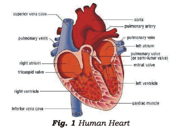
[Figure: Fig. 1 Human Heart. A longitudinal section of the heart labelled superior vena cava, aorta, pulmonary artery, pulmonary veins, pulmonary vein, left atrium, pulmonary valve (or semi-lunar valve), mitral valve, right atrium, tricuspid valve, left ventricle, right ventricle, cardiac muscle and inferior vena cava]
The auricles have comparatively thinner walls. They receive blood from different parts of the body through the veins, and pour the blood into the ventricles. The opening of the auricle into the ventricle is guarded by a valve, which controls the flow of blood.
The ventricles have thick muscular walls, which pump the blood to various parts of the body through the arteries (Fig. 1).
The right auricle and the right ventricle receive carbon dioxide rich blood through the pre-caval and post-caval veins and pour it into the right ventricle. The left and right sides of the heart are separated by a muscular wall called the septum. The left auricle and left ventricle receive oxygen rich blood from the lungs through the pulmonary veins. There is a complete separation of pure and impure blood in the heart. This is ensured by the valves present in the heart, which allow unidirectional flow of blood.
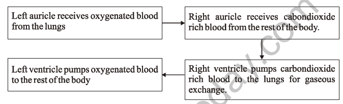
The passage of blood in the heart can be summarised as follows:
[Figure: Flow chart with four boxes. Right auricle receives carbon dioxide rich blood from the rest of the body, an arrow leads to Right ventricle, which pumps carbon dioxide rich blood to the lungs for gaseous exchange. Left auricle receives oxygenated blood from the lungs, an arrow leads to Left ventricle, which pumps oxygenated blood to the rest of the body]
When the right and left ventricles are full of blood, the heart is in an expanded condition, called diastole. Then the ventricles contract. The left ventricle sends oxygen rich blood to different parts of the body through the aorta, while the right ventricle sends carbon dioxide rich blood to the lungs through the pulmonary arteries for purification. This state of contraction is called systole.
The alternate systole and diastole produce a lub-dub sound, which is the heartbeat. A normal human heart beats 72 to 80 times per minute, while a newborn baby's heartbeat can be up to 140 beats per minute. The human heart pumps about 5 litres of blood per minute.
Thus, impure blood from the body first enters the right side of the heart and then goes to the lungs for purification. From the lungs, the blood again enters the heart from the left side, from where it is pumped to various parts of the body. This type of circulation of blood is called double circulation.
Pulse:
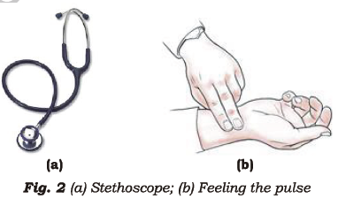
The force or pressure with which the blood is pushed out of the heart can be felt in an artery near the wrist. This is called the pulse. Doctors use a stethoscope (Fig. 2) to record the pulse or heart beat. The pulse rate increases during fever and exercise.
[Figure: Fig. 2 (a) Stethoscope; (b) Feeling the pulse, showing a person's fingers placed on the wrist of another person]
Blood Pressure:
The pressure in the arteries during systole and diastole is called blood pressure. The normal blood pressure of an adult is 120/80. Exercise, consumption of fats and anxiety are the two major and most common factors responsible for an increase in blood pressure.
BLOOD VESSELS
There are three types of blood vessels: (a) arteries, (b) veins and (c) capillaries.
Arteries:
Each heartbeat pumps blood into the blood vessels called arteries, which carry oxygen rich blood away from the heart to the various organs of the body. They lie deep in the muscles and can be felt only at the places where we feel the pulse, such as the throbbing at the hollow spot on the wrist. Arteries further divide into smaller arteries. The largest arteries are called aortae.
Veins:
Veins bring carbon dioxide rich blood from the various organs of the body to the heart. Veins are located just below the skin. The main veins are called pre-caval and post-caval veins. The pressure with which the blood rushes through the veins can be measured with an instrument called a sphygmomanometer.
Capillaries:
Capillaries are the thinnest and finest blood vessels. The arteries branch into smaller arteries and finally into capillaries. The capillaries unite to form the veins. Their thin walls help them perform their functions effectively.
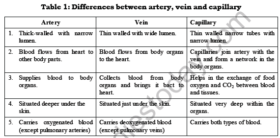
Table 1: Differences between artery, vein and capillary
| Feature | Artery | Vein | Capillary |
|---|---|---|---|
| Wall and lumen | Thick-walled with narrow lumen. | Thin walled with wide lumen. | Thin walled narrow tubes with narrow lumen. |
| Direction of blood flow | Blood flows from heart to other body parts. | Blood flows from body organs to the heart. | Capillaries join artery with vein and form a network in the body organs. |
| Function | Supplies blood to body organs. | Collects blood from body organs and brings it back to the heart. | Helps in the exchange of food, oxygen and CO2 between blood and tissues. |
| Position | Situated deeper under the skin. | Situated just under the skin. | Situated very deep within the organs. |
| Type of blood carried | Carries oxygenated blood (except pulmonary arteries). | Carries deoxygenated blood (except pulmonary veins). | Carries both types of blood. |
Importance of Capillaries:
Capillaries have very thin walls, which help them in the following ways:
(a) Picking up CO2 and other wastes from every cell and passing them into the alveoli of the lungs and the kidneys respectively.
(b) Picking up oxygen and digested food from the alveoli in the lungs and the small intestine, and passing them to each and every body cell for cellular respiration.
BLOOD
Blood is the main transporting tissue in our body. It carries food, air, waste products, hormones and other substances. Our body contains 3 to 5 litres of blood. Blood consists of plasma (the fluid part) and blood cells, of three types, which float in the plasma.
Plasma:
Plasma is a yellowish fluid medium. It contains water (about 85 to 90%), and inorganic and organic substances. The organic substances are mainly proteins. Plasma also carries dissolved excretory products, hormones, enzymes, digested food and even CO2.
Blood also contains three types of blood cells:
(a) RBC or red blood corpuscles or erythrocytes
(b) WBC or white blood corpuscles or leucocytes
(c) Platelets
RBC:
RBCs are non-nucleated, disc-shaped cells. The red colour of RBCs is due to the presence of the protein haemoglobin. There are about 5 to 5.5 million RBCs in one cubic mm of blood. They carry oxygen to the different parts of the body. Oxygen combines with haemoglobin to form oxyhaemoglobin, in a reversible reaction:
Oxygen + haemoglobin ⇌ Oxyhaemoglobin
When oxygen rich blood reaches the tissues and cells of the body, oxygen is released for use in cellular respiration. The haemoglobin is then free to carry more oxygen.
Anaemia is a very common disorder in which the amount of haemoglobin is lowered. This leads to a decrease in the oxygen carrying capacity of the blood.
WBC:
The number of WBCs in blood is less than the number of RBCs. They are comparatively bigger in size than RBCs and are meant for protecting the body against harmful bacteria and diseases. They show amoeba-like movements and are capable of ingesting foreign material. WBCs are the first cells to reach an infected or injured part of the body, by passing through the walls of the blood vessels.
Platelets:
Platelets help in blood clotting at the area of injury and block blood flow, so they prevent blood loss. They are smaller in size than RBCs. Haemophilia, or bleeder's disease, is a genetic platelet disorder. In a person suffering from haemophilia, there is no clotting of blood. Even a small wound or cut can prove fatal for such people, as they can bleed to death.
Functions of Blood:
1. It is a medium for the exchange of oxygen and carbon dioxide in the alveoli of the lungs and the body cells.
2. Blood capillaries present in the villi of the small intestine help in the absorption of food.
3. Hormones secreted by different endocrine glands in the body are also circulated throughout the body by the blood.
4. Platelets present in the blood help in blood clotting and thus prevent excessive blood loss.
5. Blood circulates in the body and collects and transports waste materials to the excretory organs for excretion.
6. Blood circulation also maintains a constant internal body temperature.
7. WBCs present in the blood protect us from germs and infections.
TRANSPORT OF SUBSTANCES IN PLANTS
Different flowering plants, such as herbs, shrubs and trees, draw water from the soil through their roots. Many of the outer cells at the root extend to form root hairs.
Root Hairs:
The root hairs absorb water along with dissolved minerals and transport them to the leaves through the xylem part of the vascular system (Fig. 3). Leaves are known as the "food factories" of the plant. Food is prepared here by the process of photosynthesis. The raw materials required are water from the soil, carbon dioxide from the atmosphere, chlorophyll present in the leaves, and sunlight.
[Figure: Fig. 3 Absorption of water by root hair. Soil containing water, a root hair and a magnified view of a root cell]
Food prepared in the leaves is in the form of glucose. It is converted into a soluble sugar and transported through the phloem to storage organs such as seeds, fruits, stems and roots. Thus xylem and phloem form the vascular system of plants, which runs from roots to leaves and covers the stem and the branches.
Note: A group of cells, mostly of a similar type, performing a specialised function is called a tissue. A group of tissues makes an organ. Xylem and phloem are examples of plant tissues.
Conduction of Water and Minerals through Xylem:
The process by which water and mineral salts are transported from the xylem of the roots upwards to the stem, branches and leaves is called the ascent of sap. Two important forces are responsible for the uptake of water and minerals in plants: one arising from the roots and the other from the leaves at the top. The mode of transport in herbs, shrubs and trees is almost the same.
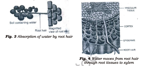
[Figure: Fig. 4 Water moves from root hair through root tissues to xylem. Labels are Epidermis, Root hair, Cortex and Vascular tissue, with water moving inwards through the layers to the xylem]
The two kinds of forces are:
1. Root pressure
2. Transpiration pull
Root Pressure:
The roots of a plant are in direct contact with water in the soil. The branching nature of root hairs provides a large surface area for the absorption of water and mineral salts. Root cells contain large vacuoles filled with a fluid called cell sap. The cell sap has a higher concentration of salts than the soil water. This feature enables the root hair cells to draw water from the soil through a process known as osmosis. The continuous absorption of water by the root hairs builds up sufficient force to push the water upwards through the xylem vessels.
Xylem is a complex tissue composed of more than one type of cell. It consists mainly of dead elements, namely tracheids, vessels and xylem fibres, and a living element, xylem parenchyma.
Transpiration Pull:
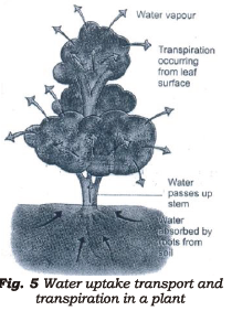
The loss of water from the aerial parts of plants in the form of water vapour is known as transpiration. Water evaporates through stomata, the tiny pores present on leaf surfaces. When water is lost from the leaves, either due to transpiration or because it is consumed in photosynthesis, the water column becomes greatly stretched and creates a pull. The continuity of the water column does not break, because of the forces operating between the water molecules and between the water molecules and the walls of the xylem vessels. The transpiration pull is sufficient to raise water in the leaves of the tallest trees.
[Figure: Fig. 5 Water uptake, transport and transpiration in a plant. Water vapour escapes from the leaves through transpiration. Water passes up the stem, and water is absorbed by the roots from the soil]
Thus, due to the combined effect of the push force of root pressure and the pull force of transpiration by the leaf cells, water moves upwards along the xylem vessels (Fig. 5).
Osmosis:
Osmosis is a physiological process that takes place when two solutions having different concentrations are separated by a semipermeable membrane. A semipermeable membrane is one that allows only water molecules to pass through. The movement of water molecules takes place across the membrane from a dilute solution to a concentrated solution. This process enables the cell-to-cell movement of water in the root cells after it is absorbed from the soil by the root hairs. Thus water finally reaches the xylem of the root.
Conduction of Prepared Food through Phloem:
Food prepared by green leaves by photosynthesis needs to be sent to storage organs and other parts of the plant body. This process is known as translocation of prepared food. The phloem, a special type of vascular tissue, is responsible for carrying the prepared food to all parts of the plant. Phloem is a complex tissue consisting of living elements (sieve tubes, companion cells and phloem parenchyma) and a dead element (phloem fibres). These are continuous vessels from the leaves to other parts of the plant.
EXCRETION
Many harmful and unnecessary substances or by-products are produced in the body of living organisms, due to the continuous synthesis (nutrition) and breakdown (respiration) of materials. The unwanted by-products or waste materials include carbon dioxide, water vapour, and nitrogenous wastes such as urea and uric acid. If they are allowed to accumulate in the body beyond a certain concentration, they poison the cells and slow down the metabolic processes. Thus, these waste products must be thrown out of the body as such or changed into harmless material before being removed from the body.
The process of removal of metabolic waste materials from the body is called excretion.
Excretion in Unicellular Organisms:
A large part of the ammonia produced remains unused in the body. It is either excreted as such, or after conversion to urea or uric acid. In unicellular organisms like Amoeba, Paramecium, Hydra and sponges, there are no special organs for excretion. The waste products are thrown out into the surrounding water through the general body surface, by diffusion directly from the cells in which they are produced.
Excretion in Animals:
In higher or multicellular organisms, direct diffusion for waste removal is too slow. Larger animals have a well-developed excretory system. For example:
1. Earthworms and leeches have specialised organs called nephridia (tubular structures).
2. Insects have Malpighian tubules.
3. Vertebrates have kidneys.
Excretion in Plants:
Plants do not have well-developed excretory organs, but they have a unique method of waste disposal.
1. Oxygen and water vapour, the waste products or by-products of photosynthesis, also diffuse out through the stomata and lenticels throughout the day, by a process called transpiration. The waste material produced by respiration is carbon dioxide. Plants need CO2 and water vapour in the presence of sunlight to perform photosynthesis, so carbon dioxide is partly used by the plant itself for the synthesis of food. The excess carbon dioxide escapes through the stomata and lenticels from the surface of the leaf and stem respectively.
2. Plants can tolerate considerable variations in the concentrations of salts present inside them. These excess salts get deposited in the form of crystals such as calcium carbonate and calcium oxalate. For example, in yam (zamikand) cells, needle-like crystals of calcium oxalate (called raphides) accumulate as waste. These crystals cause the burning sensation felt in the throat on eating yam. The raphides can be dissolved by boiling yam pieces in water with tamarind (imli) in it. If the yam is cooked after this treatment, no burning sensation is felt.
3. In older plant cells, harmless excretory products may remain trapped in the large vacuoles, and may stay inside the cell throughout its life.
4. Plants also get rid of wastes by shedding their leaves in autumn. Some plants also shed bark at regular intervals.
5. Not all plant wastes are useless. Some wastes are of great value, such as gum, resin, sandalwood oil, eucalyptus oil (a pain reliever used in many medicines), tannins (found in the bark of trees and used for tanning leather), latex (used for making rubber products) and alkaloids. Alkaloids are very poisonous compounds, but some of them, such as quinine and digitalis, extracted from foxglove, are used to make medicines. Digitalis is a powerful toxin used in cardiac medicine.
Excretion in Humans:
The human body has specialised organs for the removal of solid, liquid and gaseous wastes. These organs are:
1. Large intestine: The undigested food is removed from the large intestine and thrown out of the body through the anus, in the form of faeces.
2. Lungs: Carbon dioxide and water vapour are the respiratory wastes, which are exhaled out of the body through the nostrils during breathing.
3. Skin: The skin helps in eliminating excess water, urea, salts and other metabolic wastes in the form of sweat. Sweat is secreted from the sweat glands below the skin (Fig. 6). Excess salt (NaCl), nitrogenous waste (urea produced due to the breakdown of proteins in the body) and water (diffusing into the sweat glands from the blood capillaries) are removed from the body through the sweat pores. Sweating or perspiration also regulates body temperature, as it causes cooling due to evaporation.
4. Liver: Dead RBCs and excess amino acids and proteins are removed from the body in the form of urea by the liver, from the blood.
[Figure: Fig. 6 Sweat glands under the skin. Labels are Hair, Oil gland, Epidermis, Pore, Sweat duct and Sweat gland]
THE URINARY SYSTEM
Blood circulates in the body and collects water and waste products from the different body organs. The blood then needs to be filtered, so it enters the urinary system. The human urinary system consists of:
1. Kidneys
2. Ureters
3. Urinary bladder
4. Urethra
[Figure: Fig. 7 The excretory system. Two kidneys on either side, each with a cortex and medulla, connected by ureters to the urinary bladder, from which the urethra leads out]
Kidneys:
The kidneys are situated along the back of the abdominal wall, at the level of the waist on either side of the vertebral column. They are brick red in colour and bean shaped. Each kidney weighs about 150 g and is about 12 cm long, 6 cm wide and 3 cm thick. Each kidney has two layers: a thin outer covering, the cortex, forming a capsule, and an inner layer, the medulla. The basic structural and functional unit of a kidney is the nephron. There are more than a million nephrons in each kidney (Fig. 7).
Structure of Nephron:
Each nephron consists of a cup-like structure, the Bowman's capsule, and a long tubular structure. The Bowman's capsule lies in the cortex part of the kidney, and the urinary tubule lies in the medulla part. Inside the Bowman's capsule lies a network of capillaries called the glomerulus. Water and wastes such as urea and salt are filtered into the cup from the blood in the glomerulus. The clean blood then flows out of the kidney, while the filtrate flows into the urinary tubules. The glomerulus along with the urinary tubules constitutes the Malpighian body (Fig. 8).
[Figure: Fig. 8 Structure of a nephron. Labels are Bowman's capsule, Glomerulus, Tubule and Renal capillaries]
Many urinary tubules come together in the medulla to form the collecting tubules, which are in turn surrounded by a network of renal capillaries. Many collecting tubules further join to form bigger tubes, the ureters. The urine formed in the collecting tubules flows into the ureters, which carry it out of the kidney and collect it in the urinary bladder.
Ureters:
Human beings have two ureters, one coming from each kidney. Ureters are thin-walled, urine-carrying ducts. They are about 30 cm long and run downwards from each kidney to open into the urinary bladder.
Urinary Bladder:
The urinary bladder is an elastic, sac-like structure where urine is stored. Its size varies with the amount of urine present in it. The kidneys of an adult produce only about 1.5 litres of urine, whereas they filter 135 to 150 litres of fluid every day. The bladder can expand to hold up to about 0.5 litres of urine. The desire to urinate begins when the bladder is half full.
Urethra:
From the urinary bladder, urine is carried through a duct called the urethra and finally discharged from the body. A human being passes about 1.5 to 2.5 litres of urine every day.
Urine is normally light yellow in colour. It contains 95% water, 2.5% waste products (mineral salts and compounds of sodium and potassium) and 2.5% urea. The composition of urine in a normal healthy person remains fairly constant, but it may change during sickness.
Note: An examination of urine tells the physician whether various organs of the body are functioning normally. That is why doctors usually advise a urine test to diagnose diseases.
KIDNEY MALFUNCTION
The kidney functions as an excretory as well as an osmoregulatory organ. The amount of water in the blood is controlled by osmoregulation. Generally, glucose or sugar is not present in the urine. If sugar comes from the blood into the filtrate along with the urine, it indicates that the person may be suffering from diabetes. Diabetes interferes with the conversion of the unused sugar in the body into glucose and its further breakdown.
Kidney Failure or Renal Failure:
Our body needs 2 to 3 litres of water every day to flush out urea and to keep the urea concentration at a low level in the body.
The presence of blood in urine is an indication of damaged kidneys. This occurs due to the presence of a toxin produced by the Streptococcal bacteria. The toxin damages the tiny filters (nephrons) in the kidneys, due to which blood cells and albumin pass into the urine. Sometimes an infection in the kidney, restricted blood flow, or an injury can cause severe damage to the kidney, and it may stop working. In such cases, urea and other waste products accumulate in the blood, and the osmoregulatory function of the kidney is also affected. If not treated immediately, this can ultimately lead to death.
The usual treatment for kidney failure is either dialysis or kidney transplant.
Dialysis:
Dialysis is the process of cleaning the blood of a person with kidney failure, by separating the waste substances using a dialysis machine.
To perform dialysis, the blood from an artery in the patient's arm is passed through the dialysis machine. The machine contains a tank with a dialysing solution, and long cellulose tubes are coiled and dipped in the tank. The dialysing solution contains water, glucose and salts in concentrations similar to those normally found in blood.
The patient's blood has a higher concentration of glucose, salts and wastes. When it passes through the dialysing solution, the wastes present in it pass through the semi-permeable cellulose tubes into the dialysing solution. The clean blood is then pumped back into a vein of the patient's arm. Dialysis continues till all the blood in the body has been cleaned. This treatment has to be done regularly. Dialysis is an expensive and painful process, so if the condition of the patient worsens, doctors generally recommend a kidney transplant.
Note: If the kidneys fail to work or become functionless, an artificial kidney can be installed. The artificial kidney is used to filter the blood of a person whose kidneys are damaged. This process is called haemodialysis. It involves a good deal of discomfort and a risk of the formation of blood clots.
Kidney Transplant:
Kidney transplant is the best long-term solution for patients with kidney failure. The damaged kidney is replaced, by a surgical procedure, with a matching kidney donated by a healthy person. Until a suitable match is available, the patient has to undergo dialysis periodically. Nowadays, facilities for dialysis and even kidney transplantation are available in good hospitals all over the country.
Note: The first kidney transplant in India was performed on 1.12.1971 at Christian Medical College, Vellore, Tamil Nadu, on Shanmughan.
Importance of Cappillaries
Free study material for Science
Free CBSE Study Guides: Class 7 Science Chapter 11 Transportation in Animals and Plants
Quick Revision Material for Chapter 11 Transportation in Animals and Plants
Access comprehensive study material for Chapter 11 Transportation in Animals and Plants, including revision notes, concept maps, and high-probability questions. These resources are designed in alignment with the latest 2026 CBSE syllabus for Class 7 Science to support effective exam preparation.
Expert Notes and Solved Exam Questions
Built using official NCERT guidelines for Class 7 Science, these materials provide reliable academic support. Integrating past examination questions and step-by-step solutions helps students understand official CBSE grading criteria.
Additional Study Resources for Class 7
Maximize your grade potential by utilizing Class 7 Science practice papers in tandem with these structured notes. All printable assignments and interactive mock tests on our platform are updated for the 2026 session and available free of charge.
FAQs
Our advanced study package for Chapter 11 Transportation in Animals and Plants includes detailed concepts, diagrams, Mind Maps, and explanation of complex topics to ensure Class 7 students learn as per syllabus for 2026 exams.
The Mind Maps provided for Chapter 11 Transportation in Animals and Plants act as visual anchors which will help faster recall during high-pressure exams.
Yes, teachers use our Class 7 Science resources for lesson planning as they are in simple language and have lot of solved examples.
Yes, You can download the complete, mobile-friendly PDF of the Science Chapter 11 Transportation in Animals and Plants advanced resources for free.
Yes, our subject matter experts have updated the Chapter 11 Transportation in Animals and Plants material to align with the rationalized NCERT textbooks and have removed deleted topics and added new competency-based questions.