NEET Biology Human Reproduction And Embryonic Development Chapter Notes

Download NEET Biology Human Reproduction And Embryonic Development Chapter Notes in PDF format. All Revision notes for Full Course Human Reproduction have been designed as per the latest syllabus and updated chapters given in your textbook for Human Reproduction in Full Course. Our teachers have designed these concept notes for the benefit of Full Course students. You should use these chapter wise notes for revision on daily basis. These study notes can also be used for learning each chapter and its important and difficult topics or revision just before your exams to help you get better scores in upcoming examinations, You can also use Printable notes for Full Course Human Reproduction for faster revision of difficult topics and get higher rank. After reading these notes also refer to MCQ questions for Full Course Human Reproduction given on studiestoday

Revision Notes for Full Course Human Reproduction Human Reproduction

Full Course Human Reproduction students should refer to the following concepts and notes for Human Reproduction in Full Course. These exam notes for Full Course Human Reproduction will be very useful for upcoming class tests and examinations and help you to score good marks

Human Reproduction Notes Full Course Human Reproduction


 Human beings reproduce sexually and are viviparous i.e. they give birth to young ones. The reproductive units in sexual reproduction are specialized cells called gametes.
 
PRIMARY SEX ORGANS
 
 Primary sex organs are those sex organs which produce gametes. They are also called gonads.
 They also produce steroid hormones essential for reproduction as well as the growth and development of the entire body.
 
SECONDARY SEX ORGANS
 
• Sex organs, glands and ducts which do not produce gametes and hormones but are otherwise essential for sexual reproduction are known as secondary sex organs.
 Secondary sex organs of human male reproductive system are vasa efferentia, epididymes, vas deferentia, ejaculatory ducts seminal vesicles, cowper’s gland, urethra, prostate gland and penis.
 Secondary sex organs of human female reproductive system are fallopian tubes, uterus, vagina, external genitalia and mammary glands.
 
ACCESSORY OR EXTERNAL OR SECONDARY SEX CHARACTERS
 
 They are those external features which provide distinctiveness to the two sexes.
 Major accessory or external or secondary sex characters of human male are facial hair, body hair, more height, more muscles, broadening of shoulder, low pitched voice, narrow but strong pelvis
 Major secondary sex characters of human female are development of breasts, broader pelvis, rounded body contours, fat deposition in thighs, buttocks and face, high pitched voice, pubic hair and sterna breathing
 
PUBERTY
 
 Beginning of sexual maturity or ability to reproduce is known as puberty. It is the period when primary sex organs become functional and start secreting sex hormones which bring about developments of secondary sex organs and appearance of secondary sex characters.
• Puberty is characterized by rapid growth. The average age of onset for girls is 10 -14 years and for boys, 12 -15 years old
 In boys pubertal changes occur in response to testosterone, whereas in female child, estrogen is produced by ovarian follicles.
 
MALE REPRODUCTIVE SYSTEM
 
It is made of a pair of testes, scrotum, vasa efferentia, a pair of epididymes, a pair of vasa differentia, a pair of seminal vesicles, a pair of ejaculatory ducts, urethra, prostate gland, a pair of cowper’s glands and penis 
 
TESTES
 
  They are a pair of oval, pinkish primary sex organs of male reproductive system, each of with a size of 5cm ( length), 3cm ( thickness) and 2.5cm ( width), weight about 12gm and lying obliquely in scrotum.
  During early foetal life, the testes develop in the abdominal cavity just below the kidney then they descend into the scrotum.
  The testis is surrounded by three layers
(i) Tunica vaginalis, a bilayer of peritoneum with narrow coelomic cavity filled with coelomic fluid. It helps the testis in frictionless sliding
(ii) Tunic albuginea, is the actual covering of the testes. The covering is made up of dense bluish with fibrous or collangenous connective tissue
(iii) Tunica vasculosa, is delicate loose connective tissue which lines the testicular lobules inner to tunica albuginea. It has rich supply of blood vessels.
 
Tunica albuginea also projects inside testes to form a thick incomplete vertical column called mediastimum and a number of transverse septa. The septa produces 200 – 300 conicular testicular lobules and each lobules is filled with connective tissue and 1-3 yellow convoluted seminiferous tubules. A total of 900 – 1000 yelloish seminiferous tubule occur in each testis. Each tubule is about 70-80 cm long. Seminiferous tubule join to form 20-30 short straight ducts called tubulic recti. Tubuli recti enter a network of channels called rete testes.
 The connective tissue lying in between seminiferous tubules are called Leydig’s cell and it contain yellow pigment granules. Leydig cells are large polyhedral cells which have eccentric nucleus and small lipid containg vacuoles. They secrete testosterone and other androgens.
 The lining of seminiferous tubules is formed by a single layered germinal epithelium. The epithelium has two types of cells, spermatogenic ( primary germ cells) and sertoli cells ( supporting cells), Spermatogenic cells form 4-8 layers. The cells are destined to undergo spermatogenesis and form supermatozoa.
 Sertoli cells are large, elongated and pyramidal cells with bases resting on basal lamina ( basement membrane) and apics projecting into the lumen of the seminiferous tubules. They secrete spermatogonic substances for nourishing and differentiation of cells undergoing spermatogenesis. They also secrete hormone inhibin for controlling FSH secretion.
 
NEET Biology Human Reproduction and Embryonic Development Chapter Notes
 
SCROTUM
 
 It is a pouch of deeply pigmented skin arising from lower abdominal wall below the pubic symphysis and hanging between and infront of groin part of the thighs. Scrotal skin bears sebaceous glands that produce a characteristic odour, sweat glands and nerve endings.
 Wall of scrotum has three layers – outer wrinkled skin, connective tissue and smooth muscles. An internal septum scroti divides the scrotum into two sacs, each for one testis. Left testis lies a bit lower than the right one. Scrotum possesses a smooth involuntary muscle called dartos muscle. A testis rests in its chamber over pad called gubernaculum.
 The scrotum remains connected with the abdomen or pelvic cavity by the inguinal canals. The spermaticord, formed from the spermatic artery, vein and nerve, bound together with connective tissue passes into the testis through inguinal canal.
 The testes develop in the abdominal cavity during the 7th month of gestation descend permanently into the respective scrotal sacs 
 The scrotum acts as a thermoregulator, maintaining the testes at a temperature 2OC lower than that of the body. Movement of dartos muscle help in changing position of testes to keep them at proper temperature. When the body is chilled the smooth muscle contracts and brings the testes closer to the pelvic cavity to get warmth
 In some person testes fail to descend in scrotum. The condition is called cryptorchidism. It results in sterility
 
VAS EFFERENTIA
 
 Rete testies is connected to caput epididymis by 12-20 fine tubules called vasa efferentia
 Their lining epithelium is pseudostratified. It has large columnar ciliated cells and small nonciliated cells with endocytic activity, ciliated cells help in conducting sperms. Tubuli recti, rete testis and vas efferentia constitute an intratesticular genital duct system
 
EPIDIDYMES
 
• The epididymes is a mass of long narrow closely coiled tubule which lies along the inner side of each testis. coiling forms three parts – upper caput epididymes or head middle corpus epididymes or body and lower cauda epididymes or tail
•  In the head of the epididymis, the sperms undergo physiological maturation, acquiring increased motility and fertilising capacity. In the tail of the epididymis sperms are stored before entering the vas deferens
 Epididymis is lined by pseudostratified epithelium that secretes nutrients required for maturation of spermatozoa. Non-ejaculated sperms are broken down after an interval.
 
VAS DEFERENS
 
 The vas deferens is a continuation of cauda epididymis which leaves the scrotal sac and enters the abdominal cavity. After entering abdomen it loops over the urinary bladder and dilates to spindle like ampulla for temporary storage of spermatozoa. It also conducts sperms.
 
EJACULATORY DUCTS
 
 They are shorter ducts of about 2cm length where male ejaculate is produced. Each duct is formed by joining of vas deferens and duct of seminal vesicle
 Ejaculatory ducts enter the prostate gland and join the prostatic urethra to produce a single urinogenital duct. In ejaculatiory duct, the sperm mix up with secretation of seminal vesicles. The walls of ejaculatory ducts are muscular to quickly conduct the ejaculate through urinogenital duct.
 
URETHRA
 
 It arises from urinary bladder and is about 20cm long, differentiated into three regions
(i) A short proximal prostatic urethra which is surrounded by prostate glands.
(ii) A very short middle membranous urethra without any covering
(iii) A long distal penile urethra that passes through a penis
 The urethra has internal sphincter of smooth muscle fibres at its beginning and external sphincter of straited muscle fibres around its membranous part
 
PENIS
 
 It is male erectile copulatory organ which transfers semen into reproductive tract of female during sexual intercourse
 The penis contains three cyclindrical masses of erectile tissue – two dorsal corpora cavernosa and one ventral corpus spongiosum
 The corpus spongiosum, which contains the penile urethra, is enlarged at the distal end of the penis to form glans penis. It is covered with smooth skin, foreskin.
 
SEMINAL VESICLES
 
• They are a pair of lobulated contorted musculoglandular sacs of 5cm length between urinary bladder and rectum. Their ducts join the vasa deferens to form ejaculatory ducts.
• They produce an alkaline secretion which forms 60% of the volume of semen.
The secretion of seminal vesicles contains fructose, citrate, prostaglandins, inositol and clotting protein.
•  Alkaline nature of the acidic environmental of the male urethra which otherwise would inactivate and kill sperms
•  The prostaglandins stimulate uterine contractions and thus may help the sperm to be moved towards female’s oviducts, where fertilization takes place. The clotting protein help semen coagulate after ejaculate.
 
PROSTATE GLAND
 
 It is a large grayish to red pyramidal gland of 4cm with and 3cm height that encloses a part of urethra including its junction with ejaculatory ducts
 The produces a milky slightly alkaline secretion which forms 25% of the volume of semen. It possesses calcium, phosphate, bicarbonates, enzymes, clotting enzymes, prefibrolysin and prostaglandins. Secretion of the prostate gland nourish and activates the spermatozoa to swim. Prostaglandins helps in liquefying cervical mucus and helps in liquefying cervical mucus and stimulating movement in the female tract.
 
BULBOURETHRAL GLANDS
 
 A pair of small yellow pea seed sized lobulated tubualveolar glands, 4-5cm below prostate and opening into membraneous urethra by separate ducts
 The secretion has abundant mucus for lubrication of reproductive tract. It neutralizes the urethra from remains of urine. Secretion of cowper’s gland is produced before the ejaculation of semen
 
SEMEN
 
 The secretion of accessory sex glands and mucus are added to sperms to form seminal fluid, or semen or seminal plasma
 It rich in fructose, calcium and certain enzymes. It has a pH of 7.35 to 7.5.
 It is ejected from the penis during ejaculation. A single ejaculation may contain 300 million sperms. It has many functions
(i) It provides a fluid medium for transmission of sperms into vagina of the female.
(ii) It nourishes and activate the sperms to keep them viable and motile
(iii) It neutralizes the acidity of the urine in the urethra of male and vagina of female to protect the sperms
(iv) It facilitates the sexual act by lubricating the reproductive tract of the female.
 
NEET Biology Human Reproduction and Embryonic Development Chapter Notes-1
 
 
HORMONAL CONTROL OF MALE REPRODUCTIVE SYSTEM
 
 The growth, maintenance and functions of the male reproductive organs are under the hormonal control. GnRH ( Gonadotropin releasing hormone) is secreted by hypothalamus. It stimulates the anterior lobe of pituitary gland to secrete and release LH and FSH. In male, LH is called interstitial cell stimulating hormone (ICSH) because it stimulates Leydig’s cell of the testes to secrete androgens 
 Testosterone is the principle androgen. The growth, maintenance and functions of secondary sex organs and functions of secondary sex organs and accessory glands are under the control of testosterone.
 FSH stimulates sertoli cells of testis to secrete an androgen binding protein (ABP) that concentrates testosterone in seminiferous tubule and also secrete a peptide inhibin which suppresses FSH synthesis. FSH acts directly on spermatogonia to stimulate sperm production.
 
NEET Biology Human Reproduction and Embryonic Development Chapter Notes-2
 
 
COMMON DISORDERS OF MALE REPRODUCTIVE SYSTEM
(i) Prostatitis – It is inflammation of prostate generally caused by infection
(ii) Benign prostatic hypertrophy ( BPH) – This is the enlargement of prostate gland. It often causes in old age causing frequent night urination. Untreated BHP may lead to kidney damage.
(iii) Prostate carcinoma – It is cancer of prostate which may grow unnoticed up to stage of metastatis
(iv) Impotence – It is the inability of the adult male to achieve penile erection.
(v) Sterility – Sperms are unable to fertilize the ovum due to low count or less motility.
(vi) Cryptochidism – It is a failure of one or both of the testicles to descend into scrotum. It is often results in pushing of an intestinal loop into scrotum, resulting in its abnormal size and discomfort.
 
FEMALE REPRODUCTIVE SYSTEM
 
The female reproductive system consists of a pair of ovaries, a pair of fallopian tubes, uterus, vagina, external genitalia and breasts
 
OVARIES
 
 They are a pair of almond shaped, solid, greyish pink gonads of the female. Each ovary is about 2-4 cm in length, 1.5cm in width and 1cm in the thickness. It is suspended from the dorsal body wall by a fold of peritoneum called mesovarium. It is held in position by ligaments which attach it to pelvic wall and uterus.
 Ovaries are differentiated into four parts
(i) Germinal epithelium is the outermost layer of the ovary which is formed of simple squamous and cuboidal cells
(ii) Tunica albuginea is poorly differentiated sheath of dense connective tissue that lies below the germinal epithelium and outside the cortex. Tunica albuginea provides greyish colour to ovary.
(iii) Cortex is a wide layer of connective tissue having a large number of spindle shaped fibroblasts, reticular fibres and ovarian follicles
(iv) Medulla is the central part of the ovary made of less dense connective tissue. Medulla is richly supplied with blood vessels. It has elastic fibres, smooth muscles.
 The ovarian medulla contains many rounded or oval bodies called ovarian follicles at various stage of development. The number of follicles in the two ovaries of young adult female is 1,20,000 – 1,60,000
 
FALLOPIAN TUBES ( OVIDUCTS)
 
 They are pair of muscular and internally ciliated tubes of 10-12 cm length which lie horizontally over peritoneal cavity arising near and ending at uterus 
 A fallopian tube is differentiated into four parts
(i) Infudibulum is a funnel shaped fimbriated free end of the oviduct. It opens into the body cavity by an aperture called ostium. Margin of the funnel bears motile, finger like projection called fimbriae. Fimbriae have folds lined with cilia that produce a slow current towards the ostium for passage of liberated ovum towards the inside of oviduct.
(ii) Ampulla is a curved dilated part of oviduct which is also the site of fertilization of ovum
(iii) Isthmus is the very short, narrow, thick walled, straight part of the oviduct which connects ampulla with lateral wall of uterus.
(iv) Uterine part is about 1cm long part that passes into uterine wall. Oviducal wall is composed of three layers – outer serosa of visceral peritoneum, middle muscularis and inner mucosa. Mucosa has an epithelium of ciliated and secretory columnar cells. Secretory cells produce a viscous secretion for nourishing and protection of ovum. Passage of ovum is facilitated by movement of cilia and muscular contractions of the wall.
 In most vertebrates both the ovaries and oviduct are functional. In birds the right ovary and right oviduct are atrophied 
 
UTERUS
 
 It is the pyriform, hollow muscular thick walled but distensible median structure located above and behind urinary bladder that is meant for nourishing and development of foetus. For this, uterus is capable of tremendous enlargement. The empty uterus is 7.5cm, 5cm broad and 2.5cm thick
 Uterus is attached to pelvic wall by means of ligament
 Wall of uterus is differentiated into three layers
(i) Internal glandular endometrium
Endometrium has two parts, epithelium and lamina propia. Epithelium lines the luminar surface of uterus. It contains two types of columnar cell, ciliated and secretory. Lamina propia contains connective tissue with fibroblasts, tubular glands and blood vessels. Endometrium shows cyclic changes during menstrual cycle.
(ii) Myometrium is the middle layer of smooth muscles. It undergoes strong contractions during delivary of the baby
(iii) Perimetrium is an outer layer of uterus formed of either adventitia ( connective tissue only) and serosa ( connective tissue and mesothelium) 
 Uterus is differentiated into three regions
(i) Tundus: Upper dome – spahped part above the opening of fallopian tube
(ii) Body : It is broad towards fundus and narrow down towards the cervix
(iii) Cervix: 2.5cm long narrow inferior extremity of uterus which protrudes into vagina. It is connected to the body by internal child birth it becomes irregular bilipped. Upper part of uterus leans forwards. It is almost at right angle to vagina
 
VAGINA
 
 It is an elastic muscular tube of 8-10cm length which functions as female copulatory organ, pathway for menstrual flow and birth canal. It is easily stretched
 The opening of the vagina is called vaginal orifice. Vaginal orifice is partially covered by a perforate membrane called hymen. It often gets ruptured during vigorous physical exercise or during sexual intercourse. In some it persists even after coitus
 
EXTERNAL GENITALIA
 
 The area having external genitalia is characterized by mons pubis on the upper side, perineum on the lower side and vestibule or depression in the centre
 Mons pubis is an eminence formed by fat over the public symphysis bones. Vestibule has urinary meatus with urethral opening on the upper side and vaginal orifice on the lower
 A small erectile organs, the clitoris, lie at the anterior junction of labia minora. It is homologous to the penis. It consists of a short shaft with erectile tissue. its tip is round and of erectile tissue
 Vestibule is flanked by two pairs of fleshy folds of skin; the inner small, thin, moist labia minora which form clitoris in front and are connected behind by fourchette. They possess sebaceous gland. The outer larger, hair –covered labia majora. They also possess sebaceous gland.
 
NEET Biology Human Reproduction and Embryonic Development Chapter Notes-3
 
VESTIBULAR OR BARTHOLIN’S GLAND
 
 They are a pair of small tubulociner glands which open in the vestibule lateral to vaginal orifice. The secretion is thick viscid and alkaline for lubrication and counteracting urinary acidity. A number of small vestibular gland (paraurethral gland or gland of skene) are present on the either side of urethral opening.
 
MAMMARY GLAND OR BREASTS
 
 Mammary glands or breasts are modified sweat glands that lie over the pectoral muscle which develops during puberty but becomes active only after child birth
 Each breast has a broad tip called nipple for the release of milk. A circular pigmented area called areola lies below it. Each breast contains 15-20 glandlar lobes having secretory alveoli and each lobe is separated from each other by connective tissue and adipose tissue.
 The cells of alveoli produce milk which is stored in the cavities of alveoli. Alveoli opens into mammary tubules and then into mammary ducts.
 Mammary ducts forms mammary ampulla from which a lactiferous duct develops. Each lobe has a separate lactiferous duct and each opens at nipple by separate pores. 
 During pregnancy, the glands grow under the influence of oestrogen and progesterone. On the infant’s birth, the hormone prolactin stimulates the production of milk and hormone oxytocin causes release of milk as infant sucks the breast.
 
NEET Biology Human Reproduction and Embryonic Development Chapter Notes-4
 
 
HORMONAL CONTROL OF FEMALE REPRODUCTIVE SYSTEM
 
  GnRH secreted by hypothalamus stimulates the anterior lobe of pituitary gland to secrete LH and FSH. FSH stimulates the growth of ovarian follicles and also increase the development of egg(oocyte with the follicle to complete the meiosis I to form
secondary oocyte. FSH also stimulates formation of oestrogen.
  Lutenising hormone (LH) stimulates the corpus luteum to secrete progesterone.
  Rising level of progersterone inhibits release of GnRH which in turn inhibits the production of FSH, LH and progesterone.
 
NEET Biology Human Reproduction and Embryonic Development Chapter Notes-5
 
MENSTRUAL CYCLE
 
 It is a series of cyclic changes that occurs in the reproductive tract of human females with periodicity of 28 days, right from menarche to menopause except during period of pregnancy.
 The first menstrual period is called menarche. This occurs between the age of 12 to 15 years. Menopause is stoppage of menstrual period. It occurs between 45-55 years.
 Menstrual cycle is controlled by gonadotropin and ovarian hormones.
 Menstrual cycle has following phases:-
 
I. Menstrual phase
 This phase lasts for about 4 days.
 If fertilization do not occur progesterone secreted by persistent corpus luteum inhibits the release of LH from pituitary.
 Reduction of LH causes subsequent fall in progesterone level.
  The uterine lining due to deficiency of progesterone causing rupture of blood vessels and leads to discharge of blood, a serous fluid, cell debris and mucosal fragments.
  Lowered levels of progesterone causes release of FSH from the anterior pituitary. This initiates a new cycle.
 
II. Follicular phase (proliferative phase)
  In the presence of FSH released by anterior pituitary, 6-12 ovarian follicles begin enlargement through proliferation of their granulose cells to secrete estrogen. Estrogen stimulates the proliferation of the endometrium of uterine wall.
  After about a week of development only one ovarian follicle continues growth while the rest degenerates.
  Towards the end of proliferative phase the endometrium becomes about 3mm thick.
 
III. Ovulatory phase
  High level of both LH and estrogen cause rapid growth of graafian follicle.
 Graafian follicle rises to the surface of the ovary, produce a protuberance or stigma. The stigma ruptures and the ovum surrounded by corona radiate comes out in a viscous fluid. The process is called ovulation. It occurs after about 14 days.
 
IV. Luteal or secretory phase
 Under the influence of LH the empty graafian follicle continues growth.
 The follicular cells are converted into lutein cells by deposition of yellowish lipid inclusions. The phenomemnon is called lutenisation.
The rupture graafian follicle is now called corpus luteum. It secretes progersterone.
 Both LH and progesterone help in further growth and thickening of endometrium. Thickness of endometrium becomes 5-6mm.
 Progesterone inhibits uterine movements as well asproliferation of new ovarian follicles. The phase lasts for about 10 days.
 
NEET Biology Human Reproduction and Embryonic Development Chapter Notes-6
 
 If puberty starts early then it is known as precocious puberty.
 Dysmenorrhoea is painful menses.
 Menorrhagia is excessive menstruation.
 Oligomenorhoea is infrequent menstruation.
 Amenorrhoea is non-occurences of menses.
 
DISORDER OF THE HUMAN FEMALE REPRODUCTIVE SYSTEM
 
(i) Breast cancer: Breast cancer increases after menopause. The standard treatment for breast cancer is mastectomy (removal of breasts).
(ii) Cervical cancer: It is relatively slow growing cancer. Cervical cancer may be treated by radiation or surgery.
(iii) Oophorocystosis: Ovarian cysts are fluid filled tumors of the ovary. Such cysts sometimes rupture and regresses during pregnancy.
(iv) Ectopic pregnancy: It is implantation of embryo at a place other than uterus, generally oviduct.
(v) Oophoritis: It is inflammation of ovary, usually caused by an infection.
(vi) Endometriosis: It is growth of endometrial tissue outside the uterus.Treatment is usually hormone therapy or surgery.
(vii) Infertility: Infertility in women is inability to become pregnant.
 
SPERMATOGENESIS
 
  Spermatogenesis is the process of formation of haploid spermatozoa(sperms) from diploid spermatogonia inside the testis of the male.
Formation of spermatids
(i) Multiplication phase: At sexual maturity, the underdiffernetiated germ cells divide several times by mitosis to produce a large number of spermatogonia(2N) are of two types:-
Type A spermatogonia fuction as stem cells and produce more spermatogonia.
Type B spermatogonia divide mitotically to form primary spermatocytes. This process is called spermatocytogenesis.
(ii) Growth phase: Primary spermatocytes grow to become almost double in size.
(iii) Maturation phase: Each diploid primary spermatocyte undergoes meiosis I to form two haploid secondary spermatocytes. Each secondary spermatocytes divides by meiosis II, giving rise to two haploid spermatids. The spermatids becomes partially embedded in sertoli cells for nourishment and support.
(iv) Spermiogenesis: The transformation of spermatids into spermatozoa is called spermiogenesis. The different changes which occur during spemiogenesis are
a. Formation of acrosome by golgi apparatus
b. Elongation and condensation of nucleus.
c. Separation of centrioles.
 Spermatogenesis requires about 64 days.
 The number of sperms produced by adult male per day is 1012-1013.
Sertoli cells act as phagocytes. They consume the residual cytoplasm discarded during spermiogenesis.
• After maturation of spermatozoa, they get detach from sertoli cells. The process is called spermiation. The released sperms are stored in epididymis upto one month.
 
NEET Biology Human Reproduction and Embryonic Development Chapter Notes-7
 
SPERM
 
• Human sperm is dart-like flagellate structure of 60 μm length and maximum breadth of 3.5μm.
•  It has four parts:
(i) Head: Knob-like structure but flat terminal part, 4-5 μm long. It has two components acrosome and nucleus. Cytoplasm is nearly absent. Acrosome forms a covering over the anterior two third of head. It is derived from golgi apparatus from spermatid. It contains lysins like hyaluronidase and proteolytic enzymes. Surface contains compatibility protein bindin for attaching to receptors of egg. Nucleus is dense mass of chromatin having protamines.
These may be one or more less dense areas called nuclear vacuoles. On the outside is present a double membrane head cap.
(ii) Neck: It is a short narrow part between head and middle piece which contains two centrioles, unconnected proximal centriole and distal centriole attached to filament that passes into middle piece.
(iii) Middle piece: It is a cylindrical part 5-7μm long and 1μm in breadth. It has axial filament surrounded by 10-14 spiral turns of mitochondria and bearing towards the end of a ring centriole. Mitochondria provides energy for swimming. All are embedded in a thin sheath of cytoplasm.
(iv) Tail: It is narrow vibratile long part about 50μm in length. The tail is broad at beginning but narrows down gradually.
 
SIGNIFICACE OF SPERMATOGENSIS
(i) During spermatogenesis, one spermatogonium produce four sperms
(ii) Sperms have half the number of chromosomes
(iii) During meiosis “I” crossing over takes place which brings variation
 
 
NEET Biology Human Reproduction and Embryonic Development Chapter Notes-7
 
OOGENESIS
 
Oogensis is the process of formation of functional haploid ova from the diploid germinal cells in the ovary
It consists of three phase
(i) Multiplication phase
 During foetal development, certain cells of the germinal epithelium of ovary which are larger than others function as germ cells. They undergo repeated mitotic divisions to produce undifferentiated germ cells oogonia or egg mother cells (2N)
 The oogonia multiply by mitotic divisions and vecomes primary oocyte
 Other oogonia forms a regular layer, the follicular epithelium around the primary oocyte to protect and nourish it. The structure thus formed, is called primary ovarian follicle.
(ii) Growth phase
 This phase of the primary oocyte is very long. The oogonium grows into a large primary oocute by taking food from the surrounding follicle cells, it happens after puberty
 Meiosis begins in the primary oocytes soon after their formation. However, the oocytes are arrested in the early part of meiotic prophase “I”. This is the first resting stage. They undergo a round of DNA synthesis and chromosome pairing takes place, but meiosis does not proceed further until years later
(iii) Maturation phase
 After attainment of puberty, follicles and their contained oocytes resume development. Follicular sheath differentiates into layers of granulose cells. Another layer called theca, primary follicle is transformed into two layers. Granulosa cells secrete fluid that causes the development of a cavity or antrum around the primary oocyte. The stage is called tertiary follicle.
 Primary oocyte grows further and completes meiosis “I”. It produces a large secondary oocyte and a small polar body. Both are haploid. The polar body has a very small amount of cytoplasm but whole chromosome set. The bulk of nutrient rich cytoplasm is retained in the secondary oocyte.
 The follicle grows to maximum size and is called graafian follicle. Zoa pellucida develops around secondary oocyte
 The secondary ooccyte proceeds with meiosis II but division gets arrested in metaphase II stage due to accumulation of metaphase promoting factor (MPF). This is the second resting stage
 It is in this stage of oocyte that the ovum is shed during ovulation. It passes into oviduct, where in ampulla part, cell cycle will resume only after the entry of sperm. It triggers the breakdown of MPF and promotes synthesis of anaphase promoting complex(APC) meiosisII is completed.
 As secondary polar body is extruded. The first polar body is extruded. The first polar body divide to form two second polar bodies. Thus form one oogonium, one ovum and three polar bodies are formed. In human females the polar body does not divide further. The oocyte is now changed into ovum or ootid. The ovum is actual female gametes. The polar bodies take no part in reproduction and soon degenerates due to lack of cytoplasm and food. The formation of non-functional polar bodies enables the egg to get rid of excess chromosomes. The unequal cytoplasmic division enables the ovum to retain the whole of the cytoplasm of the primary oocyte in it for the development of the future
 
NEET Biology Human Reproduction and Embryonic Development Chapter Notes-8
NEET Biology Human Reproduction and Embryonic Development Chapter Notes-9
 
SIGNIFICANCE OF OOGENESIS
 
(i) One oogonium produces one ovum and three polar bodies
(ii) It helps to retain sufficient amount of cytoplasm in the ovum which is essential for the development of early embryo
(iii) During meisosis first crossing over takes place which brings about variation
 
OVUM
 
 Human egg or ovum is noncleidoic ( without shell) and alecithal ( absence of yolk) rounded female gamete having diameter of about 100μm
 The ovum possesses three coverings – inner plasma membrane, middle glycoprotein zona pellucidaa and outer cellular corona radiate with radially elongated scattered cells held in mucopolysaccharide.
 Zona pellucidaa carries compatibility receptor proteins collectively called as fertilizing. In between plasma membrane and zona pellucida is perivitelline space in which one polar body is present towards animal pole. The opposite
end is vegetal pole. Cytoplasm of ovum is called ooplasm. It has a large nucleur or germinal vesicle.
 Typical nucleus or pronucleus is formed at the time of fertilization
 Ectoplasm possesses mucopolysaccharides granules and microtubules. Mucopolysaccharides granules or cortical granules are extruded membrane and zona pellucida for preventing entry of another sperm. Endoplasm has mitochondria, golgi apparatus, ribosomes, RNA, fat droplets, glycogen particles and proteins.
 
TYPES OF OVUM
 
On the basis of amount of yolk, eggs are classified as:
(i) Alecithal – No yolk, example : human egg
(ii) Microlecithal – Small amount of yolk example : Sea urchin
(iii) Mesolecithal – Moderate amount of yolk, example frog and other amphibian egg
(iv) Macrolecithal or polylecithal – Large amount of yolk example : reptilian and avian eggs
 On the basis of distribution of yolk, eggs are classified as:-
(i) Isolecithal or Homolecithal _ Having homogenously distributed yolk, example : Protochordates and echinoderms.
(ii) Hetrolecithal – egg with unevenly distributed yolk
(iii) Telolecithal – Having yolk concerned in one half Example : amphibian eggs
(iv) Centrolecithal – yolk is concentrated in centre and cytoplasm is peripheral example – insect eggs
(v) Discoidal or Meiolecithal – Almost the whole of the egg is occupied by the yolk except a small disc, example : eggs of birds and reptiles
 On the basis of presence and absence of shell eggs are differentiated into cleidoic ( surrounded with water proof shell e.g. birds and reptiles) and noncleidoic ( shell absent)
 
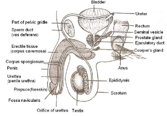
FERTILIZATION IN HUMANS
 
 It is fusion of male and female gametes to form zygote. In human being fertilisation is internal. Human beings are viviparous
 Here the embryo is retained and nourished inside the uterus of the female by means of an attachment called placenta. At one time only a single ovum is released in human females from one of the two ovaries towards the middle of ovarian/ menstrual cycle. It passes into fallopian tubes and rests inside ampulla for some time. The journey time is 12-24 hours
 Human male produces 300-400 millions sperms per ejaculation. They are deposited in vagina during coitus. The process of deposition of sperms in the female genital tract is called insemination. A number of them are demobilized or eat but a number of them remain functional and undergo capacitation ( sperm activation) that provides them the ability to fertilise an ovum.
Capacitation requires 6-10 hrs.
 Capacitation consists of three process
(i) Neutralisation of inhibitory factors present in semen
(ii) Weakening of covering membrane of acrosome head by dissolution of cholesterol
(iii) Entry of Ca2+ into sperms which changes sperm movement from undulation to whiplash motion
 The activated sperm being to pass into uterus and from there to oviducts. Viscous fluid secreted by female genital tract further enhances sperm motility. A number of sperms reach the ampulla part of oviduct where the egg rests temporarily
 Fertilization involves following steps:
(i) Approximation of sperm and ovum
Sperm can remain motile for 24-48 hrs. They swim at the rate of 1.5-3 mm/min. They are able to reach the ampulla part of female genital tract partly by contraction of uterus and fallopian tubes stimulated by prostaglandins (in male semen) and oxytocin (often formed in females).
The movements are powerful within 5 minutes. After reaching an ovum, one sperm comes to lie against it. It releases lysine from its acrosomal region. Hyalouronidase and corona penetrating enzymes as well as dissolves cells of corona radiata. The sperm head now reaches zona pellucida where receptors protein fertilizing helps in attachment to specific protein of sperm. It is compatibility reaction. 
(ii) Acrosome reaction
In contact with zona pellucida, acrosome covering degenerates. The contained enzymes are released. Acrosin or zona lysine dissolves zona pellucida in area of contact.
(iii) Egg reaction
A small protuberance or fertilization cone develops from the surface of ovum in the region of animal pole.
(iv) Penetration of sperm
Sperm head established contact with the lateral surface of fertilization cone. It produces a weak depolarization and ca2+ wave. Plasma membranes of the two, dissolve. Contents of head, neck and middle sperm enter ooplasm. Tail is left outside. Fertilization cone subsides. Cortical granules are extruded. They convert plasma membrane and deactivate sperm receptor of zona pellucida. A perivitelline space is created between it and zona pellucida. This prevents entry of a second sperm.
(v) Activation of ovum
Ovum undergoes meiosis II and extrudes a secondary polar body. It is now the actual ovum or female gametes.
(vi) Fusion of sperm and egg
The envelope of the sperm and egg pronuclei degenerates to form ‘synkaryon’. The act is called karyogamy or syngamy. The proximal centriole brought by sperm helps form the spindle for the division of synkaryon (cleavage nucleus). Fertilized egg is also called zygote. It immediately begins cleavage.
 
SIGNIFICANCE OF FERTILIZATION
 
(i) It restores the diploid number of chromosomes, characteristics of species i.e. 46 in human being
(ii) Fertilization initiates cleavage
 
CLEAVAGE
 
 Cleavage is a series of rapid mitotic divisions of the zygote, characterized by absence of growth of daughter cells, which convert the single celled zygote into a multicellular structure called blastula ( blastocyst)
 Cleavage differ from mitosis in the respect that
(i) There is no growth phase between successive division.
(ii) The size of cells gradually decreases
(iii) The metabolism becomes fast
(iv) There is rapid DNA replication
(v) High consumption of oxygen
 
 Types of cleavage
(i) Holoblastic: When whole of the egg is divided, it is found in microlecithal and mesolecithal egg. It may further be..
(a) Equal – When both the blastomers are equal Example – Amphioxus
(b) Unequal – When the blastomers are unequal in size. Example- frog
(ii) Meroblastic: When a part of the egg is divided. It is found in polylecithal eggs. It may be discoidal (e.g. birds) or superficial ( e.g. insects)
 
 Planes of cleavage include meridional, vertical, equatorial and transverse. Patterns can be radial ( sponges, coelenterates, some echinoderms like star fish) biradial, spiral ( flatworms, annelids, non-cephalopod mollusks) bilateral ( nematods, cephalopods, fishes, amphibians, reptiles, birds) and rotational ( placemental mammals)
 
MORULA
 
Early cleavage produces a solid ball of cells called morula
 
BLASTULA
 Multicellular ball like embryo produced at the end of cleavage and usually having a fluid filled blastocoels, is called blastula
 It is of the following types
(i) Stereoblastula (solid blastula): It is blastula without blastocoels. E.g. Nereis
(ii) Coeloblastula : A blastula with a prominent blastocoels. e.g. frog
(iii) Discoblastula : A blastula having a many layered disc of blastomeres above the yolk. It develops as a result of meroblastic divisions in polylecithal eggs.
E.g. Hen
(iv) Superficial blastula ( Periblastula) : A blastula having a single layer of blastomeres around the central yolk. E.g. insects
 
GASTRULATION
 
 Sum total of all the processes which convert a solid or hollow ball of cells or many layered disc of blastula into two or three germinal layers of gastrula is called gatrulation
 Gastrulation takes place by the migratory or formative or morphogentic movements of blastomers from the surface of blastula to the proper position in the gastrula.
These movements are classified into
(a) Epiboly: Growth of one part over another like prospective ectoderm over the rest except blastocoels.
(b) Emboly: Morphogenetic movements like migration of ectoderm, mesoderm and notochord cells from surface to interior. The emboly may occur by way of
(i) Involution : Rolling of cells into interior
(ii) Invagination : Infolding
(iii) Ingression: New cells migrating into blastocoels
(iv) Delamination : Formation blastocoels is obliterated and a new cavity archenteron is formed which in future alimentary canal of the animal. Blastopore is opening of archenteron.
 
ORGANOGENESIS
 
The development of tissues and organs from the three germ layer is called organogenesis
 
MORPHOGENESIS
 
The assumption of shape, size and other morphological features by embryo is called morphogenesis
 
DIFFERENTIATION
 
It is the formation of different types of cells, which become different in size, form, chemical composition and perform different functions
 
FATE OF THREE GERMINAL LAYERS
 
Ectoderm
 
Central nervous system, nerves, retina, lens, cornea of eyes. Conjunctiva, ciliary and iridial muscles, lining of nasal chambers. Labyrinth, epidermis, cutaneous, glands, hair, nails, claws, hypophsis, adrenal medulla, salivary glands and enamel of teeth
 
Mesoderm
 
Dermis of skin, connective tissue, muscles, notochord, skeleton, blood, heart, blood vessel, adrenal cortex, urino – genitals system except part of urinary bladder, lining of coelom, spleen and eyes.
 
Endoderm
 
Digestive glands, liver, pancreas, middle ear, Eustachian tubes, lining of urinary bladder, respiratory system, adenophypophysis thymus, parathyroid and thyroid glands, lining of vagina and urethra, prostate.
 
FOETAL MEMBRANES
 
They are extra embryonic membranes that provide protection and nourishment to foetus. Foetal membranes are of four types
(i) Chorion – Outer foetal membrane that also takes part in formation of placenta.
(ii) Amnion – Inner foetal membrane that invests the embryo and forms a space called amniotic cavity. It is filled with fluid called amniotic fluid. Amnion protects the foetus from shock
(iii) Allantois – sac – like, develops from gut of embryo, supplies blood vessels to placenta. In reptiles and birds, it helps in respiration nutrition and excertion
(iv) Yolk sac – Membraneous sac attached to embryo near allantois, having yolk in egg laying animals and forms corpuscles in mammals till liver takes over
 
EMBRYO FORMATION IN HUMAN BEINGS
 
MORULA FORMATION
 
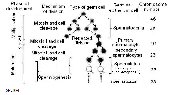
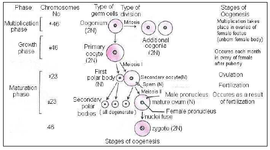
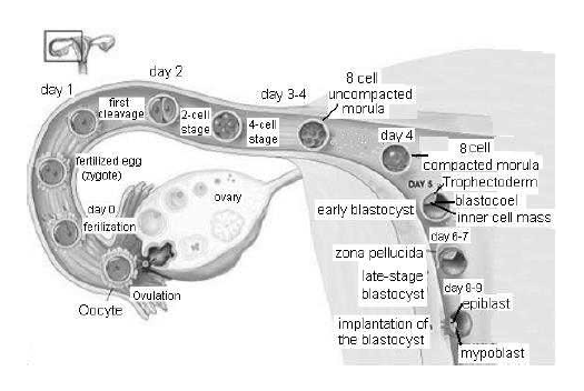
• Soon after fertilization, the zygot begins cleavage or segmentation. Cleavage consists of early mitotic divisions of fertilized egg without involving growth of daughter cells. There is rapid synthesis of new DNA and increased oxygen consumption. Surface –volume and nucleo-cytoplasmic ratio increases. The cells formed after cleavage are called blastomers. Cleavage is simple and holoblastic in humans as there is no yolk.
•  The first cleavage is animal – vegetal axis or primary axis. It is slow and is completed within 30 hours of pertilization
•  One of the two blastomers, is however slightly layer. Hnece, the first cleavage is holoblastic and unequal. Second cleavage is at right angle to the first one. It takes about 30 hours and completed slightly earlier in the larger blastomere so that a transitional 3-celled stage appers. Subsequent divisions are rapid and occur in different planes. They produce a solid ball of blastomers called morula. Phase of compaction ensues in 8-celed stage. Morula has almost the same size as the of fertilized egg due to presence of zona pellucida 
•  Morula has 16-32 cells. Th cells are compacted and of two types, outer slightly smaller peripheral cells with tight junctions than the inner mass of cells with ggap junctions. During the cleavage oung embryo descends in the fallopian tube slowly due to feeble fluid produced by epithelial secretion and cilia. Ultimately it reach the uterus. It takes 4-6 days. Corona radiate dissolved away during this period.
 
BLASTULATION
 
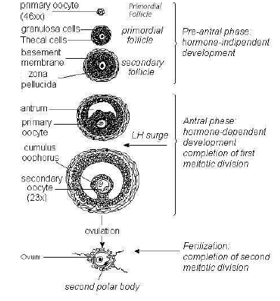
•  In uterus, the endometrial cells become full of nutrients which are also secreted into uterine cavity. As the young embryo reaches uterus, its outer cells begins to absorb nourishment and grow while covered by zona pellucida
•  The outer cells enlarge, flatten and form trophoblast. Trophoblast pours as fluid towards interior producing a cavity called blastocoels or blastocyst. It is equivalent to blastula of othe animals. The size of blastocyst is roughly three times the size of morula.
•  Trophoblast then separates from inner cells except at one point called embryonic pole. The inner cells now occur at one side and called inner cell now occur at one side and called inner cell mass or embryonal knob as the latter is to form the body of embryo. Trophoblast cells in contact with inner mass are called cell of Rauber. Embryonic pole is also called animal pole. The opposite end of blastocyst is called abembryoic pole
•  Blastocyst stage is completed after abour 5 days of fertilization. Trophoblast later becomes two – layered, outer synctiotrophoblast and inner cytophoblast. It secretes hCG ( human chorionic gonadotrophin). Forms villi for implantation and later on produces chorion, amnion and foetal part of placenta 
 
IMPLANTATION
 
• It is a choring or embedding of blastocyst into endomerium of uterus. Implantation begins about 7th day after fertilization of ovum. It takes about process to be completed
 Blastocyst comes in contact with the endometrium in the region of embryonal knob or embryonic disc
 The surfacea cells of trophoblast secrete lytic enzymes which causes corrosion of endometrial linning. They also give rise to figure – like outgrowths called chorionic villi and uterine tissue becomes interdigitated. Villi not only help in fixation but also absorption of nourishment
 The blastocyst sink in the pit formed in endometrium then get completed buried in the endometrium that grows around it. The embedded blastocyst forms villi all around it to obtain nourishment.
 Implantation causes nutrient enrichment, enlargement of cells and vascular endometrium. Vascular endometrium ( deciduas of pregnancy) are stomal cells which have accumulated glycogen and lipid in their distended cytoplasm. The interglandular tissue increases in quantity.
 They may offer nutrition which is engulfed by the syntial trophoblast but they have been regulated as defensive mechanism 
 Trophoblast covering secretes hormone called human chorionic gonadotropin ( hCG). The hormone can be detected in the urine of woman within day after implantation 
 hCG maintain the corpus luteum beyond its normal life. It continues to secrete pregnancy which prevents menstruation and maintains uterine lining in nutrient rich state.
 Progesterone induces cervical glands to secrete viscous mucus for filling the cervical canal to form a protective plug. By the 16th week of pregnancy, placenta produces enough progesterone and the corpus luteum regresse.
 
NEET Biology Human Reproduction and Embryonic Development Chapter Notes-10
 
PLACENTA
 
 Placenta is a temporary organ found only in mammals during gestation period and it is composed of cells derived from two different organisms, the foetus and mother
 Placenta is connection between the foetal membranes and uterine wall.
 
Formation of placenta
 
 The outer surface of the chorion in human develops a number of finger like projections known as chorionic villi, which grow into the tissue of the uterus. These villi, penetrate the tissue of the uterine wall in which they are embedded to make up the organ known as placenta by means of which the developing embryo obtains nutrients, oxygen and gets rid of carbon dioxide and metabolic wastes
 A fully formed human placenta is reddish – brown disc. It foetal surface is smooth and has the umbilical cord. The allantois gives rise to umbilical cord. Umbilical cord has two umbilical arteries (small diameter) and two umbilical veins ( large diameter)
 Umbilical arteries convey oxygen poor blood from the foetus to placenta and umbilical veins carries oxygen rich blood from the placenta to the foetus 
 The blood of foetus in the capillaries of the chorionic villi comes in close contact with the mothers blood in the tissue between the villi, but are always separated by a membrane, through which substances must diffuse
 The maternal and foetal blood are not in direct contact in the placenta because
(i) Two may be incompatible
(ii) The pressure of maternal blood is far too high for the foetal blood vessels
(iii) There must be a check on passage of harmful material into foetal blood
 
Functions of placenta
(i) Nutritive organs – Food materials from the mother’s blood into the foetal blood through the placenta
(ii) Digestive organ – Trophoblast of placenta digests proteins before passing them into the foetal blood.
(iii) Respiratory organ – Oxygen diffuses from the maternal blood into the foetal blood through the placenta. Carbon dioxide diffuses from the foetal blood into the maternal blood also through the placenta for elimination by the mother’ lungs. Foetal haemoglobin has a greater affinity for oxygen than adult haemoglobin
(iv) Excretory organ – Nitrogenous waste such as urea, pass form the foetal blood into the maternal blood via placenta for elimination by mother’s kidney
(v) Endocrine organ – Placenta secretes some hormone such as estrogens, progesterone, human chorionic gonadotropin ( hCG), human placental lactogen ( hPL) chorionic thyrotropin, chorionic corticotrophin and relaxin.
The hCG stimulates and maintains the corpus luteum to secrete progesterone until the end of pregnancy. The hPL stimulates the growth of the mammary glands during pregnancy. Relaxin facilitates parturition by softening the connective tissue of pubic symphysis
(vi) Storage organ – The placenta stores glycogen for the foetus before liver is formed 
 
PARTURITION
 
 It is the process of giving birth to a baby . The physical activities in parturition like uterine and abdominal contractions dilation of cervix and passage of baby are collectively called labour
 Labour is accompanied by localized sensation of discomfort or agony called labour pains.
 Parturation is controlled by complex neuroendocrine mechanism. Signals originate from fully formed foetus and placenta. They cause mild uterine contraction called foetal ejection reflex. It is accompanied by rise in estrogen to progesterone ratio, increase in oxytocin receptors in uterine muscles, increase in level of oxytocin secretion by both mother and foetus and stretching of uterian musculature
(i) Dilation stage
Uterine contractions begin from top. They occur once every 30 minutes. Contractions forces the baby towards cervix. The intervals between successive contraction decreases about every 1-3 minutes. Contractions are accompanied by pain caused by compression of blood vessels and uterine muscles Oxytocin induce contraction and more oxytocin secretion. The strength of uterine contraction continues to increase due to stimulatory reflex. As the baby is pushed down in uterus, its head come to lie against cervix which therefore gets dilated and stretched, A similar dilation also occurs in vagina. The first stage of labour continues for 6-12 hours. It culminates in rupturing of amniotic membrane. The amniotic fluid flows out
(ii) Expulsion stage
The intensity of uterine and abdominal contractions increases. The foetus passes out through cervix and vagina with head in forward direction in normal deliveries. It causes intense labour pain. Expulsion stage takes about 20-60 minutes. Umbilical cord is tied and cut off close to navel
(iii) After birth
Within 10-45 minutes of the delivery of baby the placenta separates from uterus and is expelled out due to series of strong uterine contractions In neonate there is a change in respiratory and circulatory system. The switch over is initiated by gaseous hormone nitric oxide (NO). Lungs expand and infant starts breathing. Blood flow through umbilical cord, formen ovale ceases. It starts passing through heart, aorta and pulmonary arteries 
 
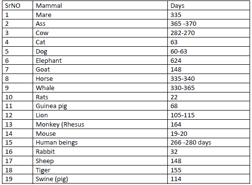
GESTATION PERIOD IN SOME MAMMALS
 
NEET Biology Human Reproduction and Embryonic Development Chapter Notes-11
 
IMPORTANT DEVELOPMENTAL CHNAGES IN HUMAN EMBRYO
1. Week 1
 Fertilization
 Cleavage starts about 24 hours after fertilization
 Cleavage to form a blastocyst after fertilization
• More than 100 cells
 Implantation 6-9 days after fertilization
2. Week 2
 The three primary germ layers develops
3. Week 3
 Woman will not have period. These may be first sign of pregnancy
 Beginnings of backbone
 Neural tube develops, the beginning of brain and spinal cord
4. Week 4
 Heart, blood vessels, blood and gut start forming
 Umbilical cord developing
5. Week 5
 Brain developing
 ‘Limb buds’ a small swelling which are the beginnings of arms and legs
 Heart is a large tube and starts to beat, pumping blood. This be seen on a ultra sound scan
6. Week 6
 Eyes and ears start to form
7. Week 7
 All major internal organ developing
 Face, forming
 Eye have some colour
 Mouth and tongue develop
 Beginnings of hands and feet
 2.5 cm long
8. By week 12
 Foetus fully formed with all organs, muscles, bone, toes and fingers
 Sex organs well developed
 Foetus reaches 7.5cm in height and about 14g weight
9. By week 20
 Hair beginning to grow, including eyebrows and eyelashes
 Fingerprints developed
 Fingernails and toenails growing
 Firm hand grip
 Between 16 and 20 weeks bay usually felt moving for first time
10. Week 24
 Eyelids open
 Foetus measures about 32 cm and weighs about 650gm
11. Week 26
Has a good chance of survival if born prematurely
12. By week 28
• Baby moving vigorously
• Responds to touch and noise
 Swallowing amniotic fluid
13. By week 30
Usually lying head down ready by birth
Foetus is about 43cm long and its weight is about 1800gm
14. By week 40
Birth
Generally child is about 50cm long and weighs about 3300gm
 
LACTATION
 
  Production of milk in the female’s breasts following the birth of a young one in mammals is called lactation
Preparation of breast ( mammogenesis)
During pregnancy, the breast enlarges due to growth of mammary glands Synthesis and secretion from the breast alveoli ( lactogenesis) Secretion and storage of milk begins after birth of the young one, usually within 24 hours under the influence of prolactin
When the estrogen and progesterone are withdrawn following delivery, prolactin begins its milk secretory activity in previously fully developed mammary glands
 
Ejection of milk
 
The actual release of milk called milk letdown, requires the presence of oxytocin, which brings about contraction of smooth muscles of the ducts within the mammary gland. Secretion of prolactin and oxytocin depends on suckling stimuli produced by the nursing infant on the nipples of breasts Maintenance of lactation ( galactopoiesis)
For maintenance of lactation, suckling is important
Milk pressure reduces the rate of production and hence periodic breast feeding is necessary to relieve the pressure which is in turn maintains the secretion.
 After birth, the breast first release is not milk, but colostrums for 2 or 3 days. It is a thin, yellowish, fluid called foremilk which is rich in protein, antibodies but low in fat.
 Human milk consists of water, fat, casein, lactose, mineral salts and vitamins. A nursing woman secretes 1 to 2 litres of milk per day 
 
DEVELOPMENTAL DISPRDERS
 
AMNIONITIS
 
It is inflammation of amnion, usually resulting from premature rupture of amnion
 
ABORTION
 
It is giving birth to an embryo or foetus at the stage of about 20 weeks of gestation
 
TERATOGENY
 
Teratogens are certain agents or drugs that cause abnormal development in developing embryo/foetus. It may cause malformation in developing embryo.
 
ECTOPIC PREGNANCY
 
 The developmental site of foetus is other than the uterus like fallopian tube or cervix
 The growth of foetus may cause tube to rupture and bleed and my lead to miscarriage
 The condition is diagnosed by ultrasound and foetus may be removed by laproscopy before damage is done to fallopian tube.
 
 
Reproduction in Flowering Plants
NEET Botany Reproductive Morphology Chapter Notes

More Study Material

NEET Full Course Human Reproduction Human Reproduction Notes

We hope you liked the above notes for topic Human Reproduction which has been designed as per the latest syllabus for Full Course Human Reproduction released by NEET. Students of Full Course should download and practice the above notes for Full Course Human Reproduction regularly. All revision notes have been designed for Human Reproduction by referring to the most important topics which the students should learn to get better marks in examinations. Studiestoday is the best website for Full Course students to download all latest study material.

Notes for Human Reproduction NEET Full Course Human Reproduction

Our team of expert teachers have referred to the NCERT book for Full Course Human Reproduction to design the Human Reproduction Full Course notes. If you read the concepts and revision notes for one chapter daily, students will get higher marks in Full Course exams this year. Daily revision of Human Reproduction course notes and related study material will help you to have a better understanding of all concepts and also clear all your doubts. You can download all Revision notes for Full Course Human Reproduction also from www.studiestoday.com absolutely free of cost in Pdf format. After reading the notes which have been developed as per the latest books also refer to the NCERT solutions for Full Course Human Reproduction provided by our teachers

Human Reproduction Notes for Human Reproduction NEET Full Course

All revision class notes given above for Full Course Human Reproduction have been developed as per the latest curriculum and books issued for the current academic year. The students of Full Course can rest assured that the best teachers have designed the notes of Human Reproduction so that you are able to revise the entire syllabus if you download and read them carefully. We have also provided a lot of MCQ questions for Full Course Human Reproduction in the notes so that you can learn the concepts and also solve questions relating to the topics. All study material for Full Course Human Reproduction students have been given on studiestoday.

Human Reproduction NEET Full Course Human Reproduction Notes

Regular notes reading helps to build a more comprehensive understanding of Human Reproduction concepts. notes play a crucial role in understanding Human Reproduction in NEET Full Course. Students can download all the notes, worksheets, assignments, and practice papers of the same chapter in Full Course Human Reproduction in Pdf format. You can print them or read them online on your computer or mobile.

Notes for NEET Human Reproduction Full Course Human Reproduction

NEET Full Course Human Reproduction latest books have been used for writing the above notes. If you have exams then you should revise all concepts relating to Human Reproduction by taking out a print and keeping them with you. We have also provided a lot of Worksheets for Full Course Human Reproduction which you can use to further make yourself stronger in Human Reproduction

Where can I download latest NEET Full Course Human Reproduction Human Reproduction notes

You can download notes for Full Course Human Reproduction Human Reproduction for latest academic session from StudiesToday.com

Can I download the Notes for Human Reproduction Full Course Human Reproduction in Pdf format

Yes, you can click on the link above and download notes PDFs for Full Course Human Reproduction Human Reproduction which you can use for daily revision

Are the revision notes available for Human Reproduction Full Course Human Reproduction for the latest NEET academic session

Yes, the notes issued for Full Course Human Reproduction Human Reproduction have been made available here for latest NEET session

How can I download the Human Reproduction Full Course Human Reproduction Notes pdf

You can easily access the link above and download the Full Course Notes for Human Reproduction Human Reproduction for each topic in Pdf

Is there any charge for the Full Course Human Reproduction Human Reproduction notes

There is no charge for the notes for NEET Full Course Human Reproduction Human Reproduction, you can download everything free of charge

Which is the best online platform to find notes for Human Reproduction Full Course Human Reproduction

www.studiestoday.com is the best website from which you can download latest notes for Human Reproduction Human Reproduction Full Course

Where can I find topic-wise notes for Full Course Human Reproduction Human Reproduction

Come to StudiesToday.com to get best quality topic wise notes for Full Course Human Reproduction Human Reproduction

Can I get latest Human Reproduction Full Course Human Reproduction revision notes as per NEET syllabus

We have provided all notes for each topic of Full Course Human Reproduction Human Reproduction as per latest NEET syllabus