
NEET Biology Animal Tissue Chapter Notes

Revision Notes for Full Course Animal Tissues: Animal Tissues

Review targeted revision notes for Full Course Animal Tissues with the NEET Biology Animal Tissue Chapter Notes. Built according to official educational guidelines for the 2026-27 academic year, these downloadable summaries for Animal Tissues support daily study and last-minute exam readiness.

Review Animal Tissues for Full Course Animal Tissues

Access the complete concept summary PDF for Animal Tissues below. Regular review of these targeted notes builds familiarity with complex Full Course Animal Tissues themes and helps secure higher marks in final school evaluations.
INTRODUCTION
 
Tissue : A organised group of cells similar in structure, function and origin.
In a tissue cells my be dissimilar in structure and function but they are always similar in origin.
 –Word animal tissue was coined by – Bichat
– N. Grew coined the term for Plant Anatomy.
 – Study of tissue – Histology
– Histology word was given by – Mayer
 – Father of Histology – Bichat 
 – Study of tissue is also called Microscopic anatomy.
– Founder of microscopic anatomy – Marcello Malpighi
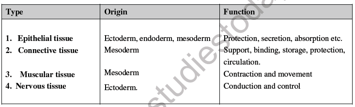
Based on functions & location tissues are classified into four types :
NEET Biology Animal Tissue Chapter Notes
 
EPITHELIAL TISSUE
 
Word epithelium is composed of two words Epi – Upon
Thelio – grows
– A tissue which grows upon another tissue is called Epithelium.
– Cover external or internal surface.
– Cells are either single layered or multilayered.
– Cells are compactly arranged and there is no intercellular matrix.
– Cells of lowermost layer always rest on a non living basement membrane.
– Cells are capable of division and regeneration throughout the life.
– Free surface of the cells may have fine hair cilia or microvilli or may be smooth.
– Epithelial tissue is non-vascularised.
Due to absence/less of intercellular spaces blood vessels, lymph vessels are unable to pierce this tissue so blood circulation is absent in epithelium. Hence cells depend for their nutrients on underlying connective tissue. 
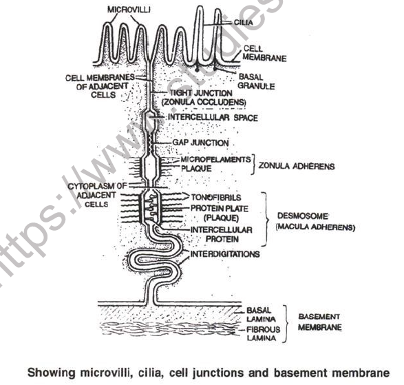
Between epithelium & connective tissue, a thin non living acellular basement membrane is present which is highly permeable.
 
Basement membrane consist of 2 layers.
 
(a) Basal lamina : made up of glycoprotein, and secreted by epithelium cells.
(b) Fibrous lamina : Formed of collagen and reticular fibres suspended in mucopoly-saccharide which is matrix of connective tissue.
– So basement membrance is secreted by both epithelium and connective tissue.
 Mucopolysaccharide is present in the form of Hyaluronic acid which is composed of 2 components–N acetylglucosamine & glucuronic acid. Both these components are found in alternate form.
– NAG – GA – NAG –
 
– Specialized junctions between epithelial cells : -
 
 – To provide mechanical support for the tissue plasma membrane of adjacent epithelial cells modified to form following structures called as Intercellular Junctions.
 Tight junctions (Zonula occludens) : help to prevent substances from leaking across the tissue.
Plasma membranes in the apical parts become tightly packed together or are even fused.
 
 Interdigitations : These are interfitting, finger like processes of the cell membranes of the adjacent cells.
 
 Intercellular Bridges : These are minute projections that arise from adjacent cell membrances.
They make contact with one anther.
 
NEET Biology Animal Tissue Chapter Notes1
 
→Gap Junctions : Facilitate the cells to communicate with each other by connecting the cytoplasm of adjoining cells, for rapid transfer of ions, small molecules and sometimes big molecules.
 Intermediate Junctions (= Zonula adherens) : These usually occur just below tight junctions.
 There is no intercellular filaments between the adjacent cell membranes.
 They probably serve anchoring functions.
 →Desmosomes ( =Macula adherens) : Perform cementing to keep the neighbouring cells together.
These are like zonula adherens but are thicker and stronger and are disc like junctions. They have intercellular protein. The microfilaments which extend from Plasma membrane are called
tonofibrils.
 Hemidesmosomes (single sided desmosomes) are similar to desmosomes, but the thickening of cell membrane is seen only on one side.
Hemidesmosomes join epithelial cells to basal lamina (outer layer of basement membrane).
 
– Specialised functional structures shown by Epthelial Cells : -
 
Plasma membrane of free end get modified to form 3 types of functional structures.
Microvilli .
 – Minute protoplasmic process which are non motile, non contractile.
– Help in absorption, secretion, excretion
 –Increase surface more than 20 times.
 Present in the wall of Intestine, Gall bladder, Proximal convoluted tubule etc.
 
Cillia or Kinocilia
 – Long cylindrical protoplasmic process.
 – Motile and contractile
 –Movement of cilia is always in uniform direction.
 – Originated from basal granule or kinetosome.
– Diameter of cilia is same from base to apex.
 – In internal structure of cilia 9 + 2 arrangement of microtubules is present. 
– They helps in conduction
 e.g. – Fallopian tube.
– Trachea.
– Fallopian tube
– Uterus.
– Ependymal epithelium : (Inner lining of ventricles of brain & central canal of spinal cord. Function of cilia is to conduct substances in CSF.)
 
Steriocilia
– Long cytoplasmic process
– Non motile, non contractile
– Basal granule is absent
– Plasma membrane is thick & rigid.
– Base of stereocilia is broad & apical part is narrow so they are conical in shape.
– They increase surface area .
eg. Epididymis
Vasadeferens
 

Revision Notes and Key Concepts for Full Course Animal Tissues Animal Tissues

Key Concepts and Summary for Full Course Animal Tissues Animal Tissues

Review targeted chapter notes for Full Course Animal Tissues Animal Tissues. Built according to official NEET guidelines, these summaries highlight high-yield topics frequently tested in school evaluations.

NCERT-Aligned Chapter Summary and Notes

Each chapter summary is structured around standard NEET textbooks, allowing students to check their understanding and clarify complex ideas early in their revision.

Next Steps in Your Exam Preparation

Explore our broader library of printable assignments, chapter notes, and mock tests designed to support continuous revision and secure higher marks in NEET assessments.

FAQs

Where can I download the latest PDF for NEET Biology Animal Tissue Chapter Notes?

You can download the teacher prepared revision notes for NEET Biology Animal Tissue Chapter Notes from StudiesToday.com. These notes are designed as per 2026-27 academic session to help Full Course students get the best study material for Animal Tissues.

Are these Animal Tissues notes for Full Course based on the 2026 board exam pattern?

Yes, our NEET Biology Animal Tissue Chapter Notes include 50% competency-based questions with focus on core logic, keyword definitions, and the practical application of Animal Tissues principles which is important for getting more marks in 2026 NEET exams.

Do these Full Course notes cover all topic-wise concepts for Animal Tissues?

Yes, our NEET Biology Animal Tissue Chapter Notes provide a detailed, topic wise breakdown of the chapter. Fundamental definitions, complex numerical formulas and all topics of NEET syllabus in Full Course is covered.

How can I use NEET Biology Animal Tissue Chapter Notes for quick last-minute revision?

These notes for Animal Tissues are organized into bullet points and easy-to-read charts. By using NEET Biology Animal Tissue Chapter Notes, Full Course students fast revise formulas, key definitions before the exams.

Is there any registration required to download Full Course Animal Tissues notes?

No, all study resources on StudiesToday, including NEET Biology Animal Tissue Chapter Notes, are available for immediate free download. Full Course Animal Tissues study material is available in PDF and can be downloaded on mobile.