1.1. INTRODUCTION
The cell biology or cytology is a branch of science which deals with the study of cells from morphological, physiological, anatomical, developmental, genetically and evolutionary points of view. Cytology is relatively a young science and has gained individual recognition only by the end of 19th century; 19th century has been referred to as the classical era of cytology as most of the discoveries in this field of science were made during this period. Cells can be called the building blocks of life. Just like the bricks are units of a wall, cells are units of living organisms.
Bricks are non-living things, identical in size and shape, whereas cells are active; vary enormously in shape, size and function. All living organisms are made up of cells; diverse forms exhibited by various organisms have arisen from specializations during the course of evolution.
In short, cells are defined as structure and functional units of living organisms.
1.2. HISTORY OF CELLS
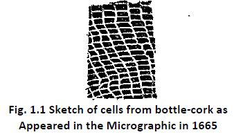
The history of cell began with publication by Robert Hooke in 1665. In 1665, Hooke observed a honey comb like pattern in a slice of cork under his primitive microscope (incidentally he designed one of the earliest optical microscopes) and observed that the cork was composed of box like compartments which he called ‘cells’. Although he coined the term cell, he did not realize their real significance. He considered those cork cells as structures forming passages for conducting fluids. We now know that what he observed were the cell walls enclosing the vacant spaces left by dead cork cells (Refer Flg. 1.1).
Before Hooke, several scientists like Marcello Malpighi (1628-1694) and Nehmiah Grew made attempts to study plants and reported that certain parts of plants were made of “utricles”.
“Sacs” and “vesicles”. They did not use the term cell. Malpighi studied a variety of animal tissues too and generally he is considered to be the “Father microscopic anatomy”. A.V.
Leeuwenhoek (1632 – 1723) discovered animalcules, certain protozoa, spermatozoa, RBC, muscles, nerves, skin, teeth and certain plants.
Later, M.J. Schleiden (1804 – 1881) and Theodor Schwann (1810 – 1882) formulated the cell theory.
1.3. (A) THE CELL THEORY
The cell theory may be summed up as follows:
♦ All living thins are composed of cells and their products.
♦ All cells arise from pre-existing cells. This observation was summarized by Rudolf Virchow in 1885. Virchow stated that where a cell exists, there must have been a pre-existing cell just as the animal arises only a from on animal and plant from only a plant.
♦ All cells are basically alike in chemical composition and metabolic activities and metabolic processes are carried out within the cells.
♦ Growth of organisms occurs by cell growth and cell multiplication.
♦ Each cell can act independently but they function as an integral part of a complete organism.
♦ The cells contain genetic material which is passed on from one generation to the next.
Exceptions to cell theory: Various cytological investigations hove shown that all living organisms are not cellular as stated in cell theory. Some are without true cell. Cell is defined as a mass of protoplasm having a distinct nucleus and cytoplasm. Bacteria and blue green algae have no true cellular organisation. They are called Prokaryotes. These lack cellular organelles and the genetic material s not limiter by nuclear membrane. Cells all higher plants and animals exhibit cellular organelles and their genetic material is enclosed within nuclear membrane. Such cells are called Eukaryotes. Certain fungi like Rhizopus have a body made of an unlimited mass of protoplasm in which many nuclei remain scattered called coenocytic condition. Viruses have neither protoplasm nor nucleus, They have only DNA/RNA as their genetic material. These
examples cannot be explained on the basis of cell theory.
(1.3(b) TOOLS AND TECHNIQUES FOR STUDY OF CELL
The cells of most animal and plants are too small to be seen by the naked eye. Their size ranges from 1 micrometer (μm) to 1 millimeter (mm). The human eye cannot resolve objects as separate entitles if they are closer together than 0.1 mm or 100 μ m, besides, the living cells are transparent in ordinary light. Hence, it becomes difficult to discriminate among various cellular components. To overcome these practical difficulties, early cytologists investigated various methods such as aide preparation, that included steps like cuffing tissues into thin sections, staining, mounting, magnifying and so on. A few of them we described below:
1. Simple Microscope: It is made up at a simple lens with low magnification.
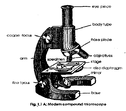
2. Compound Microscope: Made up of 3 lens systems – condenser, objective and ocular lens.
The image is observed directly in a microscope by looking into the eyepiece. Coarse and fine focus control permit the up and down movement of the body tube. This facilitates precise focus. The condenser lens system which occurs beneath the specimen helps to collect and focus the light rays on the object or specimen which is placed on the stage of the microscope. The objective lens system remains near and above the specimen. It product and magnifies the image of the specimen. The eyepiece lens system which remain near the eyes of the observer magnifies and forms the image (secondary) or the (primary) mage previously produced by the objective. Thus there are two images. The first image is formed by the objective system which enlarged by the
eye piece system. This second image is what we see. Light is transmitted through objects. Their visibility is usually enhanced by staining.
Study of plant and animal cells under the microscope
To study the plant cells, the epidermal layer from the lower surface of the leaf of Rhoeo sp. or from the inner layer of onion bulb is removed with the help of a forceps. The tissue which is one cell thick is placed on a drop of water and stained with safranin and spread flat on a glass slide. The cells are observed under the low and high power of compound microscope to study the size, shape and structure of the cells. Similarly a very thin slice of bottle cork is obtained a using a singe edged razor blade. The cells are observed after mounting the slice n a drop of water on a glass (similar observation on cork cells was done by Robert hook in 1665)
To study the animal cells, epithelial cell from the inner side of the cheek is obtained using a sterile tooth pick or clean spatula. The cell scrapings are mounted on a glass slide and stained with methylene blue and covered with a slip. The mounted specimen is observed under the microscope (both under low and high power). Similarly blood cells of human being or frog is made into a smear using two glass slides and the blood smear is stained with Leishman’s stain, to study the nucleus and their details of the cells.
The study/observation of cells under the microscope reveal that plant cells have a prominent cell wall and inner portion of the cells appear transparent. The cells are compactly arranged without any intercellular spaces between them. The tissue tom the leaf of Rhoeo appear purple in colour and show presence of stomata. Observation of blood cells reveal that they are ovoid and cheek cells are cuboidal with prominent nuclei.
3. Electron Microscope: In the last few decodes our entire concept of the cell has been revolutionized by the development of the electron microscope (Figure 1.1B).
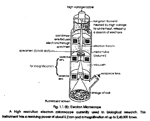
A high resolution electron microscope currently used in blological research. This Instrument has a resolving power of about 0.2 mm and a magnification of up to 2, 40,000 times.
This instrument uses an electron beam instead of light and electro-magnets instead of glass lenses. The electron beam has a much shorter wavelength than light, with result that a modem electron microscope has a resolving power thousand times greater than the optical microscope.This means that objects can be magnified much more without loss of clarity A good optical microscope can only magnify on object effectively about 1.500 times. The electron microscope can give clear pictures that are magnified as high as 1,00,000 times. It is important to appreciate what this means in practice: with the electron microscope an object the size of a pinhead can be enlarged to the point at which it has a diameter of well over a kilometer: a cell with a diameter of 10 micrometers finishes up with a diameter of five metres. It is impossible to exaggerate the impact which this instrument has had on biology. Materials which were formerly described as structure less have been shown to have an elaborate internal organization, and so-called homogeneous fluids are now known to contain a variety of complex structures. The electron microscope has opened up a whole new world o structure whose existence was barely realized forty years ago.
Stains: Cells are fixed and stained before studying it for better differentiation and observation of its various parts. The more commonly used stains are – eosin, congo red, picric acid, safranin, methylene blue, methyl green, crystal violet etc.Some stains such as Janus green B and methylene blue are used to study various parts in living cells. Such stains are called vita stains.