Significance of cell reproduction
• All cells are formed by division of pre-existing cells
• Each new individual begins its life as a new single cell commonly the fertilized egg or zygote
• A large number of cells are being torn or killed every moment in the body of a multicellular organism through skin peelings, lining of digestive tract, old red blood corpuscles, e.g. 25 million/sec in an adult human body. They are being continuously replaced through formation of new cells.
• An injury is healed through formation of new cells by healthy cells around the area of injury.
• It is mode of multiplication in unicellular organisms. In multicellular organisms, cell reproduction is required to form propagules and gametes.
• The mechanism of cell reproduction or cell division is fundamentally similar in all the organisms showing kinship and unity of life
Factor controlling cell reproduction
• A number of factors are known to induce cell division. The important ones are as follows:
- Minimum growth: A newly formed cell does not divide immediately. Some amount of minimum growth in cell and its component is required before a cell attains the ability to undergo division.
- Surface –Volume ratio : Increase in cell size results in decrease of surfacevolume ratio. This disturbs efficiency of surface exchange required for maintaining optimum metabolism. As it reaches a critical stage the cell undergoes division.
- Nucleocytoplasmic or kernplasma ratio: Cell functions are controlled by nucleus. The size of nucleus does not change while that of cytoplasm increases during cell growth. As nucleocytoplasmic artio decreases, the cell is stimulated to divide
- Mitogens: they are substances or factors which bring about cell division. Cytokinin is a plant hormone which functions as mitogen. There are several mitogenic substances known in human beings. E.g. EGF ( Epidermal growth Factor), PDGF ( platelet derived growth factor) and lymphokines.
- There are some agents which inhibit cell division. They are called mitotic poisons. Example, azides, cyanides, chalones, colchicines. Colchicines is obtained from atucrocus (Colchicum autumnale) . It arrests cell division at metaphase due to non-formation of spindle.
CELL CYCLE
• Cell cycle ( Howard and Pelc, 1953) is genetically controlled series of changes that occur in a newly formed cell by which it supplicates ins contents, undergoes, growth and division to form two daughter . it consists of two states or periods, a long nondividing growth I-phase and a short dividing Mphase. Both have substages. I-phase represents interphase.
• The regular sequence of G1, S, G2 ( interphase) and M phase ( mitotic phase) is called the cell cycle.
• Interphase is called ‘resting stage”, but it is in fact a period of great activity.
Three important process, which are preparatory to cell division, take place during interphase. Thus it is also known as preparatory phase. These processes are
- Replication of DNA along with the synthesis of nuclear proteins such as the histones
- In animal cells, duplication of centriole takes place by the outgrowth of daughter centrioles from the parent centrioles, which are at right angle to each other.
- Synthesis of embryo rich compounds, which provide energy for mitosis, and synthesis of proteins at the end of interphase
• Interphase ( L.inter-in between, Gk-phase-stage) is intermitotic stage of cell division in which a series of changes occur in newly formed cell and nucleus undergoes certain changes to be fit for division. Non dividing state of mature cell or nucleus is called interphase. It is also called energy phase. Interphase of dividing cell has been classified into three subphases - G1- Phase, S-phase and G2-phase
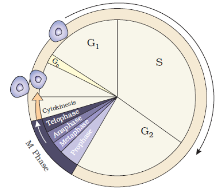
G1-phase
♦ G1 phase is also known as first growth phase or post mitotic gap phase. It is the longest phase of cell division. In this phase different types of RNA (mRNA, tRNA, rRNA) and proteins are synthesized.
♦ All cell organelles ( ER, mitochondria, Golgi complex, ribosomes, plasmid in plant cell) multiply. The duration of G1 Phase varies from cell to cell. It is shorter in frequently dividing cells. G1 phase cell has three options. A) Continuous cycle and enter S phase b) Stops cell cycle and enter quiescent phase or g0 phase c) Stops cell cycle and undergoes cell differentiation
♦ The deciding factor for above option are availability of mitogen and energy rich compounds. This point is called check point . Once the check point of G1 – phase is crossed, cell reaches a state called ante phase where by it will divide even under unfavorable condition. Cell cycle will go on further division till completion
S-phase
♦ S-phase is known as synthetic phase. In this stage replication of DNA takes place by the synthesis of histones. As a result each chromosome under goes replication producing two chromatids. Each chromosome carries a duplicate set of genes. A haploid cell (n) becomes diploid (2n) and a diploid cell (2n), thus becomes tetraploid (4n) at the end of S-phase. Repairing of damaged DNA also takes place.
G2-phase
♦ G2 is also called second growth phase or pre-mitotic gap phase. In this phase synthesis of DNA stops and synthesis of RNAs and proteins continues. All cell organelles multiply and spindle formation takes place. It lasts for 2-5 hours in most cells. Some proteins formed in this phase cause condensation of chromosomes to initiate mitosis
G0-phase
♦ The phase in which cells fail to divide further ( do not undergo S-phase after G1-phase) and undergo differentiation is known as G0 phase or quiescent stage. It occurs due to non-availability of mitogen and energy rich compounds. The cells remain metabolically active, grow in size and differentiate for particular function after attaining a particular shape.
♦ However some cells remain in undifferentiated state as reserve cells. They may proceed with cell division when required e.g. fibroblasts; it helps in healing of wounds and grow and divide again.
M-phase
♦ The process of cell division is found to be essentially the same in all living organism and the events are chiefly centered in the nucleus. Three type of cell divisions have been distinguished:
- Amitosis or direct cell division
- Mitosis or indirect cell division
♦ Mitosis and meiosis are the two major types of cell division. The basic stage in both the types of divisions are almost identical.
♦ Amitosis is a direct division characterized by the splitting of nucleus followed by that of cytoplasm.
♦ Mitosis is a somatic cell division which takes place in vegetative cells. It maintains the chromosome number.
♦ Meiosis is a reduction division, occurring in the reproductive cells. The chromosome number are reduced to half.
AMITOSIS
♦ Amitosis ( greek, a-without, mitos – thread, osis-state). It is a method of direct cell division in which the nucleus constricts into two daughter without showing differentiation of chromosomes and development of spindle. Nuclear division is followed by cytokinesis ( division of cytoplasm)
♦ Amitosis was first described by Robert Remak (1855 ) in red blood corpuscles of chick embryo. The term was coined by Flemming ( 1882 )
Occurrence
♦ It occurs through cleavage or constriction example cartilage cell degenerate cells meganucleus of Paramecium, cells of foetal membranes of vertebrates
♦ Moneran cell division is sometimes included under amitosis due to absence of spindle.
♦ Drawbacks
As Amitosis does not distribute chromatin equitably, it results in structural and functional abnormalities in the cell.
MITOSIS
• Mitosis is a type of cell division in which chromosomes of parent cells are duplicated ( by replication of DNA) and equally distributed ( quantitatively and qualitatively) into two daughter nuclei. Term mitosis is derived from Greek word “Mitos” means thread or fibril
• Mitosis was first observed by Strassburger in plant cells (1870) and Boveri and Flemming in animal cell ( 1879 ). The term was coined by Flemming in 1882. It is also known as equational division due to equal distribution of chromosomes in daughter nuclei. It is often known as somatic cell division due to occurrence in somatic cells. It is about 1-5% of the total duration of cell cycle. On the basis of different types of cells and the species, mitosis takes 30 minutes to 3 hours for completion.
• In plants all meristematic regions are the sites of mitosis e.g. root ape, shoot apex, intercalary meristem, lateral meristem, leave, flowers, fruits, embryo, seeds etc.
• In animals embryo, skin, bone marrow etc are the sites of mitosis
• Mitosis is completed in two steps karyokinesis and cytokinesis.
Karyokinesis
• Mitosis starts with the nuclear division of parent cell known as karyokinesis ( Gk, karyon -nucleus, kinesis – movement) . The four phases of karyokinesis are prophase, metaphase, anaphase and telophase
Prophase
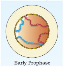
Prophase ( Gk Pro-first, phase –stage ) is often divided into three substages – early prophase, mid prophase and late prophase. It is the first stage of mitosis proper. It is the longest phase of Karyokinesis
Early prophase
• In this sub-stage nucleus and cell become spheroid and nucleus appears as boll of wool. Chromatin fibre condense to form elongated chromosome and this increases viscosity and refractivity of cytoplasm
• In animal cells duplicated centrioles. ( S phase of interphase) start to move towards opposite poles of the cell. Each centriole radiates out fine microtubular fibrils called astral rays. In animal cells and cells of lower plants, fibrils appears like spokes of a wheel around each centriole to form an aster.
Mid prophase
• In mid prophase chromosomes shift towards the periphery and leave a clear central area. It becomes shorter and thicker. Each chromosome consists of two longitudinal threads called chromatids. Both chromatids are attached to each other by centromere and are known as sister chromatids
Late prophase
• In this substage spindle fibres start appearing around the nucleus. The size of chromosomes is much reduced as compared to early prophase . Spindle poles are formed without asters in plant cells and with asters in animal cells.
• Nucleolus and other cell organelles ( like mitochondria, Golgi complex, ER, vacuoles etc) disappear. The presence of the spindle is essential for mitosis. If cells are treated with colchicines, which inhibits spindle formation, anaphasic movement of the two groups of chromosomes to the poles does not take place.
Prometaphase
• Prometaphase ( Gk Pro- before, meta-second, phase – stage) is intermediate stage of prophase and metaphase and hence acts as connecting link between them. Nuclear membrane completely degenerate in this stage. So mixing of cytoplasm with nucleoplasm occurs. It is known as extranuclear mitosis or eumitosis.
• In many protozoan, fungi and some animal cells, nuclear membrane does not degenerate throughout cell division known as intranuclear or premitosis.
• A spindle fibres consists of 4-20 microtubules formed of protein tubulin. Spindle fibre converge at two end or pole. It has the maximum diameter in the middle known as equator.
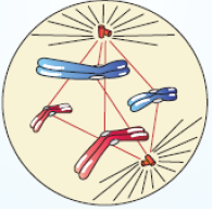
Metaphase
• In metaphase ( Gk meta-after or second phase – stage) discontinuous fibres radiate out from two poles and get connected to the disc shaped structure at the surface of the cenromere called kinetochores. A kinetochore is complex protein structure that is analogus to ring for the microtubule hook; it is the point where microtubules attach themselves to the chromosome. Chromosomes or kinetochore fibres contract and bring chromosome over equator this phenomenon is called congression.
• Smaller chromosomes directed towards the centre while larger ones are peripheral in position on equator. The centromeres of all the chromosomes lie on the equator forming an apparent plate called metaphasic or equatorial plate while arms are directed towards the poles
• The kinetochores have two functions. The main function apparently is that they serve for the attachment of microtubules of the chromosomal spindle fibres. They might also be involved in the formation of the chromosomal spindle fibres during prometaphase and metaphase by serving as centres for polymerization of the protein of microtubules.
• Metaphase is the best phase to count total number of chromosomes in any species and details study of morphology of chromosomes. Idiogram (arrangement of chromosomes in a series of decreasing length) can be drawn in this stage.
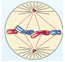
Anaphase
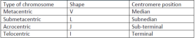
• In anaphase ( Gk ana – up, phase – stage) chromosomes are arranged on the equatorial plate for a short period. The centromeres of chromosomes starts to divide into two, forming daughter chromosomes with centromere in each. Daughter chromosomes are repulsive so, migrate towards opposite poles. Spindle fibres attached to the centromeres shorten and pull the chromosomes to the poles. The velocity of anaphasic movement does not depend on the size of the chromosomes. In anaphasic movement of chromosomes, the centromeres lead the path while the limbs trails behind. So anaphasic chromes, the centromeres lead the path while the limbs trail behind. So anaphasic chromosomes appear as V-, L-, J- and I- shaped
• At the end of anaphase two groups of chromosomes are formed, one at each pole. The number and types of chromosomes at each pole is the same as in the parent nucleus.
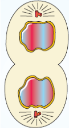
Telophase
• During telophase ( Gk. Telos-end, phase –stage) of mitosis viscosity of cytoplasm decrease. A new nuclear membrane is formed ( either from older nuclear envelop or ER) around each set of chromosomes. Chromosomes overlap one another forming chromatin. The nuclear organizer region of satellite chromosomes produce nucleolus for each daughter nucleus. Nucleoplasm surrounds in the area of chromatin. The gel state of spindle is converted into sol state and disappears.
• In this way two daughter nuclei are formed at the poles of spindle. Hence this phase is just reverse of prophase. Golgi complex and endoplasmic reticulum are reformed. Cytokinesis starts either by cleavage or constriction.
Cytokinesis
• Mitosis ends with division of cytoplasm known as cytokinesis. It is derived from greek word “cytos” means hollow or cell, “kinesis: means movement. It starts towards the middle of anaphase and is completed with the telophase. It is different in animal and plants. If nuclear divison takes place without cytoplasmic division, a syncytium is formed.
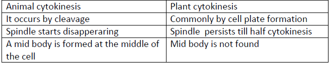
Animal cytokinesis
• The spindle gets charged into dense fibrous and vesicular structure on equator called mid body. In the middle region of cell, microfilaments starts to collected which induces the cell membrane to invigilate. The furrow forms and deepens centripetally and finally cleaves the parent cell into two daughter cells. This method of cytokinesis is known as cleavage method. All cell organelles ( mitochondria, Golgi complex, lysosomes., ER, ribosomes etc) are also distributed between two daugheter cells.
Plant cytokinesis
• It differs from animal cytokinesis due to presence of rigid cell wall. In lower plants cytokinesis occurs by cleavages method ( like animal cell) and in higher plants it takes place by cell plate method.
• In this method small vesicles of Golgi complex are collected at the equator. Here spindle persists for some time called phragmoplast. All vesicles fuse to form two sheets whih enclose a matrix or film. This film becomes solidified to form cell plate or middle lamella. It grows centrifugally and finally phragmoplast disappear. Cellulose, hemicelluloses and pectin are deposited on either side of cells plate. It forms primary wall.
Difference between animal and plant cytokinesis
Significance of mitosis
• Growth and development - A single cell zygote grows into full blown baby ( 6 ×10²² cells) by repeated mitosis. Plants are able to grow throughout their life due to mitotioc division in their apical and lateral meristems. Increases in tissue mass, results from increase in cell number called hyperplasmic. Hnece, mitosis is essential for growth and development of a multicellular organism.
• Maintenance of cell size: An overgrown somatic cell is induced to divide so that mitosis helps in maintaining a proper surface volume ratio. It has also a high nucleocytoplasmic ratio which is brought back to efficient level through divisions. These ratios are important for proper functioning of cell
• Genetic stability – All the daughter cells of a multicellular organism have the same number and type of chromosome as parent cells due to equitable distribution of all the chromosomes. This helps in proper co-ordination among daughter cells.
• Healing and regeneration – For healing of wounds new cells are produced by mitosis. Some organisms are able to regenerate missing part of body or also whole organism though mitosis
• Reparing – the mechanism for replacing old or worn out cells is called repairing. In human body roughly 5 ×10⁹ cells are lost from skin surface, lining of alimentary canal, blood cell etc. these are replaced by new cells formed through mitosis.
MEIOSIS
• Meiosis is a process of reductional division in which the number of chromosomes per cell is cut in half. In animals, meiosis always results in the formation of gametes, while in other organism it can give rise to spores. The word “meiosis” comes from greek world meioun, means “to make small”, since it results in a reduction of the chromosome number.
• The term meiosis was coined by Farmer and Moore (in 1905). The division was first studied by Van Benedin (1887), Strassburger (1888), Sutton (1900) and Winiwater (1900). Meiosis I &II were differentiated by Gregoire. In 1911 the American geneticist Thomas Hunt morgan ( 1866 – 1945) observed cross-over in Drosophila melanogaster meiosis and provided the first genetic evidence that genes are transmitted on chromosomes
• Meiosis is essential for sexual reproduction and therefore occurs in all eukaryotes ( including single –celled organisms) that reproduce sexually. Meiosis does not occur in archaea or bacteria, which reproduce via asexual process such as binary fission.
• Evidence of basic relationship – The mechanism of mitosis are similar in the majority of organism, showing basic similarity and relationship among them
• During meiosis, the genome of diploid germ cell, which is composed of long segments of DNA packed into chromosomes, undergoes two rounds of division, resulting in four haploid cells. Each of these cells contain one complete set of chromosomes, or half of the genetic content of the original cell. If meiosis produces gametes, these cells must fuse during fertilization to create a new diploid cell, or zygote before any new growth can occur. Thus the division mechanism of meiosis is a reciprocal process to the joining of two genomes that occurs at fertilization. Because the chromosomes of each parent undergoes enetic recombination during meiosis, each gamete and thus each zygote, will have a unique genetic blue print encoded in its DNA.
Together meiosis and fertilization constitutes sexually in the eukaryotes, and generate genetically distinct individuals in population.
• In lower plants, and in many protists, meiosis results in formation of haploid cells that can divide vegetatively without undergoing fertilization, referred to as spores. In these groups, gametes are produced by mitosis Biochemically, meiosis uses some of the same mechanism employed during mitosis to accomplish the redistribution of chromosomes. There are several features unique to meiosis, most importantly the pairing and recombination between homologous chromosomes, which enable then to separate from each other.
• The cells of a particular species have a constant number of chromosomes. In sexually reproducing organisms male and female gametes fuse together to form the zygote. If the gamete has the same number of chromosomes number remains constant from generation to generation. This is because of meiotic division which reduces the chromosome number to half, and counteracts the effect of fertilization. Thus fertilization and meiosis are compensating events.
Types of meiosis
• The cells in which meiosis takes place are called meiocytes. In animals, meiocytes are of two types, spermatocytes and oocytes. In higher plants, meiocytes are differentiated into microsporocytes and megasporocytes. Depending upon the stage when meiosis occurs, the latter is of three types - gametic, zygotic and sporic meiosis.
Gametic meiosis
• Meiosis is most of the animal take place during the formation of gametes (gametogenesis). It is termed as genetic meiosis. When two gametes fuse in fertilization, a diploid zygote is formed. Gametic meiosis results in diplontic life cycle
Zygotic meiosis
• In some lower plants meiosis takes place in the zygote and the resulting organism are haploid. It is called zygotic meiosis. Organism having zygotic meiosis have haplontic life cycle.
Sporic meiosis
• In plants, meiosis generally occurs at the time of sporogenesis ( formation of spore or microspores and megaspores) It is called sporic meiosis or intermediate meiosi. Spores produce a new gametophytic phase in the life cycle. Gametes are formed by gametophytes. Because of the presence of two distinct multicelluar phase, diploid and haploid, life cycle of plant is diplohaplontic .
Phases of meiosis
• Because of meiosis is “a one-way” process, it cannot be said to engage in a cell cycle as mitosis does. However, the preparatory steps that lead up to meiosis are identical in pattern and name of the interphase of the mitotic cell cycle.
• Meiosis is a type of cell division that is vital for sexual reproduction. Meiosis takes place in the reproductive organs. It results in the formation of gametes with half the normal chromosomes number. Therefore, haploid sperms are made in the testis and haploid eggs are made in the ovaries. In flowering plants, haploid gametes are made in the anthers and ovules.
• Meiosis involves two divisions of the cell. These two division are termed meiosis I nad meiosis II. Each one includes prophase, metaphase, anaphase and telophase.
• Meiosis I consists of separating the pairs of homologous chromosomes, each made up of two sister chromatids, into two cells. One entire haploid content of chromosomes is contained in each of the resulting daughter cells; the first meiotic division therefore reduces the ploidy of the original cell by a factor of 2.
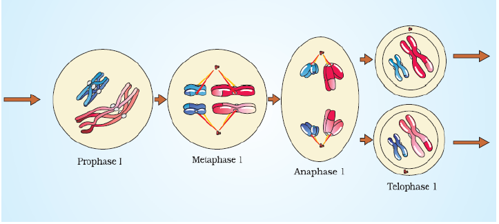
Meiosis I
• In meiosis I, the homologous pairs in a diploid cell separate, producing two haploid cells, so it is also referred to as a reduction division. Like mitosis, it is studied under for stages – prophase, metaphase, anaphase and telophase.
Prophase I
• Prophase I is more complicated and prolonged as compared to the siliar stage of mitosis. For the sake of convenience, prophase I is divided into five subphases: Leptotene, zygotene, pachytene, diplotenme and diakinesis. Another sub-phase called preleptoneme is sometimes recognized prior to leptonema. In this phase chromosomes are not distinguishable because of their thinness but sex chromosomes (if present) are often seen as heterochromatic ( heteropyknotic) bodies.
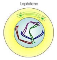
Leptotene
• Leptotene also known as leptonema is a first stage of prophase I during which individual chromosomes begin to condense into long strands within the nucleus which are loosly interwoven. However the two sister chromatids are still so tightly bound that they are indistinguishable from one another.
• Leptotene chromosomes may be irregularly arranged or may be polarized towards the centrioles forming a ‘bouquet’. Electron microscope studies have shown that bouquet formation results when a group of chromosomes is attached close together on the nuclear membrane . In plant cells the chromosomes may sometimes form a tangle of threads, called the synizetic knot, on one side of nucleus.
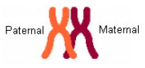
• There are two sets of chromosomes in a diploid cell undergoing meiosis, one set contributed by the male parent and other by the female parent. These are always two similar chromosomes, having the same size, form and structure. They are called homologous chromosomes. One of them is paternal chromosome and the other maternal chromosome.
Zygotene
• During zygotene or zygonema the chromosomes become shorter anad thicker. The homologous chromosomes come to lie side by side in pairs . ( G. zygon = yolk; tene = thread). This pairing of homologus chromosomes is known as synapsis or syndesis. A pair of homologous chromosomes lying together is called a bivalent. The chromatids are still not visible. A fibrillar, somewhat ladder-like , organelle, called synaptonemal complex, develops between the synapsed homologous chromosomes. It is thought to stabilize the paired condition of chromosomes till crossing over is completed.
• Pairing of two homologous chromosomes begins when their corresponding ends come together on the nuclear matrix. Pairing may occur in one of the following three ways-
(i) Proterminal pairing : It starts at the ends and proceeds towards the middle
(ii) Procentric pairing: it begins at the centromeres and progresses towards the ends.
(iii) Random ( intermediate) pairing: It commences at many point towards the ends.
• The synaptonemal complex is attached at both ends through its lateral element to the inner surface of the nuclear membrane. The central elements is not attached directly . Also arising from the lateral elemnent is another series of smaller loops. These loops fuse in the middle line to make up the central element.
Pachytene
• Zygotene is followed by pachytene or pachynema. It is said to begin when synapsis is completed. It lasts from the completion of the synaptonemal complexes to the stage where their breakdown begins
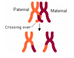
• The synapsed chromosomes continue to become short and thick ( G. pachus = thick, tene = thread). The chromatids of each synapsed chromosome slightly separate and become visible. The two visible chromatids of a chromosome are referred to as a dyad. A group of four homologous chromatids (two dyads) is called sister chromatids and those of two homologous chromosomes (bivalent) are termed non-sister chromatids.
• Crossing over ( recombination) occurs during pachytene. Recombination involves mutual exchange of the corresponding segments of non-sister chromatids of homologous chromosomes. It takes place by breakage and reunion of chromatid segments. Breakage, called nicking, is assisted by an enzyme endonuclease and reunion, termed annealing, is aided by an enzyme ligase.
• It has been found that crossing over is a common event. Normally, each tetrad undergoes at least one recombination.
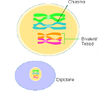
Diplotene
• During diplotene or diplonema the synaptic forces keeping the homologous chromosomes together come to an end. The homologous chromosomes start separating ( G. diplos = double; tene = thread). This is called disjunction. It makes chromatids more distinct and the tetrads very clear. Separation of homologous chromosomes does not take place at the points called chiasmata (singular, chiasma). The chiasmata make the sites where crossing over occurred during pachytene ( Gr . chiasma = crosspiece). They help in holding homologous chromosomes together.
• The number and position of chiasmata varies with the length of the chromosomes and with the species. Chiasmata are found in the meiosis of almost all eukaryotic organism. However, achiasmatic meiosis ( meiosis without chiasma) has been reported in some organisms, e.g. males of higher dipteral ( includeing Drosophila), Panorpa ( scorpion fly), many mantids and roaches, some grasshoppers and scorpions. A chiasma formed at the ends of chromosomes is called termical chiasma. Chiasmata formed along the lengths of chromosomes are called interstitial chiasmata.
• During diplotene the chiasmata begins to be displaced along the length of the chromosome. The terminal chiasma slips off the ends of the chromosomes, and its position is taken up by an interstitial chiasma, which is now called the terminal chiasma. This process is called terminalization. As diplotene progresses the number of interstitial chiasmata becomes lesser in number. The terminalization may be due to electrostatic force or despiralization of chromosomes.
• When terminalization is completed the homologous remain in contact through the terminal chiasma. The degree of terminalization is expressed by the terminalization coefficient (T).
• The synaptonemal complexes mostly disappear during diplotene. In certain regions short segments may persist. The most common regions where the complexes persists are, near the ends of the bivalents where the lateral elements are attached to the nuclear membrane, and at the sites of chiasmata formation. With the disappearance of the synaptonemal complexes the axial filaments become unpaired.
• In diplotene, the chromosomes may unfold to nearly normal form and start transcription of mRNA and rRNA to build up food reverves in the cytoplasm. This process is most profound in the primary ooctytes of amphibians, reptiles and brids. In some species, the chromosomes enlarge greatly, assuming lampbrush form.
Diakinesis
• Diakinesis is not sharply differentiated from diplotene. The chromosomes become more conctracted. The bivalents are more evenly distributed in the nucleus and migrate towards the periphery. RNA synthesis stops. Nucleolus degenerates. A spindle begins to develop, with or without centrioles.
Prometaphase I
• The nuclear membrane disappears in prometaphase I and the chromosomes reach their maximum contraction. Spindle formation begins
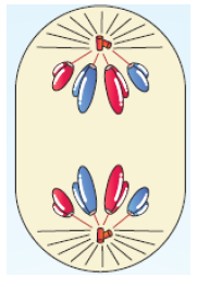
Metaphase I
• The chromosomes now become arranged on a equator of the cell, The spindle is formed. Spindle fibres becomes attached to the centromeres of the two homologous chromosomes. The two centromeres of each bivalent lie on opposite side of the equatorial plate.
• The attachment of tetrads to the spindle fibres in metaphase I is different from that of mitotic metaphase chromosomes. Each homologous chromosome has two kinetochore, one for each of its two chromatids. Both the kinetochores of a homologous chromosome connect to the same spindle pole. The two kinetochores of its homologous join the opposite spindle pole. In metaphase I of meiosis there are bivalents, each bivalent consisting of two centromeres.
Anaphase I
• During anaphase I, from each tetrad two chromatids of a chromosome move as a unit ( dyad) to one pole of a spindle, and the remaining two chromatids of its homologue migrate to the opposite pole.
• Thus, the homologous chromosomes of each pair, rather than the chromatids of a chromosome, are separated. As a result, half of the chromosomes, which appear in early prophase, go to each pole. It is here in the anaphase I that the real reduction in the poles is still double and consists of two chromatids. This is in contrast to the single-stranded chromosomes of mitotic anaphase
• The paternal and maternal chromosomes of each homologous pair segregate during anaphase I independently of the other chromosomes. Anaphase I is cytological event that corresponds to Mendel’s law of independent assortment. Although the paternal and maternal chromosomes of a
homologous pair have the genes for the same traits, either chromosome of a pair may carry different alleles of same genes. Therefore, independent assortment of homologous chromosomes in anaphase I introduces genetic variability.
Telophase I
• During telophase I the chromosomes at each pole of the spindle uncoil and elongate, but remain straight and often do not assume interphase form. The satellite chromosome develop forms around the chromosomes and nucleoli.
The spindle and the astral rays gradually disappear.
• The cytoplasm divides at its middle by cleavage ( constriction) in an animal cell and by cell plate formation in plant cell. This produces two daughter cell, each has received only one chromosome from each homologous pair. Thus it has half the number of chromosomes, but double the amount of nuclear DNA as each chromosome is still double.
• The daughter cells formed by meiosis I are called secondary spermatocytes or secondary oocytes in male and female animals.
• Cell enter a period of rest as interkinesis or interphase II. No DNA replication occurs during this stage. Protein and RNA synthesis may occur. It is important for bringing true haploidy.
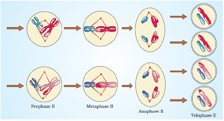
Meiosis II
• The second meiotic division is essentially similar to mitosis. In this division, the two chromatids of each chromosome separate from each other and go to separate daughter cells. With the result, the number of chromosomes remains the same as produced by meiosis I, Meiosis II is, therefore, known as homotypic division. If however, differs from mitosis in that DNA does not duplicate, while centromere do so. It has 4 phases – Prophase II, metaphase II, anaphase II, and telophase II.
Prophase II
• Prophase II takes an inversely proportional time compared to telophase I. In this process we see the disappearance of nucleoli and the nuclear envelop again as well as the shortening and thickening of chromatids. Centrioles move to the polar region and are arranged by spindle fibres.
Metaphase II
• In metaphase II the chromosomes become oriented on the equatorial plate and have the relationship to the spindle as in mitosis.
Anaphase II
• Anaphase II, where the centromeres are cleaved, allows the kinetochores to pull the sister chromatids apart. The sister chromatids by convention are now called sister chromosomes, and they are pulled towards opposite poles
Telophase II
• In telophase II the group of chromosomes at each pole of the spindle gets enclosed by a nuclear envelope. Nucleoli are laid sown, Astral rays and spindles are lost
Cytokinesis
• Cytoplasm divides at its middle by furrowing in an animal cell and by cell plate formation in a plant cell. This produces two daughter cells. The latter have half the number of chromosomes and half the amount of nuclear DNA. These cells are mature gametes in animals and spores in plants.
• Cytokinesis may occur after each nuclear division. In such cases, it is said to be of successive type. First the diploid parent cell divides by heterotypic division into two haploid cells, which then produce four haploid cells by homotypic division. The four daughter cells may form a linear or isobilateral tetrad. Often cytokinesis is delayed until both the nuclear divisions are completed, so that four cells are simultaneously formed, each with a haploid nucleus. The cytoplasmic division in such cases is said to be of simultaneous type Significance of meiosis
• Formation of gametes – Meiosis forms gametes that are essential for sexual reproduction .
• Genetic information – It switches on the genetic information for the development of gametes or gametophytes and switches off the sporophytic information
• Meiosis facilitates stable sexual reproduction – Without the halving of ploidy, or chromosome count, fertilization would result in zygotes that have twice the number of chromosomes than the zygotes from the previous generation. Successive generations would have an exponential increase in chromosome count, resulting in an unwildy genome that would cripple the reproductive fitness of the species. Most importantly, however, meiosis produces genetic variety in gametes that propagates to offspring. Recombination and independent assortment allows for a greater diversity of genotype in the population. As a system of creating diversity, meiosis allows a species to maintain stability under environment changes.
• Crossing over- It introduces new combination of traits or variations.
• Mutations – Chromosomal and genomatic nutations can take place by irregularities of meiotic divisions. Some of these mutations are useful to the organism and are perpetuated by natural selection.
• Evidence of basic relationship of organisms – Details of meiosis are essentially similar in the majority of organisms showing their basic similarity and relationship.
ABNORMAL CELL GROWTH
• Cell division is a gene controlled process. The telomere of chromosomes contains repetitive sequence of six nucleotide. These regions code for an enzyme telomerase which control cell division. As cells go on dividing with each division the number of nucleotide decreases and ultimately cells stop dividing.
• Uncontrolled cell division may lead to the formation of undifferentiated aggregate of cells termed tumor or neoplasm.
• Uncontrolled cell division leads to hyperplasis, hypertrophy, metaplasis, neoplasia, and He La cell.
• The increased production and growth of normal cells in a tissue or organ is termed hyperplasia. It is an accelerated rate of cell division resulting from an increased level of cell metabolism. This generally results in an enlargement of tissue mass and organ size. It occurs only in tissues capable of mitosis such as the epithelium of skin, intestine and glands. Some cells do not divide and thus can not undergo hyperplasia, for example nerve and muscle cells.
• An increase in the size of a tissue or organ brought about by the enlargement of its cells is termed hypertrophy. When cells hypertrophy, components of the cell increase in number with increased functional capacity to meeting increased cells needs. Hypertrophy generally occurs in situations where the organ or tissue can not adapt to an increased demand by formation of more cells. This is commonly seen in cardiac and skeletal muscles cells, which do not divide to form more cells.
• The process of conservation of normal tissue cells into an abnormal form in response to stress or injury or infection is termed metaplasia. It is a cellular replacement process.
• The new and abnormal development of cells that may be benign or malignant is termed neoplasia. There are two types of neoplasm – benign and malignant - Benign growth : the benign growth is restricted to a particular site of the body and the cells never spread out to different parts of the body e.g. simple tumor
- Malignant growth : in malignant growth after the cells are being formed at a particular site, the cells move out different parts of the body and initiate similar type of growth. The stage of malignant growth in which the cells spread out through the body fluid to different parts of the body is termed metastasis. Malignant growth is also termed cancerous growth.
• He La cells (an aneuploid epithelial cells) are cell line culture of first human cancerous cells donated by Henrietta Lacks from their uterine carcinoma cells since 1952. These cells are maintained for use in studying cellular processes.