through another set of capillaries instead of sending blood into the heart. There are three types of portal systems – hepatic, hypophysial and renal.
of the colon), superior mesenteric (small intestine, caecum and proximal part of the colon) and gastroepiploic( from stomach and pancreas). Hepatic portal vein enters liver and breaks into capillaries. The system function as a short circuit for
(i) Removal of glucose, amino acids, and other nutrients.
(ii) Deamination of extra amino acids and conversion of harmful ammonia into urea
(iii) Separation of toxic chemicals and their detoxification
(iv) Direct pouring of liver products into venous blood
Hypophyseal portal system
It is a minor portal system that occurs in higher vertebrates. The system consists of a single hypophysial portal vein. The portal vein is formed by capillaries in the hypothalamus. It passes into the anterior lobe of pituitary glands and breaks up into capillaries there. The hypophyseal portal system is meant for pouring hormones secreted by hypothalamus directly into the anterior part of the pituitary.
Renal portal system
It occurs in lower vertebrates (fish and amphibians), reduced in reptiles and aves and is absent in mammals. It consists of renal portal veins that bring blood from a posterior part of the body directly into kidneys for removal of waste products.
Lymphatic system
It comprises lymph, lymphatic capillaries, lymphatic vessels, lymphatic nodes and lymphatic ducts.
Lymph
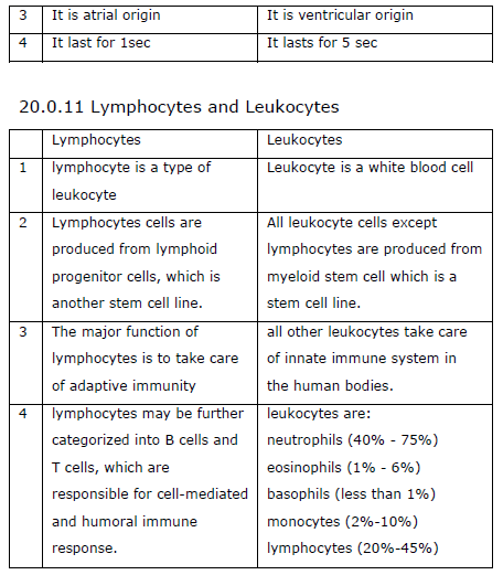
Lymph, a colourless fluid is a part of tissue fluid, which in turn, is a part of blood plasma. So the composition of tissue fluid and lymph is same as that of blood plasma but it lacks RBCs and large plasma proteins. As compared to the tissue fluid, the lymph contains very small amount of nutrients and oxygen but contains abundant carbon dioxide and other metabolic wastes. Amoeboid shaped white blood corpuscles may be present in the lymph.
Lymphatic capillaries
Lymphatic capillaries lie close to the blood capillaries but differ from them to extent that they end blindly. Moreover, they have extremely thin walls. They are composed of a single layer of endothelial cells. The lymphatic capillaries of intestine absorb the
digested fats. They are milky in appearance and are, therefore, called the lacteals.
Lymphatic vessel
The lymphatic capillaries unite to form large lymphatic vessels. They are composed of an outer coat of fibrous tissue, middle coat of muscular tissue and an inner lining of endothelial cells. The lymphatic vessels have numerous valves.
Lymph node
• These are small oval or bean-shaped structures located along the length of lymphatic vessels. Lymph nodes are most numerous in the thoracic mediastinum on the posterior abdominal wall in the abdominal mesenteries and in the pelvis neck and proximal ends of the limbs.
• Lymphatic nodes perform the following main functions.
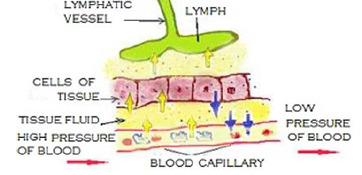
Both B-lymphocytes and T-lymphocytes are produced here.
• Macrophages of lymph nodes remove bacteria, foreign material and cell debris from the lymph.
• B-lymphocytes change to plasma cells that produce antibodies against invading antigens, while T-lymphocytes attack cells that are ‘foreign’ to the host body.
Thoracic duct
The lymphatic vessels of left side unite to form a thoracic duct. This duct begins at the cisterna chyli, which is a sac-dilation situated in the front of the first and second number vertebrate. The thoracic duct contains several valves. It discharges its lymph into the left subclavian vein.
Right lymphatic duct
The lymphatic vessel of the right side of the thorax, head, and neck unite to form the right lymphatic duct. It is about 1 cm in length. It discharges its lymph into the right subclavian vein.
Lymph movement
The lymph flows in lymphatic vessels very slowly. Forcing out of fluid from the blood capillaries sets up some pressure in the tissue fluid. This establishes a pressure gradient in the lymphatic, causing the flow of lymph in the latter. Movements of viscera and
contractions of the body muscles help considerably in squeezing the lymph along. The valves present in lymphatic vessels prevent its backflow. Movement of villi assists flow of lymph in the lacteals. Gravity helps in moving the lymph down the lymphatic vessels of head and neck.
Functions of lymph
The lymph or lymphatic system serves functions as:
• It drains excess tissue fluid from the extracellular spaces bin to the fluid.
• Some of the fluid from the digestive tract is absorbed into the lymph. The lymphatic vessels store this fluid temporarily and release it gradually so that the kidney does not face a sudden pressure of urine execration.
• It carries carbon dioxide and nitrogenous waste materials that diffuse into the tissue fluid to the blood.
• It takes lymphocytes and antibodies from lymphatic nodes to the blood.
• It transported fat that is digested and absorbed in the intestine to the blood in the form of chylomicron droplets.
• It destroys the invading microorganisms and foreign particles in the lymphatic nodes.
• It maintains quality and quantity of the blood by restoring the fluid and solute that leaves it.
• It brings plasma protein macromolecules synthesized in the liver cells and hormones produced in the endocrine glands to the blood.
Spleen
The spleen is the largest component of the lymphatic system. It is large (7-10 cm in diameter), bean-shaped, vascular, dark red organ located in the abdomen just below the diaphragm at the tail of the pancreas behind the stomach.
The spleen is composed of red pulp (reticular tissue rich in RBCs) having small patches of white pulp (lymphatic nodes) scattered in it. The red pulp is enclosed by a capsule of white fibrous tissue. The capsule sends trabeculae into the pulp and is surrounded by visceral peritoneum.
Functions
i) Destruction of worn-out red corpuscles
ii) Reservoir of red corpuscles
iii) Formation of agranulocyte
iv) Production of antibodies
v) Storage of iron
vi) Erythropoiesis
vii) Disposal of foreign elements
Thymus
The thymus is also a lymphatic organ. It lies in the upper chest near the neck. It is prominent in children but begins to degenerate in early childhood. It educates the lymphocytes in the foetus to distinguish cells from foreign cells.
Tonsils
Tonsils too are lymphatic tissues. They are located in the throat. They do not filter lymph. They are thought to protect against infection.
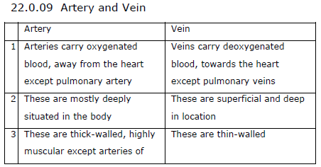
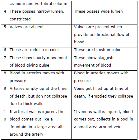
Some common cardiovascular defects
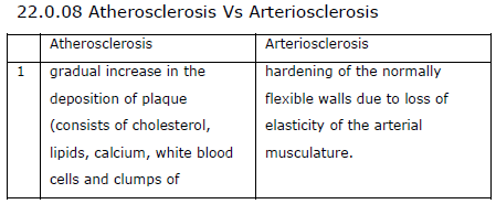
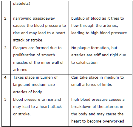
1. Arteriosclerosis:
Sclerosis and hardening of walls of generally smaller arteries and arterioles are called arteriosclerosis. The common cause is deposition of calcium in tunica media cholesterol may get calcified. The walls of arteries become stiff and rigid. There is a loss of elasticity. The phenomenon is called hardening of arteries. Limb arteries are usually the first to undergo arteriosclerosis. Lesions develop at branch points. It ultimately leads to distal obstruction causing pain, numbness of extremities, Peripheral edema, cyanosis etc. rupturing of some vessels also occur. It forms a blood clot and blocks the flow of blood.
2. Atherosclerosis:
It is wall thickening and narrowing of the lumen of medium and large arteries. In atherosclerosis, yellowish plaques (atheromas) of cholesterol and other lipids are deposited within tunica intima and inner part of tunica media were smooth muscles abound. They are mostly caused by lowdensitylipoproteins or LDL which can pass through the endothelium. Plaques grow. The smooth muscles also proliferate probably caused by the release of plateletderived growth factor (PDGF). This occurs due to the
roughness of inner arterial lining. Thickening of arterial wall reduces the lumen size. In extreme cases, thegrowth of plaques may completely block an artery. Atherosclerosis leads to hypertension, reduced blood supply to limbs and other organs resulting in their dysfunctioning.
Atherosclerosis in coronary arteries results in reduced O2 supply to heart walls causing angina, myocardial infection or heart attack or stroke.
3. Coronary artery disease (CAD)
Coronary arteries undergo atherosclerosis. There is deposition of calcium, fat and fibrous tissue which results in narrowing of the arterial lumen. The flow of blood in the affected arteries is reduced. The cardiac muscles supplied by the affected arteries will begin to deteriorate. There are a thoracic pain, nausea, perspiration and E.C. G changes. The defect can be treated through angioplasty (breaking of arterial blockage by balloon catheter) and bypass surgery.
4. Angina or Angina pectoris
It is recurrent, spasmodic suffocating thoracic (or heart) pain which often radiates to the left arm. Angina is generally caused by the deficient blood supply to heart muscles. It is precipitated by excitement or strenuous physical activity. Angina pectoris can occur in all types of individuals, Both men and women of any age. However, it is more common in middle-aged and elderly persons.
Reduced blood supply to myocardial muscles occurs either due to constriction or obstruction of blood vessels.
5. Heart Failure
It is the inability of the heart to supply blood in adequate quantities to all parts of the body. Heart failure is a syndrome of ventricular dysfunction. The person suffering from heart failure has reduced exercise capacity. The health of different muscles of the body would also be affected. Heart failure should not be confused with a heart attack (heart muscle damaged due to inadequate blood supply) or cardiac arrest in which case there is a stoppage of the heartbeat.
6. Cardiomegaly: hypertrophy of the heart. Inflammation of heart is carditis.
7. Cardiomyopathy: aNoninflammatory disease of heart muscle.
8. Ischaemic heart: Heart with degenerate or defective components due to rheumatic disorder or fever in childhood.
9. Rheumatic heart: Heart with degenerate or defective components due to the rheumatic disorder of fever in childhood.
10. Embolus: Mass of clotted blood, other formed elements, fragments, air, calcium etc. coming from a larger blood vessel is forced into a smaller or narrow blood vessel resulting in its blockage and hence obstruction of blood circulation.
11. Myocardial Infarction: Complication due to reduced blood supply to heart wall-pain, pallor, perspiration, nausea, ECG changes.
12. Heart Burn (Pyrosis). The sensation of burning occurring in waves in esophagus tending to rise upward towards neck often with reflux into the mouth. It has nothing to do with the heart.
13. Varicose Veins: Unnatural permanently distended veins.
14. Hematoma: Localized collection of usually clotted blood in a tissue or organ due to injury and rupturing of the blood vessel.
Distinguish
Blood and Lymph
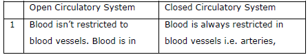
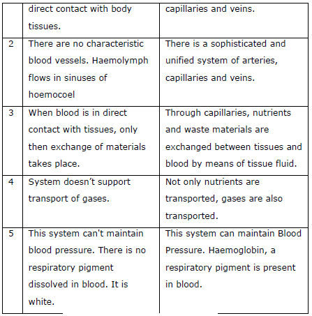
Open and closed circulatory System
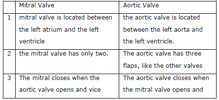
Mitral Valve and Aortic Valve
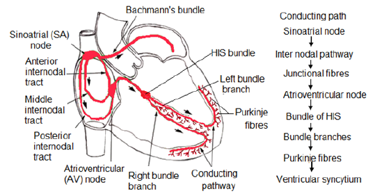
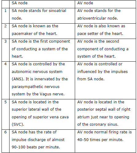
SA node Vs AV node
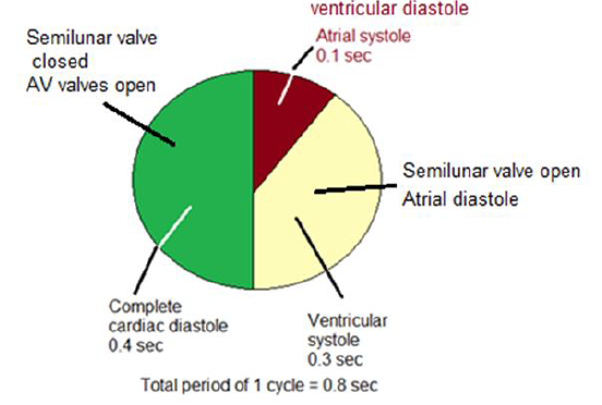
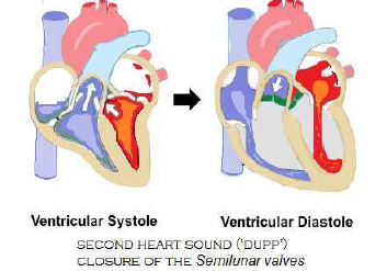
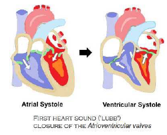
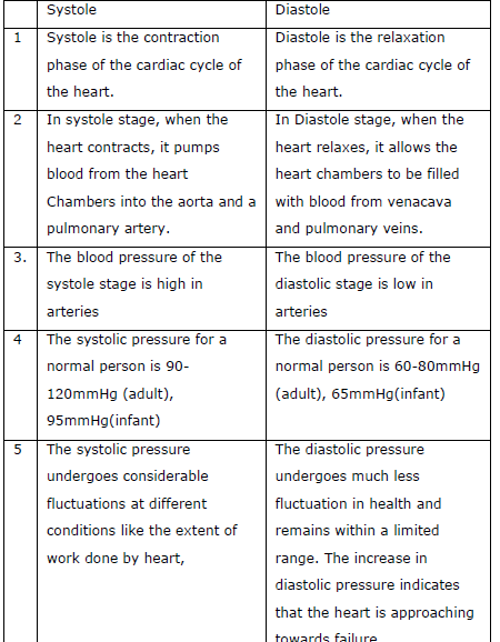
Systole Vs Diastole