Digestion and Absorption MCQ Questions with Answers Class 11 Biology
Question. The digestion of which food component is affected if pancreas is removed?
(1) Carbohydrates
(2) Proteins
(3) Fats
(4) All of these
Answer (4)
Question.Enzyme trypsinogen is a component of
(1) Mucus
(2) Saliva
(3) Pancreatic juice
(4) Intestinal juice
Answer (3)
Question.Pyloric sphincter regulates the opening of
(1) Pharynx into oesophagus
(2) Oesophagus into stomach
(3) Stomach into duodenum
(4) Ileum into large intestine
Answer (3)
Question.Which carbohydrate splitting enzyme initiates the chemical process of digestion in the oral cavity?
(1) Lysozyme
(2) Salivary amylase
(3) Pepsin
(4) Rennin
Answer (2)
Question.The type of cells present in the gastric glands which secretes intrinsic factor?
(1) Peptic cells
(2) Chief cells
(3) Parietal cells
(4) Both (1) & (2)
Answer (3)
Question.Mark the odd one out
(1) Gastrin
(2) Trypsin
(3) Secretin
(4) Enterocrinin
Answer (2)
Question.The pancreatic juice contains various enzymes, except
(1) Pepsinogen
(2) Trypsinogen
(3) Chymotrypsinogen
(4) Procarboxypeptidase
Answer (1)
Question. The food that enters into intestine from stomach is
(1) Alkaline chyle
(2) Fundus
(3) Acidic chyme
(4) Bolus
Answer (3)
Question. Select the incorrect option
(1) Bilirubin and biliverdin are the bile pigments
(2) Emulsification is the breakdown of the fats into very small droplets
(3) Rennin is a proteolytic enzyme found in the pancreatic juice of infants which helps in the digestion of milk protein
(4) Mucus and bicarbonates protect mucosal epithelium from excoriation by highly conc. HCl
Answer (3)
Question. The structural and functional unit of liver is
(1) Cystic duct
(2) Hepatic lobule
(3) Hepatopancreatic duct
(4) Sphincter of Oddi
Answer (2)
Question. Select the incorrect option regarding digestion and absorption of substances in different parts of digestive system
(1) In large intestine, absorption of water, some minerals and drugs takes place
(2) Absorption of water, simple sugars and alcohol takes place in stomach
(3) Small intestine is the principal organ for absorption of nutrients
(4) The digestion is completed in large intestine
Answer (4)
Question. The main enzymes present in the gastric juice are
(1) Trypsin, pepsin and lipase
(2) Pepsin, amylase and trypsin
(3) Pepsin, rennin and carboxypeptidase
(4) Pepsin, lipase and rennin
Answer (4)
Question. At which site the emulsification of fat takes place?
(1) Pancreas
(2) Gall bladder
(3) Liver
(4) Duodenum
Answer (4)
Question. The proteolytic enzyme found in the gastric juice of infants which helps in the digestion of milk proteins is
(1) Renin
(2) Rennin
(3) Salivary amylase
(4) Lysozyme
Answer (2)
Question. Which of the following is a modification of mucosa of alimentary canal?
(1) Villi
(2) Microvilli
(3) Rugae
(4) All of these
Answer (4)
Question. Upper molars in human dentition have
(1) Four roots
(2) Three roots
(3) Two roots
(4) Single root
Answer (2)
Question.Bile can be prevented to release into duodenum by
(1) Sphincter of Oddi
(2) Cardiac sphincter
(3) Pyloric sphincter
(4) Ileo-caecal valve
Answer (1)
Question.Which of the following can be taken as true stomach in ruminants?
(1) Rumen
(2) Reticulum
(3) Omasum
(4) Abomasum
Answer (4)
Question.Oblique muscle layer is present in
(1) Stomach
(2) Duodenum
(3) Colon
(4) All of these
Answer (1)
Question. One of the following ions is used for activation of ptyalin
(1) Sodium ions
(2) Potassium ions
(3) Chloride ions
(4) None of these
Answer (3)
Question. A prolonged constipation may cause
(1) Hemorrhoids
(2) Ulcers
(3) Cholera
(4) Dysentery
Answer (1)
Question. When a piece of bread is chewed it tastes sweet because
(1) The sugar contents are drawn out
(2) Saliva converts starch into maltose
(3) It does not taste sweet
(4) The taste buds are stimulated by chewing
Answer (2)
Question. In acute constipation, purgatives that are used to stimulate intestinal peristalsis and evacuation of fluid faeces contain salts of
(1) Sodium
(2) Magnesium
(3) Potassium
(4) Calcium
Answer (2)
Question. When breast feeding is replaced by less nutritive food low in proteins and calories; the infants below the age of one year are likely to suffer from:
(1) Rickets
(2) Kwashiorkor
(3) Pellagra
(4) Marasmus
Answer (4)
Question. One of the constituents of the pancreatic juice while poured into the duodenum in humans is
(1) Trypsin
(2) Enterokinase
(3) Trypsinogen
(4) Chymotrypsin
Answer (3)
Question. Which one of the following statements is true regarding digestion and absorption of food in humans?
(1) Fructose and amino acids are absorbed through intestinal mucosa with the help of carrier ions like Na+.
(2) Chylomicrons are small lipoprotein particles that are transported from intestine into blood capillaries.
(3) About 60% of starch is hydrolysed by salivary amylase in our mouth.
(4) Oxyntic cells in our stomach secrete the proenzyme pepsinogen.
Answer (1)
Question. Carrier ions like Na+ facilitate the absorption of substances like
(1) Fructose and some amino acids
(2) Amino acids and glucose
(3) Glucose and fatty acids
(4) Fatty acids and glycerol
Answer (2)
Question. Jaundice is a disorder of
(1) Excretory system
(2) Skin and eyes
(3) Digestive system
(4) Circulatory system
Answer (3)
Question. A young infant may be feeding entirely on mother's milk which is white in colour but the stools which the infant passes out is quite yellowish. What is this yellow colour due to?
(1) Bile pigments passed through bile juice
(2) Undigested milk protein casein
(3) Pancreatic juice poured into duodenum
(4) Intestinal juice
Answer (1)
Question. If for some reason the parietal cells of the gut epithelium become partially non-functional, what is likely to happen?
(1) The pancreatic enzymes and specially the trypsin and lipase will not work efficiently
(2) The pH of stomach will fall abruptly
(3) Steapsin will be more effective
(4) Proteins will not be adequately hydrolysed by pepsin into proteoses and peptones
Answer (4)
Question. If for some reason our goblet cells are non-functional this will adversely affect
(1) Smooth movement of food down the intestine
(2) Production of somatostatin
(3) Secretion of sebum from the sebaceous glands
(4) Maturation of sperms
Answer (1)
Nutrition
Nutrition is a process by which animals obtain essential and nonessential substances called nutrients and utilize these produce to energy required for various life process such as growth, repair, development, reproduction and other activities from the surrounding as food.
Modes of nutrition
Autotrophic nutrition
• Preparation of organic food from the inorganic materials is called autotrophic nutrition
• It is of two types : Photoautotrophic and chemoautotrophic
a) Photoautotrophic nutrition
• All green plants, certain protists ( Euglena viridis) and some bacterial ( green sulphur bacterium, chlorobium) take carbon dioxide and water from the environment and transform these into glucose and oxygen with the help of Sun’s energy trapped by chlorophyll.
b) Chemoautotrophic nutrition
• Some bacteria utilize light as source of energy during the synthesis of food, utilize the energy obtained in the form of A.T.P
• Examples of chemoautotrophic bacteria
i) Sulphur bacteria – Beggiatoa, thiothrix and Thiobacillus thiooxidans
ii) Iron bacteria
iii) Nitrifying bacteria – Nitrosomonas,
Nitrosococcus, Nitrobacter and Nitrocystis
Hetrotrophic nutrition
• Animals, fungi etc cannot manufacture their food. They depends upon autotrophs directly or indirectly
i) Holotrophic ( Holozoic) nutrition
• Most vertebrates and invertebrates take solid or liquid food through their mouth is called holozoic nutrition
ii) Saprozoic nutrition
• It consists of obtaining food from dead and decaying organic food by first pouring digestive juices over the same and then sucking the digested food. Example – Spider, housefly
iii) Parasitic nutrition
• In this type of nutrition, food is obtained in liquid form from the body of another killing him
• Example – Plasmodium, Trypanosoma, Taenia, ascaris
Digestive system of human
• It is a system of alimentary canal and digestive glands that takes part in ingestion of food, its crushing, digestion, absorption of digested nutrients and egestion of undigested materials.
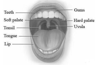
Mouth
• The mouth is a transverse slit. It is bounded by two soft movable lips
• The lips are covered with skin and mucous membrane on the inner side
• In mammals, the upper lip has a well defined cleft called median cleft, which provides exposure to the teeth
• In human, the median cleft not seen but only a depression can be seen which is called phlitrum
Vestibule
• It is a narrow space enclosed between the lips and checks externally and the gums and teeth internally
• In the vestibule, a small median fold of mucous membrane, the superior labial frenulum, connects the middle of the upper lips to the gum.
• A smaller inferior labial frenulum connects the middle of lower lip to the gum.
Oral ( Buccal) cavity
• It is a space bounded above by the palate, below by the throat and on the sides by the jaws.
• The buccal cavity is lined by stratified squamous epithelium.
Palates
• The anterior part of palate is arched and strong. It is called hard palate. This part is well supported by maxilla bone.
• It bears transverse ridges called palatine rugae. The rugae help in keeping the food in place during mastication.
• The posterior part of the palate is smooth and fleshy. It is termed soft palate. Its smooth surface makes swallowing easy.
• The soft palate contains hanging small, conical flap called uvula.
• The ulva is movable and capable of coming in contact with the posterior pharyngeal walls so as to cut off the upper nasal part of the pharynx, called nasopharynx from the lower oral part of the pharynx, termed oropharynx during swallowing.
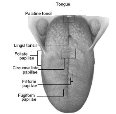
Tongue
• The tongue is a voluntary musculo-sensory and glandular structure which occupies the floor of the mouth.
• It is attached to the floor of the mouth by a fold called frenulum.
• An inverted V-shaped furrow termed the sulcus termanalis divided into the upper surface of the tongue into the anterior oral part and posterior pharyngeal part.
• Upper surface of the oral part of the tongue has a slight median groove
• The upper surface of tongue has four types of papillae , small projection which bear taste buds
i) Vallate ( surrounded by wall)
ii) Fungiform ( mushroom shaped)
iii) Foliate ( leaf –shaped)
iv) Filiform ( filament-shaped)
Function of the tongue
i) It helps in chewing the food.
ii) It plays a role in speech.
iii) It aids in swallowing the food and mixing the food and saliva.
iv) It acts as brush to clear the teeth by cleaning small food particles.
v) It is an organ of taste and can recognize four tastes, i.e. salty, sweet, bitter and sour.
Teeth
• Teeth are hard structures which are meant for holding and crushing the food.
• Most of the mammals have diphydont ( two sets of teeth-milk or deciduous and permanent), theocodont ( teeth are embedded in the socket of jaw bone) and heterodont teeth ( different types of teeth).
Types of teeth
• There are four kinds of teeth incisors, canines, premolars and molars present in the human
• Incisors: These are chisel shaped and possess sharp cutting edges
• They are usually specialized for cutting
• Canines: They lie immediately behind the incisors Canines are well developed in carnivores and may be absent in herbivores leaving a gap called diastema ( which is used to separate the chewed and unchewed food in the mouth cavity)
They have long, sharp pointed end for piercing, killing and tearing off flesh
• Premolars and molars: these are called cheek teeth which are broad, strong crushing teeth.
Last molars in human beings are called wisdom teeth.
Structure of teeth
• Teeth are embedded in the jaws man has fixed upper jaw and movable lower jaws. Each tooth consists of three parts: crown, root, neck.
a) Crown: It is the exposed portion of tooth above the gums ( gingiva)
• The gingiva is a specialized region of the oral mucosa surrounding the neck of the teeth
• Crown is covered with the hardest substances called enamel that protects the crown
• Beneath the enamel is dentine which is made up of hard substances but are not tough as enamel and they can decay
• Dentine forms the bulk of the tooth. There is a pulp cavity inside the dentine. It is jelly like substance and carries the nerves fibres, blood vessels and sensory cells.
• The nerves supply to the upper teeth is by the branches of maxillary nerves and to the lower teeth by the branches of mandibular nerves. These both are branches of 5th cranial nerve called trigeminal nerve.
b) Neck: It is a narrow portion at the gumline.
c) Root: It is embedded in the jaw bone and holds the tooth securely in place.
• The root is fixed in alveolus of the jaw bone by periodontal membrane and cementum. Cement holds a tooth in its socket and periodontal membrane covers the cement.
Number of teeth
• The milk or deciduous teeth are 20 in number, 10 in upper and 10 in lower jaw
• The deciduous teeth begin to erupt when the child is about 6 months old and should all be present by the end should all be present by the end of 24 months.
• The permanent teeth begin to replace the milk teeth in the 6th year of age. These teeth are 32 and usually complete by the 20th year
Dental formulae
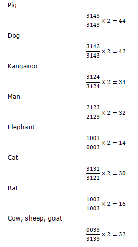
• The number and kinds of teeth in mammals are represented by an equation called dental formula. This equation looks like

I = Incisors, C = Canines Pm = Premolars
• Total number of teeth = Number of teeth in dental formula × 2
Dental formula of some animals
Dentition of animals
1. Aerodont
Part of bone, not embedded in sockets eg. Reptiles, except crocodiles
2. Theocodont
Embedded in deep socket of some jaw bones e.g. mammals, crocodiles
3. Monophycodont
Teeth grow only once in life e.g. platypus, toothed whale.
4. Diphyodont
All teeth, except molar, grow twice in life. Eg. Mammals
5. Polyphyodont
Fallen or wornout teeth can be replaced many times throughout life. Eg. Frog
6. Isodont
All teeth are similar Eg. Tooth whale
7. Heterodont
More than one type of teeth E.g mammals, crocodiles
Pharynx
The mouth leads to funnel shaped pharynx. The pharynx is about 12cm long vertical canal beyond the soft palate. The food and air passages cross here. The pharynx may be divided into three parts
i) Nasopharynx: Upper part of Pharynx Have internal nares
in the root, oval opening of Eustachion tubes
ii) Oropharynx Middle part of pharynx Has palatine tonsils
iii) Laryngopharynx
• Lower part of pharynx
• Leads to oesophagus and to pharynx through glottis ( opening to larynx) and epiglottis ( leaf like cartilageous flap)
• The function of the pharynx as a part of digestive tract is merely to serve as passage way for the food from oral cavity to oesophagus
• The lymphatic tissues of pharynx and oral cavity are arranged in a ring which are collectively called Waldeyer’s ring. All these lymphoid tissue are active in production of immunoglobin A which forms an important part of our immune system
Waldeyer’s ring
All lymphoid tissues are active in production of immunoglobulin
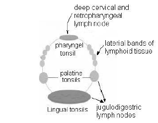
The ring mainly consists offollowing
(i) Nasopharyngeal tonsils
• In children nasopharyngeal tonsil become enlarged and is then referred to as adenoids
• The resulting swelling causes obstruction to normal breathing
(ii) Tubal tonsils
Lymphoid tissue found around the opening of each Eustachian tube
(iii) Palatine tonsils
• Located in the lateral wall of oropharynx
• The palatine tonsils is often infected leading to sore throat and surgical removal of such enlarged tonsils become necessary
(iv) Lingual tonsils
• Lymphoid tissue present at the base of tongue
Oesophagus
• The oesophagus is 25 cm long, narrow muscular straight tube lined by stratified squamous epithelium containing mucous glands
• The opening of oesophagus is called gullet
• It runs downward through the neck behind the trachea and through behind the heart and passes through diaphragm into abdomen. Here it sharply bends to open into the stomach. This bend is one of the device to check the backflow of the stomach content into the oesophagus
• Longitudinal folds keep its cavity almost closed, except during swallowing of food.
• The oesophagus serves to convey the food by peristalsis ( a series of waves of contraction that passes through ends and is meant for pushing food) from pharynx to stomach
• The nerve supply to oesophagus includes parasympathetic and sympathetic branches seen in the form of myeteric and messner’s plexus namely
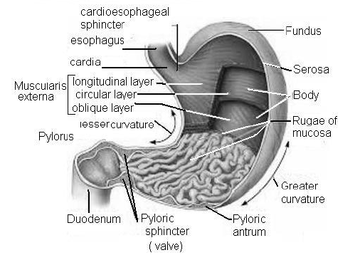
Stomach
• Stomach is the widest organ about 30 cm long and 15cm wide of alimentary canal.
• It is a hollow, J- shaped organ lying between the oesophagus and the small intestine. The exact positioning of the stomach is in the upper (5/6)th part and right (1/6)th part of abdominal cavity
• The lesser curvature is short and lies on the posterior surface of stomach. The greater curvature is on anterior surface of stomach.
• The fold of peritoneum which attaches the stomach to the posterior abdominal wall extends beyond the greater curvature. This is called the greater omentum which stores fat
• Inner surface of the stomach has numerous folds, the gastric rugae. These folds, by unfolding let the stomach expand to accommodate a large meal
• Unlike other parts of the digestive tract, the stomach wall contains there smooth muscle layers, outer of longitudinal, middle of circular and inner of oblique fibres, to churn the food and to mix it with the gastric secretion
• The stomach has four parts: cardiac part, fundus, body and pyloric part
(i) Cardiac part: It is so called because it is present near the heart. The cardic sphincter lies in the opening between oesophagus and stomach. It is a functional valve. Cardiac gland secrete mucus
(ii) Fundus: It extends superiorly from the cardiac part. The fundus is commonly filled with gas or air. Fundic glands secretes HCl and intrinsic factor
(iii) Body :It is main part stomach
(iv) Pyloric part : It is the distal part of stomach. The pyloric region is divided into the pyloric antrum and pyloric canal. The pyloric sphincter guards the opening between the stomach and duodenum and periodically permits partially digested food to leave the stomach and ether
duodenum. Pyloric glands secrete gastrin produced by their G-cells
Functions
(i) Storage: the stomach allows a meal to be consumed and the material to be consumed and the materials released incrementally into the duodenum from digestion
(ii) Chemical digestion: Pepsin begins the process of protein digestion cleaving large polypeptides into shorter chains
(iii) Mechanical digestion: The churning action of muscularis causes liquifaction and mixing of the contents to produce acid chyme
(iv) Some absorption: Water, electrolyte monosaccharides and fat soluble molecules including alcohol are all absorbed in stomach to some degrees.
Ruminant Stomach
In ruminants, the stomach is differentiated into chambers
(i) Large rumen for churning, breaking of food by cornified surface of villi, fermentation of cellulose by symbiotic microorganism
(ii) Recticulum
(iii) Omasum for mechanical churning and breaking of food, absorption of fluid
(iv) Abomasum for mixing gastric juice, ruminates the chew cud.
This is done by breaking small part of food present in rumen and sending it to buccal cavity for chewing. Omasum is absent in camel. Here rumen and reticulum have water pockets for temporary storage of food. Abomasum functions as true stomach.
Small intestine
• The small intestine is a narrow tube , about 6 metres long in a living adult. It is longest part of alimentary canal. It comprises of alimentary canal. It comprises of three parts: duodenum, jejunum and ileum
(i) Duodenum: It is a C-shaped structure and about 25 cm long. It receives the hepatopancreatic duct formed by union of bile duct (from liver) and pancreatic duct ( from pancreas) and whose opening is guarded by sphincter of oddi. Small nodules of lymphoid tissue are seen along the entire length of small intestine. These nodules are call Payer’s patches
(ii) Jejunum: It is slightly coiled moderately wide ( 3.5 – 4 cm) middle part of small intestine. Its wall is thicker and more vascular than that of illum. Jejunum is rich in digestive glands villi are rounded. Its length is about 0.8 – 1.5m
(iii) Ileum: the ileum forms the lower part of small intestine. It is about 3.5 m long, and opens into large intestine. It is characterized by club-shaped villi and Peyer’s patches. Villi increases internal surface of ileum by about 10 times. Peye’s patches contain higher concentration of white blood cells that helps protect the body from infection and disease
Functions
(i) The small intestine completes digestion of proteins, carbohydrate, nucleic acids and fats.
(ii) It absorbs nutrient materials into the blood and lymph and also lubrication of food.
(iii) It secretes certain hormones such as cholecystokinin, secretin, enterogastrone, duocrinin, enterocrinin and villikinin.
Most nutrient absorption occurs in the small intestine including minerals, vitamins, proteins, and fats. Iron, calcium, magnesium, and zinc are absorbed almost immediately after leaving the stomach – ie, in the 8 feet of the duodenum and the jejunum. Sugars and vitamin C, as well as thiamin, riboflavin, pyridoxine, and folic acid, are absorbed in the upper third of the small intestine. Protein is absorbed approximately midway through the ileum. Water and lipids are absorbed by passive diffusion throughout the small intestine.
Sodium bicarbonate is absorbed by active ransport and glucose and amino acid co-transport.Vitamins A, D, E, and K, fats, and cholesterol are absorbed in the lower third of the ileum. Vitamin B12 is absorbed just before the small intestine joins the large intestine. Bile salts are reabsorbed in the distal ileum and the ascending colon.
Large intestine
• Its diameter varies from one region to another but it is always larger than that of small intestine. It is about 1.5 metres long and is divisible into there parts caecum, colon and rectum
(i) Caecum and vermiform appendix:
The caecum is a pouch – like structure which is about 6cm. the caecum is called ileocoecal junction guarded by ileocaecal valve which prevent back flow. The vermiform appendix is an outgrowth of the caecum. It is a slightly about 8 cm long. Its wall contains prominent lymphoid tissue. Appendix is thought to be vestigial. The inflammation of vermiform appendix due to decay of food or warm infection is called appendicitis and rupture of appendix leads to spilling of faecal matter onto the peritoneum leading to infection and inflammation known as peritonitis.
The caecum is well developed in herbivorous mammals like, horse, rabbit, etc.
(ii) Colon: The caecum leads into the colon, which is divided into four region, the ascending, transverse, descending and sigmoid colon. The sigmoid colon is S-shaped and enters the pelvis and joins the rectum. Ascending colon is shortest part of the colon
The right colic flexure marks the boundary between the ascending and transverse colon; the left flexure ( splenic flexure) marks the boundary between the transverse and descending colon
(iii) Rectum: the sigmoid colon opens into the rectum. The rectum comprises the last 20cm of the digestive tract and terminates 2cm long anal canal. The mucosa of anal canal is folded into several vertical folds, called anal columns, supplied with arteries and veins
• The opening of anal canal is called anus. The anus has internal anal sphincter composed of smooth muscle fibres and external anal sphincter comprised of striped muscle fibres
• Structures formed due to enlargement of venis of anal columns in anal canal as well as anus are called haemorrhoids or piles
Functions
(i) The large intestine does not secrete enzymes. It plays a minor role in the absorption of nutrients. It stores unabsorbed food remnants temporarily, concentrates the contents by absorbing water to form faeces and the movement of the colon helps to avoid faecus through anus
(ii) It also play role in digestion and excretion
(iii) The colon bacteria ( E. Coli) produce vitamins B and K which are absorbed
Histology of human gut
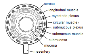
It is a hollow tube having a central cavity or lumen surrounded by a distensible wall, wall of gut is differentiated into four regions which starting from outside to inside are serosa, muscuaris, externa, submucosa and mucosa
(i) Serosa: It is fibrous outer coat or covering layer of gut.
Serous consists of two parts, visceral peritoneum and a sheet of loose connective tissue called subserosa
(ii) Muscularis externa( muscular coat): It is quite thick in the region of stomach and proximal part of intestine muscularis externa has an outer layer of longitudinal smooth fibres followed by a thick layer of circular smooth muscles. Another layer of oblique muscle fibres occurs in stomach inner to circular muscle fibres. On contraction, circular muscular muscle fibres bring about narrowing of gut while longitudinal muscle fibres cause shortening of gut
(iii) Submucosa. It is a layer of connective tissues that lies below the muscularis externa. Submucosa contains nerves, blood vessels and lymph vessels. The tissue has a lot of collagen and elastin fibres for permitting changes in transverse and longitudinal dimensions
(iv) Mucosa: It is the innermost which is thrown into circular folds or plical circulars ( valves of kerking or valvulae conniventes) in the region of intestine. Longitudinal folds or rugal occur in case of empty stomach. Circular folds slow down the movement of food through the intestine. The intestinal wall also bears millions of microscopic figure like projections called villi. Mucosa is differentiated into three layer, namely epithelium, lamina propria and muscularis mucosae
(a) Epithelium
These are columnar cell and possess upto 3000 microvilli on their free surface.Goblet or mucous cells are columnar shaped glandular cells which secrete mucus. Mucus functions as lubricant. It protects the wall of gut from action of proteolytic enzymes. Mucus further provides protection to the epithelium against abrasion or excoriation
Epithelium bears a number of pits. The pits bears deep tubular glands in stomach and upper duodenum. They are called crypts of Lieberkuhn in the region of intestine
(b) Lamina propia
It is a layer of aerolar reticular connective tissue which contains some scattered smooth muscles cells, blood, capillaries, lymph vessels and non-medullated nerve fibres. It posses Peyer’s patches
(c) Mucularis mucosae: It forms the outer boundary of mucosa that possesses smooth longitudinal muscle fibres externally and smooth circular muscle fibres internally.
Digestive glands
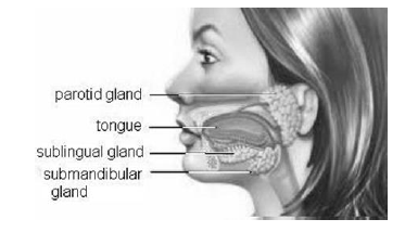
Salivary glands:
There are three pairs of salivary glands : parotid, sublingual and submaxillary or submandibular.
(i) Parotid glands: These are the largest salivary glands. They lie on the sides of the face, just below and in front of ears. Viral infection of parotid glands causing swelling and pain, is the disease called mumps.
(ii) Sublingual glands: These lie under the front part of the tongue
(iii) Submaxillary glands: These lie at the angles of the lower jaw. The submaxillary ducts, also known as wharton’s ducts open under the tongue
Saliva
• The salivary gland secrete a viscous fluid called saliva. It contains water, salts, an enzyme salivary amylase or ptyalin
• Its pH is neutral, being 6.7
• About 1.0 – 1.5 litres of saliva is produced daily
• The salivary gland secretion is very rich in salivary amylase.
The secretion from sublingual and submaxillary glands are rich in mucin. Saliva contains two important enzyme, amylase and lysozyme.
• Amylase is a starching digesting enzyme, breaking starch into maltose and triose. Lysozyme causes lysis of several common bacteria that may be present in mouth. Mucin in saliva helps to lubricate the food for swallowing
Function
(i) It moistens and lubricates the buccal cavity, tongue and lips, thus making speech possible.
(ii) It also moistens food and changes it to semi-solid mass for easy swallowing
(iii) Saliva washes mouth and tongue clear of food debris
(iv) Its enzyme help in digestion
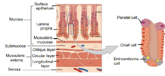
Gastric glands
• Fundic glands ( oxyntic glands): They secrete HCl, pepsinogen and soluble mucin
• Pyloric glands: Secretion is rich in mucin and does not contain HCl
• Cardiac glands : Secrete mucin and very little pepsinogen
• Types of cells present in the epithelium of gastric glands are –
(i) Peptic cells are usually basal in location and secrete gastric digestive enzymes as proenzymes, pepsinogen and prorenin. The chief cells (zymogenic or peptic cell ) also produce small amount of gastric amylase and gastric lipase, pepsinogen and chymosin
(ii) Oxyntic cells are large and are most numerous on the side walls of the gastric glands. They secrete hydrochloric acid (HCl) and castle’s intrinsic factor
(iii) Mucous cells are present between other and types of cells and secrete mucus. It protects stomach wall against HCl action and produce protein digestive enzyme
(iv) Endocrine glands are usually present in the basal parts of the gastric glands. They secrete serotonin, somatostatin and histamine, gastrin
• Serotonin is a vasoconstrictor and stimulates the smooth muscles
• Somatostatin suppresses the release of hormones from the digestive tract
• Histamine dilates the walls of blood vessels
• Gastrin stimulate the gastric glands to release gastric juice
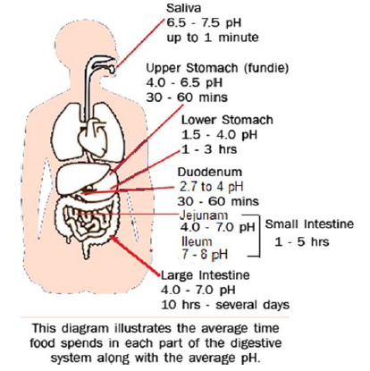
Gastric juice
The secretion of the cells of gastric glands from gastric juice with pH 2 – 3.7. About 2,000 to 3,000 ml of gastric juice is secreted per day
Liver
• It is the largest internal gland of the body which is reddish brown in colour.
• The liver lies in upper right side of the abdominal cavity just below the diaphragm.
• It is heavier in males than females. In males it is generally 1.4 -1.8 kg and in females 1.2 – 1.4kg
• It has both endocrine and exocrine parts
• Liver is differentiable into small left lobe and large right lobe.
The right lobe is further made of right lobe proper, quadrate lobe and caudate lobe. A pyriform yellow green muscular sac or gall bladder lie on inferior surface of right lobe. It is 8cm long and 2cm wide
• The two lobes of liver produce right and left hepatic ducts. They join to form common hepatic duct. A cystic duct comes from gall bladder. The two join to form bile duct that attaches with pancreatic duct before opening in duodenum.
• The opening of bile duct into pancreatic duct is controlled by sphincter of Boyden
• Amount of bile produced per day is 600 ml. Bile from gall bladder is 5-10 times more concentrated. Its pH is 7.6 compared to 8.6pH of hepatic bile
• Bile is yellowish green alkaline solution with 89-98% water, no digestive enzymes but four types of chemicals and some nondigestive enzymes
(a) Inorganic salts: Bicarbonates, chlorides, carbonates and phosphates of sodium, potassium and calcium
(b) Bile salts: Sodium taurocholate and sodium glycocholate,. They are partly water soluble and partly fat soluble
(c) Fatty substances: Cholesterol, lecithin and other phospholipids
(d) Bile pigments: Bilirubin ( yellowish) and biliverdin (greenish).
Other minor bile pigment are bilicyanin, bilipresin, bilifusun, bilihumin and bilifulvin
Functions of liver
(i) Secretion of bile
(ii) Deamination – It is a process by which the amino group (-NH2) is removed from amino acids resulting the production of ammonia which is converted into urea
(iii) Excretion
• Liver synthesis urea with the help of ammonia and carbon doxide urea is passed out through excretory system
• The bile contains bile pigments ( bilirubin and biliverdin) are excretory products
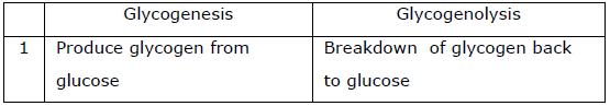
• The liver cells also eliminate certain other waste products like cholesterol, metal ions and waste products of haemoglobin (iv) Glycogenesis : It is the conversion of the excess of glucose into glycogen by liver cells with the help of insulin secreted by
pancreas
(v) Glycogenolysis: It is the conversion of glycogen into glucose by the liver cells with the help of glucagon secreted by pancreas
(vi) Lipogenesis: It is the conversion of glucose and amino acids into fats which takes place in liver
(vii) Gluconeogenesis: It is the formation of glucose or glycogen from non-carbohydrate sources like amino acids, fatty acids etc.
(viii) Detoxification: Liver converts toxic substances into harmless substances
(ix) Haemopoiesis: The process of formation of blood corpuscles is called haemopoiesis. The liver produces red blood corpuscles in the embryo
(x) Synthesis of blood protein: The liver produces blood protein such as prothrombin and fibrinogen that help in the clotting of blood
(xi) Secretion of heparin- Liver secretes heparin ( anticoagulant)
(xii) Lymph formation: Liver is important for lymph formation
(xiii) Synthesis of vitamin-A from B-carotene
(xiv) Destruction of R.B.C. : The old worn out red blood corpuscles are broken down in the liver cells. Their haemoglobin is changed into bile pigments
(xv) Phagocytosis: the Kupffer’s cells of the liver engulf the disease causing microorganisms, dead cells and foreign matter
(xvi) Osmoregulation: Liver produces angiotensinogen which helps kidneys in maintaining body fluid osmoregulation through the action of renin on angiotensin.
Pancreas
The pancreas is soft, lobulated grayish – pink gland which weighs about 60 grams. It is about 2.5cm wide and 12-15 cm long, located posterior to the stomach in the abdominal cavity 5.4.01 External structure of pancreas
• The main pancreatic duct ( duct of Wirsung) is formed from smaller ducts within the pancreas. The main pancreatic duct opens into hepatopancreatic ampulla.
• An ancessory pancreatic duct ( duct of Santorini) is also present in pancreas and open directly into the duodenum
Internal Structure of Pancreas
(i) Exocrine part: the exocrine part of the pancreas consists of rounded lobules (acini) that secretes with pH 8.4. About 500-800 ml of pancreatic juice is secreted out per day. The pancreatic juice is carried by the main pancreatic duct into the abdomen through hepatopancreatic ampulla.
The pancreatic juice contains sodium bicarbonate, three proenzymes; trypsinogen, chynao-trypsinogen, chymotrypsinogen anad procarboxy peptidase and some enzymes such as pancreatic amylase DNAase, RNAase pancreatic lipase. The pancreatic juice
helps in the digestion of starch, protein, nucleic acids and fats, therefore pancreatic juice is also called complete digestive juice
(ii) Endocrine part: the endocrine part of pancreas consists of groups of islets of Langerhans Each islet is separated from the surrounding alveoli by a thin layer of reticular tissue. The islets are richly supplied with blood through a dense capillary plexus. The following three types of cells occur in pancreatic islets.
a) The alpha cells (α) – Secreting hormone glucagon
b) The beta cells (β) – Secreting hormone insulin
c) The delta cells (δ) – Producing hormone gastrin
In the islets of pancreas the alpha cells are arranged towards the periphery of the islets, the beta cells near the centre and the delta cells are placed peripherally
Intestinal glands
• The intestinal glands are numerous and microscopic. These are 2 types; crypts of Lieberkuhn and Brunner’s gland
(i) The crypts of lieberkuhn are simple tubular glands and occur throughout the small intestine between the villi. They have two types of cells
(a) Paneth cells are found particularly in the duodenum. These cells are rich in zinc and contain acidophilic granules. They secrete lysozyme ( antibacterial substance)
(b) Argentaffin cells is located among epithelial cells and is common in duodenum. It secretes serotonin. Serotonin is a powerful stimulant of smooth muscle, resulting in contraction and play a role in stimulating peristaltic activity of the intestine
(ii) The Brunner’s gland are branched tubular glands and are confined to the duodenum. They secrete alkaline watery fluid, little enzyme and mucus. The mixture of secretions is called intestinal juice or success entericus
About 2 to 3 litres of succus entericus is secreted every day. It is alkalin ( ph = 8.3) and contains many enzyme amino peptideases, dipeptidases, intestinal amylases, maltase, isomaltase, nucleo-sidases
Process of digestion
• Digestion is the breakdown of large complex insoluble organic food molecules into small, simpler soluble and diffusible particles by the action of digestive enzymes
Digestion in mouth
• The tongue helps in tasting the food, mixes it well along with saliva. The saliva is continuously released and secreted into the buccal cavity by salivary glands under the influence of autonomic nervous system
• Mucus in saliva helps in lubricating and adhering the masticated food particles into a bolus. The bolus is then conveyed into pharynx and then into oesophagus by swallowing or deglutition.
• Saliva contains about 99% water along with K+ and HCO3.
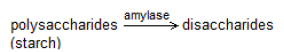
It contains a carbohydrate splitting enzyme called salivary amylase. About 30% of starch is hydrolysed into a disaccharide – maltose
Digestion by Stomach
• The bolus of food remains in the stomach for 3-4 hours and gradually converted into semifluid mixture called chyme
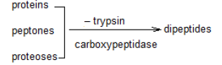
• The proenzyme pepsinogen, on exposure to hydrochloric acid gets converted into active enzyme pepsin. Pepsin converts proteins into protesoses and peptones ( peptides).
• The mucus and bicarbonates play important role in lubrication and protection of mucosal epithelium
• Renin is proteeolytic enzyme found in gastric juice of infants which helps in the digestion of milk proteins
Digestion by intestine
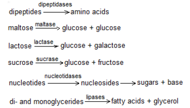
• Trypsinogen is activated by an enzyme, enterokinase, secreted by intestinal mucosa into active trypsin, which in turn activates the other enzymes. Proteins, proteases and peptones in chyme reaching the intestine are converted into dipeptides
• Bile helps in emulsification of fats i.e. breaking down of fats into very small micelles. Bile also activates lipases. Fats are broken down by lipases with the help of bile into di- and monoglycerides.
• Carbohydrates in the chime are hydrolysed by pancreatic amylase into disaccharides
• Nucleases in the pancreatic juice acts on nucleic acids to form nucleotides and nucleosides
• The enzymes in the succus entericus acts on the end products to form respective simple absorbable forms
Neural control of digestion
• The gastro intestinal tract innervated, by intrinsic nerves as well as extrinsic nerves
• The intrinsic nerves controls gastrointestinal functions like secretion
• The extrinsic nerves which can modify the activity of intrinsic neural system
• The sight, smell and presence of food in the gastrointestinal tract act as a stimuli for secretion of saliva. This happens by stimulation of vagus nerve
Digestive hormones in gastrointestinal tract-
Summary
1) Hormone Gastrin
Produced by Gastric antrum, duodenum ( Gcells)
It stimulatesecretion of gastricacid and intrinsic factor from parietal cells, stimulate secretion of pepsinogen from chief cells also promotesgastric and intestinal motility , mucosal growth
2. Cholecystokinin (CCK)
Produced by Duodenum, jejunum ( I cells)
It stimulates gall bladder contraction, stimulates release of pancreatetic enzyme, relaxes sphincter of Oddi for release of bile and enzymes, role is inducing satiety
3. Secretin
Produced by Duodenum, jejunum ( S cells)
It stimulates secretion of HCO3 frompancrease, inhibits gastrin and gastric acid secretion
4. Vasoactive intestinal peptide (VIP)
Produced by Enteric nerves
It increases water and electrolyte secretion from pancreas and gut, relaxes smooth muscles ( via nitric oxide)of gut
5.Gastric inhibitory polypeptide (GIP)
Produced by Duodenum, jejunum ( K cells),
Reduces gastric acid secretion and intestinal motility,stimulates insulin release
6. Somatostatin
Produced by Stomach, small intestine and pancreas (D cells)
It inhibits secretion and action of many hormones, including all of the above
Absorption of digested food
• Only some drugs are absorbed in mucosal lining of sublingual area
• Water, alcohol, some drugs and sugar are absorbed in stomach
• In large intestine absorption of Na+, Cl- and vitamins ( vit K, B12, thiamine, riboflavin) produced by intestinal bacteria is undertaken. Solidification of indigestible matter also occurs here due to withdrawal of water
• Simple diffusion helps in passage of alcohol, fatty acids, monoglycerides, cholesterol and fat soluble substances. Osmosis is involved in passage of water from lumen to epithelial lining
• Fructose and few amino acids are absorbed through facilitated diffusion Na+ is main ion absorbed actively. Others involved in active transport are Ca2+, Fe2+, K+, Mg2+
• Co-transport occurs in case of glucose and most amino acids. Most of the absorbed materials are passed into para cellular spaces
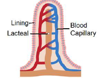
• Most monoglycerides and fatty acids are converted into triglycerides which along with cholesterol, phospholipids and some protein forms chylomicrons inside Golgi apparatus. Chylomicrons are passed into lymph vessels or central lacteals.
Nutritional requirement
Carbohydrates, proteins and fat
1. Carbohydrates
• The sources of carbohydrates in our diet are cereals ( rice, wheat, maize), potato, grams, fruits ( banana, mango, melon), sugar, honey, sugarcane, beet, jam and milk
• An adult man of average weight and doing moderate work needs about 350-500gms of carbohydrates daily
2. Proteins
• The sources of proteins in our diet are cereals, pulses, oil, seeds and nuts, fish, egg, milk, soyabean, cheese, leafy vegetable.
• About 70 – 100 gms of protein are needed daily
3. Fats
• The sources of fats in our diet are vegetable cooking oil, ghee, butter, chese, eggs.
• About 50gms of fat is needed daily
Minerals
1. Sodium
• The sources are table salt, beef, spinach
• It maintains electric potential across the membrane. It maintains pH balance of our body
• Deficiency of sodium causes cramps, diarrhea and dehydration, low blood pressure
2. Potassium
• The sources are banana, orange, milk, spinach
• It helps to retain water in cells. Maintains osmotic pressure
• Deficiency causes muscle disorder low BP
3. Chlorine
• The sources are table salt, cabbage, cheese
• It helps in regulation of osmotic pressure. It helps in formation of HCl in stomach
• Deficiency of loss of appetite, causes cramps
4. Calcium
• The sources are milk, green leafy vegetables, cheese, cereals
• Helps in clotting of blood. It helps in building of bones and teeth
• Deficiency causes rickets, muscle spasms, retarded body growth
5. Phosphorous
• The sources are cereal grains, milk, meat, peas, cheese
• It required for tooth and bone formation. It helps regulation of heart beat
• Deficiency causes metabolic disorders
6. Iron
• Sources: Oatmeal, honey, dates, eggs, spinach, pulses
• Importance : Formation of haemoglobin
• Deficiency causes: Weakness and weak immunity
7. Iodine:
• Sources: Onion, table salt
• Causes goiter ( Deficiency)
8. Sulphur
• Sources : Meats, daily products
• Importance: It helps to keep hair skin and nails healthy
• Deficiency causes unhealthy growth of skin and hair
9. Magnesium
• Sources: Dairy products, green leafy vegetable, chocolate
• It helps in muscle relaxation
• Deficiency causes hallucinations, irregularities of metabolism
10. Copper
• Sources: Peanuts, beet, barley, soyabean, onion
• It helps in development of blood vessels and connective tissue
• Deficiency causes anemia
11. Chromium
• Sources : Black gram, soyabean, carrot
• It promotes insulin action in the metabolism of sugar
• Deficiency of nitrogenous waste products
12. Fluorine
• Sources: tea, city water supply
• It maintains normal enamel and check dental caries
• Deficiency causes weak teeth which are prone to decay
13. Zinc
• Sources: Barley, soyabean, groundnut, egg, almond
• It hastens healing of wounds. It is needed for healthy skin and hair
• Deficiency causes retarded growth rough skin and weak immunity
14. Cobalt
• Sources: Milk
• Deficiency causes anemia
15. Manganese
• Sources: Nuts, legumes
• It is important for reproduction and synthesis of haemoglobin
• Deficiency causes infertility, menstrual problems
16. Molybdenum
• Sources: Onion, potato, bajra
• It is important for synthesis of haemoglobin and absorption of iron
• Deficiency causes irregular excretion of nitrogenous waste products
17. Selenium
• Sources: Barley, garlic, orange juice
• It acts with vitamin E for slowing down ageing process
• Deficiency causes higher risk of cancer, cardiovascular disease
Fat soluble vitamins
(1) Vitamin – A
• Sources: Milk, butter, maize, carrot, papaya, spinach
• Deficiency causes night blindness drying of eyeball etc
(2) Vitamin – D
• Sources: Sunlight, cod liver oil, eggs
• Deficiency causes rickets
• Increases intestinal absorption of calcium and phosphorous
(3) Vitamin – E
• Sources : green vegetables, vegetable oil
• Deficiency causes reproduction failure, slow growth, anemia
(4) Vitamin – K
• Sources: Leafy vegetables
• Deficiency causes profuse and prolonged delay in blood clotting
Water soluble vitamins
(1) Vitamin B1
• Sources : yeast, sprouted beans, nuts, pulses, cereal
• Deficiency causes beriberi, loss of appetite, accumulation of keto acids in blood
(2) Vitamin B2
• Sources: yogurt, milk, green vegetables
• Deficiency causes skin disorder, headache, cheilosis ( sores at corner of mouth)
(3) VitaminB3
• sources: pulses, liver, fish
• Deficiency causes pellagra, gum diseases, skin problems
(4) Vitamin B5
• Sources: eggs, milk, ground nut and tomatoes
• Deficiency causes fatigue, muscle cramp, poor motor coordination
(5) Vitamin B6
• Sources: All plant and animal tissues
• Deficiency cause anemia, diarrhea, nausea and vomiting
(6) Vitamin B12
• Sources: Meat, fish, egg, milk
• Deficiency causes nervous disorder
(7) Vitamin C
• Sources: All citrus fruit
• Deficiency causes scurvy, bleeding from small vessels
Disorders of digestive system
1. Jaundice: The liver is affected , skin and eyes turn yellow due to deposit of bile pigment
2. Vomiting: It is the ejection of stomach contents through the mouth. This reflex action is controlled by vomit centre in the medulla
3. Diarrhoea: the abnormal frequency of bowel movement and increased liquidity of faecal discharge is known as diarrhea
4. Constipation: in constipation, the faeces are retained within the rectum
5. Indigestion: in this condition, the food is not properly digested leading to a feeling of fullness
Distinguish
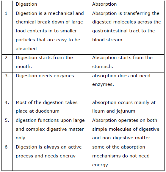
Digestion Vs Absorption
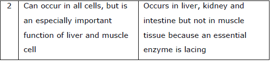
Glycogenesis Vs Glycogenolysis
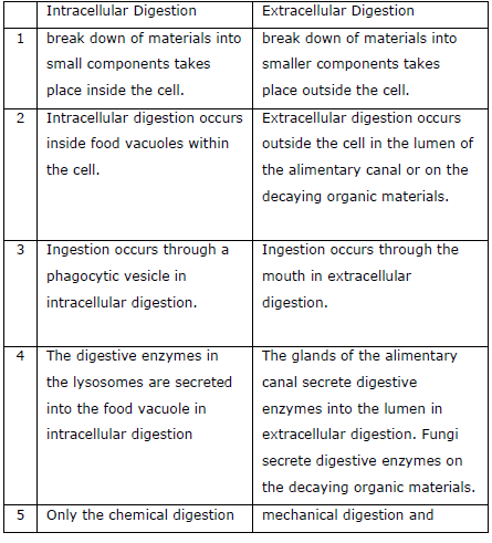
Intracellular Digestion Vs Extracellular
Digestion
Gluconeogenesis Vs Glycogenolysis
Enzyme summary
Mouth
Enzymes & Function
Salivary Amylase - break down starches convert to maltose sugarpH
neutral
Lingual lipase - starts the digestion of the lipids/fats.
Stomach
Enzymes & Function:
Pepsin- Breaks protein into small peptides
Gastric amylase- Degradation of starch
Gelatinase- Degradation of gelatin and collagen
Rennin- Conversion of liquid milk to solid particles Gastric lipase- Degradation of butter fat
Pancreas
Enzymes & Function:
Pancreatic lipase- Degrades triglycerides into fatty acids and glycerol
Phospholipase- Hydrolyzes phospholipids into fatty acids Trypsin- Converts proteins to basic amino acids
Steapsin- Breakdown of triglycerides to glycerol and fatty acids
Chymotrypsin- Converts proteins to aromatic amino acids
Carboxypeptidase- Degradation of proteins to amino acids
Pancreatic amylase- Degradation of carbohydrates to simple sugars
Elastases- Degrade the protein elastin
Nucleases- Conversion of nucleic acids to nucleotides and nucleosides
Small Intestine
Enzymes & Function:
Sucrase- Converts sucrose to disaccharides and monosaccharides
Maltase- Converts maltose to glucose
Lactase- Converts lactose to glucose and galactose
Isomaltase- Converts maltose to isomaltoses