

Official CBSE Assignments for Class 11 Biology
Access comprehensive school assignments for Chapter 18 Neural Control And Coordination using the CBSE Class 11 Biology Neural Control and Coordination Assignments. Designed to align with the 2026-27 CBSE academic guidelines, these practice sets help Class 11 Biology students reinforce core concepts and improve their problem-solving accuracy.
Solved Practice Assignments for Biology
Access the complete assignment PDF for Class 11 Biology below. Regular practice with these targeted academic tasks builds familiarity with standard question patterns and helps secure higher marks in final school evaluations.
Ques. In a man, abducens nerve is injured. Which one of the following functions will be affected?
(a) Movement of the eyeball
(b) Movement of the tongue
(c) Swallowing
(d) Movement of the neck
Answer: A
Question. Intercellular communication in multicellular organism occurs through
(a) digestive system only
(b) respiratory system only
(c) nervous system only
(d) both nervous and endocrine system.
Answer. D
Question. Nerve fibre gets depolarised when it acquires
(a) +ve charges on inner side
(b) +ve charges on outer side
(c) –ve charges on inner side
(d) Na+ and K+ from outside.
Answer. A
Question. Which of the following statement is correct about node of Ranvier of neuron?
(a) Neurilemma is discontinuous.
(b) Myelin sheath is discontinuous.
(c) Both neurilemma and myelin sheath are discontinuous.
(d) Covered by myelin sheath.
Answer. B
Question. Pre-synaptic membrane is part of
(a) dendron
(b) axon hillock
(c) telodendria
(d) soma.
Answer. C
Question. Column I lists the parts of the human brain and column II lists the functions. Match the two columns and select the correct option.
Column I Column II
A. Cerebrum p. Controls the pituitary
B. Cerebellum q. Controls vision and hearing
C. Hypothalamus r. Controls the rate of heart beat
D. Midbrain s. Seat of intelligence
t. Maintains body posture
(a) A-t; B-s; C-q; D-p
(b) A-s; B-t; C-q; D-p
(c) A-t; B-s; C-p; D-q
(d) A-s; B-t; C-p; D-q
Answer. D
Question. Four healthy people in their twenties got involved in injuries resulting in damage and death of few cells of the following. Which of the cells are least likely to be replaced by new cells?
(a) Osteocytes
(b) Malpighian layer of the skin
(c) Liver cells
(d) Neurons
Answer. D
Question. Which labelled part in the given diagram controls the process of breathing? (Img 185)
(a) A
(b) B
(c) C
(d) D
Answer. C
Question. Which part of neuron is highly branched and afferent?
(a) Axon
(b) Dendrite
(c) Cyton
(d) Both (a) and (b)
Answer. B
Question. Select the incorrect statement.
(a) The Nissl’s granules probably synthesise fats for the cell
(b) Ageing neuron contain a pigment lipofuscin
(c) Nissl’s granules are irregular masses of rough endoplasmic reticulum
(d) None of these
Answer. A
Question. By which nervous system, the blood is supplied into visceral organs?
(a) Sympathetic nervous system, voluntary
(b) Sympathetic nervous system, involuntary
(c) Parasympathetic nervous system, involuntary
(d) Both (b) and (c)
Answer. D
Question. The given diagram shows axon terminal. Select the option that correctly matches the steps in transmission of impulses (list i – vii) with the labellings (A – C) in diagram.
(i) Chemicals called neurotransmitters are released in the synaptic cleft through ion channels. (Img 185)
(ii) When an impulse arrives at the axon terminal, it stimulates the movement of synaptic vesicles.
(iii) Neurotransmitters are endocytosed into the neurons.
(iv) The ion channels close with the binding of neurotransmitters to their specific receptors vesicles.
(v) Synaptic vesicles move towards the membrane where they fuse with the plasma membrane.
(vi) Neurotransmitters are released in the synaptic cleft.
(vii) The released transmitters bind to their specific receptors on post-synaptic membrane.
A B C
(a) (ii) (iii) (i)
(b) (v) (vi) (iv)
(c) (ii) (vi) (vii)
(d) (v) (iii) (iv)
Answer. C
Question. Read the given statements and select the correct option.
I. The continuity between the pre-synaptic and post-synaptic neurons is provided by gap junctions in chemical synapses.
II. Release of calcium ions from pre-synaptic knob stimulates the release of neurotransmitter into synaptic cleft.
III. More energy is required for impulse conduction through myelinated neurons due to saltatory conduction.
IV. After transmission of impulse, neurotransmitteris hydrolysed by an enzyme present at synapse.
(a) I and II are correct but III and IV are incorrect.
(b) I and IV are correct but II and III are incorrect.
(c) I, II and III are correct but IV is incorrect.
(d) I, II and III are incorrect, but IV is correct.
Answer. D
Ques. Injury to vagus nerve in humans is not likely to affect
(a) tongue movements
(b) gastrointestinal movements
(c) pancreatic secretion
(d) cardiac movements.
Answer: A
Ques. Which cranial nerve has the highest number of branches?
(a) Vagus nerve
(b) Trigeminal nerve
(c) Facial nerve
(d) None of these
Answer: B
Ques. Sympathetic nervous system induces
(a) secretion of digestive juices
(b) heart beat
(c) secretion of saliva
(d) all of these.
Answer: B
Ques. The vagus nerve is the cranial nerve numbering
(a) 7
(b) 5
(c) 10
(d) 9.
Answer: C
Ques. By which nervous system and of what type, the blood is supplied into visceral organs?
(a) Both SNS and PNS, involuntary
(b) Para-sympathetic nervous system involuntary
(c) Sympathetic nervous system, involuntary
(d) Sympathetic nervous system, voluntary
Answer: A
Ques. The sympathetic nerves, in mammals, arise from
(a) sacral nerves
(b) 3rd, 7th, 9th and 10th cranial nerves
(c) thoraco-lumbar nerves
(d) cervical nerves.
Answer: C
Ques. Afferent nerve fibres carry impulses from
(a) effector organs to CNS
(b) receptors to CNS
(c) CNS to receptors
(d) CNS to muscles.
Answer: B
Ques. Vagus nerve is
(a) X
(b) IX
(c) VII
(d) V.
Answer: A
Ques. One function of parasympathetic nervous system is
(a) contraction of hair muscles
(b) stimulation of sweat glands
(c) acceleration of heart beat
(d) constriction of pupil.
Answer: D
Ques. Which of the following cranial nerves can regulate heart beat?
(a) X
(b) IX
(c) VIII
(d) VII
Answer: A
Ques. Nissl’s bodies are mainly composed of
(a) proteins and lipids
(b) DNA and RNA
(c) nucleic acids and SER
(d) free ribosomes and RER.
Answer: D
Ques. Myelin sheath is produced by
(a) astrocytes and Schwann cells
(b) oligodendrocytes and osteoclasts
(c) osteoclasts and astrocytes
(d) Schwann cells and oligodendrocytes.
Answer: D
Ques. Receptor sites for neurotransmitters are present on
(a) pre-synaptic membrane
(b) tips of axons
(c) post-synaptic membrane
(d) membranes of synaptic vesicles.
Answer: C
Ques. The most abundant intracellular cation is
(a) H+
(b) K+
(c) Na+
(d) Ca++.
Answer: B
Ques. When a neuron is in resting state i.e., not conducting any impulse, the axonal membrane is
(a) comparatively more permeable to Na+ ions and nearly impermeable to K+ ions
(b) equally permeable to both Na+ and K+ ions
(c) impermeable to both Na+ and K+ ions
(d) comparatively more permeable to K+ ions and nearly impermeable to Na+ ions.
Answer: D
Question. Compare the central nervous system (CNS) with peripheral nervous system (PNS).
Answer. Central Nervous system (CNS) comprises of brain and the spinal cord. It is the site of information processing and control. Peripheral nervous system (PNS) consists of neurons. Some peripheral neurons (called afferent neurons) collect information from the tissue/organs and transmit it to the CNS and other neurons transmit regulatory information from the CNS to the concerned tissue or organ.
Question. What is the difference between electrical transmission and chemical transmission?
Answer. Differences between electrical and chemical transmission are as follows : (Table 195)
Question. Fill in the blanks at (A), (B), (C) and (D). (Img 190)
Answer. (A) – Neurotransmitter
(B) – Specific receptors
(C) – Ion channels
(D) – Postsynaptic neuron
Question. Briefly describe the thirst centre located in human brain.
Answer. Hypothalamus is the thirst centre present in human brain. It is located in the forebrain. It links nervous system to endocrine system (via hypothalamus - hypophyseal axis) and
exercises a regulatory control on the functioning of endocrine glands by secreting neurohormones. It contains higher centres of autonomic nervous system controlling hunger, thirst, sleep, fatigue, emotions, satisfaction, anger, pleasure, etc. It also controls carbohydrates and fat metabolism, body temperature, blood pressure and water balance.
Question. If someone receives a blow on the back of neck, what would be the effect on the person’s CNS?
Answer. If someone receives a blow on the back of the neck, then his medulla oblongata would get hurt, as this is the place where it is located. The person might die as medulla controls respiration, cardiovascular reflexes and gastric secretions.
Question. What does nervous system of earthworm comprise of?
Answer. Earthworm has a nervous system consisting of well developed ventral nerve cord, paired segmental ganglia and segmental nerves.
Question. Name the following.
(a) The part of the human brain that is most developed.
(b) Part of our central nervous system that acts as a master clock.
(c) The scattered masses of grey matter.
(d) The additional space lying above the duramater.
Answer. (a) Cerebrum (cerebral hemispheres). (b) Pineal gland (c) Basal ganglia (d) Epidural space
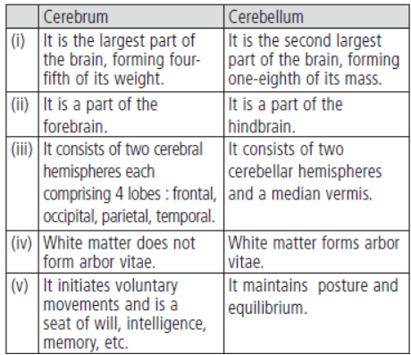
Question. Distinguish between cerebrum and cerebellum.
Answer. Cerebrum and cerebellum can be differentiated as follows:
Long Answer Type Questions
Question. Explain the detailed structure of a neuron with a suitable diagram.
Answer. A neuron (nerve cell) is the structural and functional unit of the neural system. Neurons with longer processes (projections) are the longest cells in the body. Fully formed neurons never divide and remain in interphase stage throughout life. Shortly after birth, new neurons do not develop. A neuron is a microscopic structure composed of three major parts, namely, cell body, dendrites and axon. (i) Cell body (Cyton or Soma) : Like a typical cell, it consists of cytoplasm, nucleus and cell membrane. It has abundant cytoplasm called neuroplasm and a relatively large spherical central nucleus with a distinct nucleolus. The cytoplasm has mitochondria, Golgi apparatus, rough endoplasmic reticulum, ribosomes, lysosomes, fat globules, pigment granules, neurofibrils, neurotubules and Nissl’s granules. Cyton is concerned with metabolic maintenance and growth. The processes of neurons are called neurites that are divided further into dendrites and axons. (ii) Dendrites are usually shorter, tapering and much branched processes that may be found here one to several. They conduct nerve impulse towards the cell body and are called afferent processes. (iii) Axon is a single, usually very long process of uniform thickness. The part of cyton from where the axon arises is called axon hillock. It is the most sensitive part of neuron. The axon contains neurofibrils and neurotubules but lacks Nissl’s granules, Golgi complex, ribosomes, pigment granules, fat globules, etc. The cell membrane of the axon is called axolemma and its cytoplasm is known as axoplasm. The axon ends in a group of branches, the terminal arborisations which when meet the dendrites of another neuron to form a synapse, form synaptic knobs or end plates. The synaptic knobs contain mitochondria and secretory vesicles. The part of the sarcolemma (muscle plasma membrane) that lies beneath the axon terminal/nerve endings, is called motor end plate. The axon conducts nerve impulses away from the cell body, therefore, called an efferent process. There are two types of nerve fibres namely myelinated and non-myelinated. The myelinated nerve fibres are enveloped with Schwann cells, which form a myelin sheath around the axon. The gaps between two adjacent myelin sheaths are called nodes of Ranvier. Non-myelinated nerve fibre is enclosed by a Schwann cell that does not form a myelin sheath around the axon. For labelled diagram of a neuron refer to answer 26.
Question. Explain the process of the transport and release of a neurotransmitter with the help of a labelled diagram showing a complete neuron, axon terminal and synapse.
Answer. Chemicals called neurotransmitters are involved in the transmission of impulses at chemical synapses. The axon terminals contain vesicles filled with these chemicals. When an impulse arrives at the axon terminal, it stimulates the movement of the synaptic vesicles towards the membrane where they fuse with the plasma membrane and release their neurotransmitters in the synaptic cleft. The released neurotransmitters bind to their specific receptors, present on the post-synaptic membrane. This binding opens ion channels allowing the entry of ions which can generate a new potential in the post-synaptic neuron. The given diagram shows neuron, axon terminal and synapse. (Img 199)
NEURAL CONTROL AND COORDINATION
POINTS TO REMEMBER
Action potential: A sudden change in the electrical charges in the plasma membrane of a nerve fibre.
Aqueous humour: The thin watery fluid that occupy space between lens and cornea in eye.
Blind spot: A spot on retina which is free from rods and cones and lack the ability for vision.
Cerebrospinal fluid: An alkaline fluid present in between inner two layer of meninges.
Cerebellum: A part of hind brain that controls the balance and posture of the body.
Cochlea: A spirally coiled part of internal ear which is responsible for hearing.
Corpus callosum: A curved thick bundle of nerve fibres that joins two cerebral hemisphere.
Depolarisation: A condition when polarity of the plasma membrane of nerve fibre is reversed.
Endolymph: The fluid filled within membranous labyrinth.
Eustachian tube: A tube which connect ear cavity with the pharynx.
Fovea: A area of highest vision on the ratina which contain only cones.
Meninges: Three sheets of covering of connective tissue wrapping the brain.
Grey Matter: This shows many convolutions which increase the amount of vital nerve tissue.
Medulla oblongata: Posterior most part of the brain which is continuous with spinal cord and control respiration, heart rate, swallowing, vomiting.
Pons: Thick bundles of fibres on the ventral side of brain below cerebellum.
Foramen magnum: A big aperture in the skull posteriorly through which spinal cord emerges out.
Spinal cord: A tubular structure connected with medulla oblongata of brain and situated in the neural canal of the vertebral column, covered by meninges.
Synaptic cleft: A narrow fluid filled space which separates two membranes of the two neurons at the synapse.
Synaptic vesicles: These are membrane bound vesicles in the axoplasm of the axon terminal and these store neurotransmitter.
Neurotransmitter: These are chemicals stored in synaptic vesicles, diffuse to reach the membrane of next neuron for its stimulation.
Synapse: A physiological junction between axon of one neuron and dendrite of next neuron.
CNS: Central neural system
PNS: Peripheral neural system
ANS: Autonomic neural system
Cell body: Cytoplasm with nucleus, cell organelles and Nissl's granules
Dendrites: Short fibres which branch rapidly and project out of cell
Axon: Single, long fibre, branched at distal end
Multi polar: One axon and two or more dendrites. Found in cerebral Cortex
Bipolar: One axon and one dendrite. Found in retina of eye
Unipolar: Cell body with axon only. Found usually in the embryonic stage
Conduction of nerve impulse along axon
Polarised membrane/Resting Potential: In resting phase when neuron is not conducting an impulse, the axonal membrane is called polarised. This is due to difference in concentration of ions across the axonal membrane.
At Rest:
- Axoplasm inside the axon contain high concentration of \(\text{K}^+\) and low concentration of \(\text{Na}^+\).
- The fluid outside the axon contain low concentration of \(\text{K}^+\) and high concentration of \(\text{Na}^+\).
As a result the outer surface of axonal membrane is positively charged and inner surface is negatively charged. The electric potential difference across the resting plasma membrane is called resting potential.
Action Potential: When a nerve fibre is stimulated, the permeability of membrane to \(\text{Na}^+\) is greatly increased at the point of stimulus (rapid influx of \(\text{Na}^+\)) and hence polarity of membrane is reversed and now membrane is said to be depolarised. The electric potential difference across the plasma membrane at that site is called action potential, which in fact termed as nerve impulse.
Depolarisation is very rapid, so that conduction of nerve impulse along the entire length of axon occurs in fractions of second.
Transmission of Impulses at Synapse
(i) At electrical synapses: Here the membrane of pre and post-synaptic neuron are in very close proximity. Electric current can flow directly from one neuron into other across these synapses, like impulse conduction along a single axon.
(ii) At chemical synapses: Here the membrane of pre and post-synaptic neuron are separated by fluid filled space called synaptic cleft. Neurotransmitter are involved here.
When an impulse arrives at the axon terminal, it stimulates the movement of the synaptic vesicles towards membrane and they fuse with the plasma membrane and release their neurotransmitter in the synaptic cleft. These chemicals bind to specific receptors, present on the post-synaptic membrane. Their binding opens ion channels and allow the entry of ion which generate new potential in post synaptic neuron.
Human brain: Human brain is the major portion of central neural system. Which is well protected by the skull.
The brain is surrounded by three cranial meninges:
i) Duramater - outer layer
ii) Arachnoid - middle layer
iii) Piamater - inner layer-remain in contact with brain
Parts of Brain:
Fore brain: (a) Cerebrum, (b) Thalamus, (c) Hypothalamus
Mid brain
Hind brain: (a) Cerebellum, (b) Pons, (c) Medulla oblongata
Functions of parts of brain:
Cerebrum: Centre of intelligence, memory and imagination, reasoning, judgement, expression of will power.
Thalamus: Acts as relay centre to receive and transmit general sensation of pain, touch and temperature.
Hypothalamus: Centre for regulation of body temperature, urge for eating and drinking.
Mid brain: Responsible to coordinate visual reflexes and auditory reflexes.
Cerebellum: Maintains posture and equilibrium of the body as well as coordinates voluntary movements.
Pons varoli: Relays impulses between medulla oblongata and cerebral hemisphere and between the hemisphere of cerebrum and cerebellum.
Medulla oblongata: Centre that control heart beat, breathing, swallowing, salivation, sneezing, vomiting and coughing.
Reflex action: It is a spontaneous, autonomic and mechanical response to a stimulus that occurs at the level of spinal cord, with involvement of brain.
Reflex arc: The flow of nerve impulse along the specific pathway during reflex action. It consist of:
a) A receptor
b) An Afferent neuron (sensory neuron)
c) An interneuron
d) An efferent neuron (motor neuron)
e) An effector organ
Organ of Sight - Eye
| Layer | Component | Function |
|---|---|---|
| 1. External layer | Sclera | Protects and maintain shape of the eye ball. |
| Cornea | Absorb \(\text{O}_2\) from the air, helps to focus light rays. | |
| 2. Middle layer | Choroid | Absorb light and prevent light from being reflected within the eye ball. |
| Ciliary body | Holds lens, regulate shape of the lens. | |
| Iris | Control amount of light entering. | |
| 3. Inner layer | Retina | Vision in dim light, colour vision, vision in bright light. |
Organ of Hearing - Ear
| Portion of the ear | Component | Function |
|---|---|---|
| 1. External ear | Pinna | Collect sound waves. |
| External auditory canal | Direct sound waves toward ear drum, ear wax prevents the entry of foreign bodies. | |
| 2. Middle ear | Tympanic membrane | Acts as resonator that reproduces the vibration of sound. |
| Ear ossicles | Transmit sound waves to internal ear. | |
| Eustachian tube | Helps in equalising the pressure of either side of ear drum. | |
| 3. Internal ear | Cochlea | Hearing. |
| Vestebular apparatus | Balancing of body. |
QUESTIONS
Very Short Answer Questions (1 mark each)
Question 1. Name the fluid present in membranous labyrinth.
Answer: Endolymph
In simple words: Endolymph is the fluid found inside the membranous labyrinth of the inner ear.
Exam Tip: Remember that perilymph fills the bony labyrinth, while endolymph fills the membranous labyrinth.
Question 2. Name the area of retina where only cones are densely packed.
Answer: Fovea
In simple words: The fovea is the central pit in the retina responsible for sharpest daylight and colour vision.
Exam Tip: Fovea contains only cones and lacks rods entirely, providing high visual acuity.
Question 3. Name the inner most meninges of the brain.
Answer: Piamater
In simple words: Piamater is the delicate innermost protective layer covering the brain directly.
Exam Tip: Recall the sequence from outside to inside: Duramater -> Arachnoid -> Piamater.
Question 4. To which part of the brain communication and memory are associated?
Answer: Cerebrum
In simple words: The cerebrum handles higher mental functions like memory, intelligence, and communication.
Exam Tip: Cerebrum is the main thinking part of the forebrain.
Question 5. Name the bundle of fibres that connect two cerebral hemisphere in human being.
Answer: Corpus callosum
In simple words: Corpus callosum is a thick tract of nerve fibres linking the left and right cerebral hemispheres.
Exam Tip: This is a frequent one-mark question in board examinations.
Question 6. Name the photo pigment present in the rod cells.
Answer: Rhodopsin (Visual purple)
In simple words: Rhodopsin is the light-sensitive pigment found in rods used for dim-light vision.
Exam Tip: Do not confuse rhodopsin (rods) with iodopsin (cones).
Question 7. Why can impulses flow only in one direction?
Answer: Because neurotransmitters are released only from the axon terminals into the synaptic cleft to bind with receptors on the post-synaptic membrane.
In simple words: Synapses act as one-way valves because chemical signals travel solely from presynaptic to postsynaptic neurons.
Exam Tip: Mention synaptic vesicles and neurotransmitters for complete credit.
Question 8. Where is hypothalamus located in the brain?
Answer: At the base of the thalamus.
In simple words: The hypothalamus sits right below the thalamus in the forebrain.
Question 9. Distinguish between electrical synapses and chemical synapses.
Answer: 1. At electrical synapses, pre- and post-synaptic neurons are in very close proximity allowing direct electric current flow. At chemical synapses, membranes are separated by a fluid-filled space called the synaptic cleft.
2. Electrical transmission is faster than chemical transmission, which requires neurotransmitters.
In simple words: Electrical synapses use direct current flow, while chemical synapses use chemical messengers across a gap.
Exam Tip: Present the differences in tabular form or clear points to score full marks.
Question 10. What is iris? Give the function of iris.
Answer: The pigmented, opaque continuation of the choroid that forms a visible colored portion of the eye is called the iris.
Function: It controls the amount of light entering the eye by adjusting the size of the pupil.
In simple words: The iris is the colored part of the eye that regulates how much light gets inside.
Exam Tip: Clearly mention the regulation of the pupil aperture.
Question 11. What is organ of corti? Where is it located?
Answer: The organ of Corti is a structure containing hair cells that act as auditory receptors.
Location: It is located on the basilar membrane within the cochlea of the inner ear.
In simple words: The organ of Corti is our hearing organ situated inside the cochlea.
Exam Tip: Mention both its definition and exact anatomical location.
Question 12. Differentiate between cerebrum and cerebellum.
Answer: 1. Cerebrum is the main part of the forebrain controlling intelligence, memory, and reasoning. Cerebellum is part of the hindbrain controlling posture, equilibrium, and muscle coordination.
2. Cerebrum consists of two cerebral hemispheres joined by corpus callosum. Cerebellum has a highly convoluted surface to provide additional space for neurons.
In simple words: Cerebrum handles thinking and memory, while cerebellum coordinates balance and movement.
Exam Tip: Contrast their major locations and primary physiological functions.
Question 13. Fill in the blanks in the different columns A to D:
Answer:
(A) To collect sound waves
(B) Eustachian tube
(C) Colour vision (or vision in bright light)
(D) Iris
In simple words: Matching ear and eye anatomical components with their correct physiological functions.
Exam Tip: Write the corresponding alphabet clearly along with the filled term.
Question 14. Why are grey matter and white matter contained in human nervous system named so?
Answer: Grey matter appears grey because it contains cell bodies, dendrites, and synapses of neurons which lack myelin sheaths. White matter appears white due to the abundant presence of myelinated axons reflecting light.
In simple words: Unmyelinated cell bodies look grey, while myelinated nerve fibers look white.
Exam Tip: The key differentiator is the presence or absence of myelin sheaths.
Short Answer Questions (3 marks each)
Question 15. Observe the diagram given right and answer the following questions: (i) Label the parts A and B. (ii) Give the function of C and D. (iii) Name the layers which wrap this organ.
Answer:
(i) A: Cerebrum, B: Corpus callosum
(ii) C: Balancing of body and maintenance of posture; D: Control of involuntary actions like vomiting, coughing, breathing, and heart rate (Medulla oblongata).
(iii) Meninges (Duramater, Arachnoid, and Piamater).
In simple words: Identifying brain regions, their functions, and protective membranes.
Exam Tip: Label each sub-part explicitly as (i), (ii), and (iii).
Question 16. What is a synapse? How does the nerve impulse cross the chemical synapse?
Answer: Synapse: A junction between the axon terminal of one neuron and the dendrite of the next neuron.
Mechanism: When an impulse reaches the axon terminal, synaptic vesicles fuse with the plasma membrane and release neurotransmitters into the synaptic cleft. These chemicals bind to specific receptors on the post-synaptic membrane, opening ion channels and generating a new action potential.
In simple words: A synapse is the junction where chemical messengers transmit nerve signals between neurons.
Exam Tip: Use keywords: synaptic cleft, neurotransmitters, receptors, and post-synaptic membrane.
Question 17. Give the function of the following: (i) Cerebrum (ii) Hypothalamus (iii) Mid brain
Answer:
(i) Cerebrum: Centre of intelligence, memory, imagination, reasoning, and judgment.
(ii) Hypothalamus: Regulates body temperature, urge for eating and drinking, and controls pituitary hormone secretion.
(iii) Mid brain: Coordinates visual and auditory reflexes.
In simple words: Summary of functions for cerebrum, hypothalamus, and midbrain.
Question 18. What is meant by reflex action? Name the components of a reflex arc in correct sequence from receptor upto effector. Support your answer by a diagram.
Answer: Reflex action: A spontaneous, involuntary, and mechanical response to a peripheral stimulus that occurs without conscious thought, usually mediated by the spinal cord.
Sequence of Reflex Arc:
Receptor -> Sensory neuron (Afferent neuron) -> Interneuron (spinal cord) -> Motor neuron (Efferent neuron) -> Effector organ (muscle/gland).
In simple words: A reflex action is an instant automatic response routed through a specific pathway called a reflex arc.
Exam Tip: Always write components in the exact directional sequence from receptor to effector.
Question 19. Draw a diagram of V. S. of human eye and label the following: Iris, Retina, Cornea, Blind spot, Ciliary body and Vitreous chamber.
Answer: 1. Draw a spherical outline representing the eyeball with anterior bulging for the cornea.
2. Label Cornea at the transparent front window.
3. Label Iris just in front of the crystalline lens.
4. Label Ciliary body holding the lens in place.
5. Label Retina lining the inner posterior wall.
6. Label Blind spot where the optic nerve leaves the retina.
7. Label Vitreous chamber as the large cavity behind the lens filled with vitreous humor.
In simple words: Sketch a vertical section of the human eye and clearly indicate all requested anatomical parts.
Exam Tip: Use clear, uncrossed lines for labeling diagram components.
Long Answer Questions (5 marks each)
Question 20. Describe in detail, how conduction of nerve impulse takes place through a nerve fibre.
Answer: 1. Resting Potential (Polarized State): When a neuron is not conducting an impulse, the axonal membrane is polarized. Axoplasm contains high \(\text{K}^+\) and low \(\text{Na}^+\), while extracellular fluid has high \(\text{Na}^+\) and low \(\text{K}^+\). The outer surface is positively charged and the inner surface is negatively charged.
2. Action Potential (Depolarization): When a stimulus is applied, membrane permeability to \(\text{Na}^+\) increases rapidly, causing a massive influx of sodium ions. This reverses the polarity (outer surface becomes negative, inner becomes positive), creating an action potential or nerve impulse.
3. Repolarization: Shortly after, permeability to \(\text{Na}^+\) declines and \(\text{K}^+\) efflux increases, restoring the resting negative charge inside the membrane.
In simple words: Nerve conduction involves resting polarization, stimulus-induced sodium influx (depolarization), and potassium outflow (repolarization).
Exam Tip: Mention ion distribution (\(\text{Na}^+\) and \(\text{K}^+\)), resting potential, and depolarization steps clearly to score full marks.
Free study material for Biology
Chapter Assignment & Practice Material for Class 11 Biology Chapter 18 Neural Control And Coordination
Chapter Practice Questions for Class 11 Biology
Explore reliable practice questions for Chapter 18 Neural Control And Coordination tailored for Class 11 learners. Use these structured worksheets to evaluate preparedness and strengthen core problem-solving skills.
Maximize Exam Scores with Chapter Practice Sets
- Exam Alignment: Questions strictly follow contemporary CBSE sample papers and grading schemes.
- Comprehensive Coverage: Features objective drills, case studies, and structured descriptive problems for Chapter 18 Neural Control And Coordination.
- Pacing & Precision: Regular problem-solving builds critical calculation speed and test-taking accuracy.
Effective Strategy for Class 11 Biology Assignments
- Textbook Review: Always study the core NCERT book for Class 11 Biology prior to beginning the assignment.
- Independent Attempt: Solve Chapter 18 Neural Control And Coordination questions on your own initially before cross-checking with expert solutions.
- Error Tracking: Record challenging concepts in a dedicated notebook and practice online MCQ tests for revision.
FAQs
You can download free PDF assignments for Class 11 Biology Chapter 18 Neural Control And Coordination from StudiesToday.com. These practice sheets have been updated for the 2026-27 session covering all concepts from latest NCERT textbook.
Yes, our teachers have given solutions for all questions in the Class 11 Biology Chapter 18 Neural Control And Coordination assignments. This will help you to understand step-by-step methodology to get full marks in school tests and exams.
Yes. These assignments are designed as per the latest CBSE syllabus for 2026. We have included huge variety of question formats such as MCQs, Case-study based questions and important diagram-based problems found in Chapter 18 Neural Control And Coordination.
Practicing topicw wise assignments will help Class 11 students understand every sub-topic of Chapter 18 Neural Control And Coordination. Daily practice will improve speed, accuracy and answering competency-based questions.
Yes, all printable assignments for Class 11 Biology Chapter 18 Neural Control And Coordination are available for free download in mobile-friendly PDF format.