
Download the latest CBSE Class 11 Biology Neural Control And Coordination Notes Set 01 in PDF format. These Class 11 Biology revision notes are carefully designed by expert teachers to align with the 2026-27 syllabus. These notes are great daily learning and last minute exam preparation and they simplify complex topics and highlight important definitions for Class 11 students.
Revision Notes for Class 11 Biology Chapter 18 Neural Control and Coordination
To secure a higher rank, students should use these Class 11 Biology Chapter 18 Neural Control and Coordination notes for quick learning of important concepts. These exam-oriented summaries focus on difficult topics and high-weightage sections helpful in school tests and final examinations.
Chapter 18 Neural Control and Coordination Revision Notes for Class 11 Biology
In human body the neural system and the endocrine system jointly coordinate and integrate all the activities of the organs so that they function in a synchronised fashion. Co-ordination is the process through which two or more organs interact & complement the functions of one another. The neural system provides an organised network of point-to-point connections for a quick coordination. The endocrine system provides chemical integration through hormones.
☞ Nervous system and endocrine system are called Integrative system of the body.
☞ Nervous system carries informations in the form of impulses to the different parts of body. High speed services are offered by this system.
NEURAL SYSTEM
☞ The neural system of all animals is composed of highly specialised cells called neurons which can detect, receive and transmit different kinds of stimuli.
☞ The neural organisation is very simple in lower invertebrates. For example, in Hydra it is composed of a network of neurons.
☞ The neural system is better organised in insects, where a brain is present along with a number of ganglia and neural tissues.
☞ The vertebrates have a more developed neural system.
HUMAN NEURAL SYSTEM
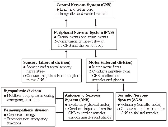
The human neural system is divided into two parts :
(i) Central neural system (CNS)
(ii) Peripheral neural system (PNS)
The CNS includes the brain and the spinal cord and is the site of information processing and control. The PNS comprises of all the nerves of the body associated with the CNS (brain and spinal cord). The nerve fibres of the
PNS are of two types :
(a) Afferent fibres
(b) Efferent fibres
☞ The afferent nerve fibres transmit impulses from tissues/organs to the CNS and the efferent fibres transmit regulatory impulses from the CNS to the concerned peripheral tissues/organs.
☞ The PNS is divided into two divisions :-
(A) Somatic neural system (SNS)
(B) Autonomic neural system (ANS)
☞ The somatic neural system relays impulses from the CNS to skeletal muscles while the autonomic neural system transmits impulses from the CNS to the involuntary organs and smooth muscles of the body.
☞ The autonomic neural system is further classified into sympathetic neural system and parasympathetic neural system.
NERVOUS TISSUE
Nervous tissue originates from ectoderm and is specialized for receiving stimuli (Excitability), transmit message (con-ductivity)
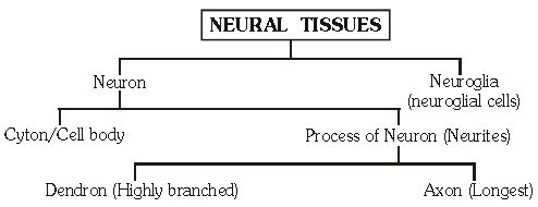
NEURON (NERVE CELL)
It is the functional and structural unit of nervous system. It generates and transmits nerve impulses. It is the longest cell of the body.
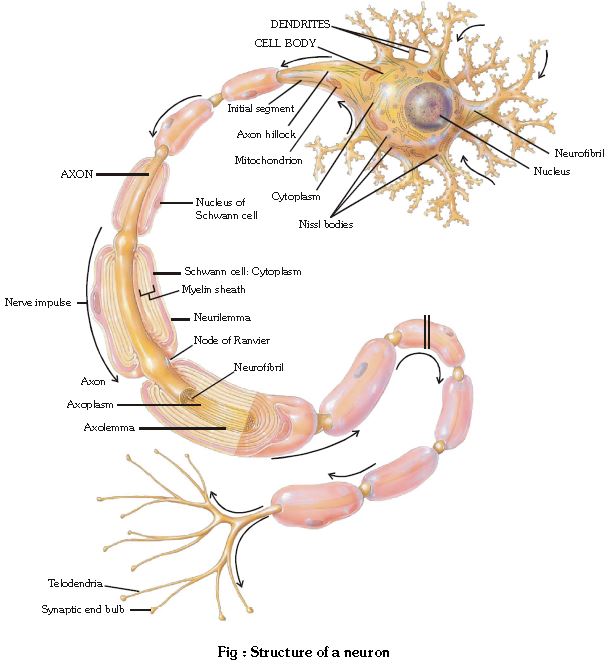
Structure of a neuron : A nerve cell is made up of cell body & cell process - (Dendron and Axon = Neurites)
(A) Cell body or Cyton or soma or perikaryon:-
☞ It contains uninucleated cytoplasm.
☞ Except centriole, all cell organelles are found in cytoplasm.
☞ Centriole is absent in the nerve cell thus cell division is absent.
☞ Some other cell organelles like Nissl's granule and neurofibril are also found in nerve cell.
(i) Nissl's granules :
☞ Endoplasmic reticulum & ribosome form granules like structure called as Nissl's granules or Tigroid body.
☞ These are the centre of protein synthesis.
☞ Site - Cyton & dendron
(ii) Many small fibrils are found in the cytoplasm called neurofibrils, these help in internal conduction in the cyton.
(B) Cell processes :
(i) Dendron :-
☞ It is small cell process. It's fine branches are called dendrites. Some receptor's are found on the dendrites, so dendron receive the stimuli & produce centripetal (towards the cell body) conduction.
(ii) Axon (Long process = Axon = Nerve fibre)-
☞ It is longest cell process of cyton, its diameter is uniform.
☞ Axon is covered by Axolemma. Part of cyton where axon arises called Axon hillock.
☞ Cytoplasm which contains in axon is axoplasm.
☞ Nissl's granules are absent in the axoplasm.
☞ Axoplasm of axon contains only neurofibrils and mitochondria.
☞ The axon hillock is the neuron's trigger zone, because it is the site where action potential are triggered.
☞ The terminal end of axon is Telodendria and button shape structure are called as Synaptic knob, which possess synaptic vesicles containing chemicals called neurotransmitters. The axons transmit nerve impulses away from the cell body to a synapse or to a neuro-muscular junction.
☞ More mitochondria are found in the telodendria which synthesize neurotransmitters like Acetylcholine (Ach) with the help of Acetyl-choline transferase enzyme.
☞ Axon is the functional part of nerve cell, therefore term nerve fibre usually refer to Axon.
Differences between Axon & Dendron
Axon
1. It is always single in a neuron.
2. It has no Nissl's granules.
3. It is long
4. Nerve impulse travels away from the cell body. (centrifugal)
Dendron
1. One or more.
2. Nissl's granules present.
3. Short.
4. Nerve impulse travels towards the cell body.(centripetal)
MYELINOGENESIS
Myelin is a fatty material with a high electrical resistance and acts as an electrical insulator in the same way as the rubber and plastic covering of electrical wiring. Peripheral nervous system (PNS) :-
☞ Axon is covered by a layer of phospholipids/sphingomyelin which is called as medulla or myelin sheath.
☞ Medulla is covered by thin cell membrane, which is called as neurilemma or sheath of schwann cells.
☞ The neurilemma is composed of schwann cells.
☞ In the peripheral nerves, myelinogenesis begins with the deposition of myelin sheath in concentric layer around the axon by schwann cells.
☞ Myelin sheath is discontinuous around the Axon. Those interruptions where Axon is uncovered by myelin sheath are called nodes of Ranvier
☞ Schwann cell takes part in the deposition of myelin sheath (myelinogenesis).
☞ Myelin sheath acts as insulator and prevent's leakage of ions.
Central nervous system (CNS) :-
* Neurilemma or schwann cells are not present in CNS, therefore myelinogenesis process occurs with the help of oligodendrocytes (Neuroglia).
* Neurons in which myelin sheath is present, are called medullated or myelinated neurons. In some nerve cells where myelin sheath is absent, called as non medullated or non myelinated neurons.
Myelinated nerve fibres are found in spinal and cranial nerves. Unmyelinated nerve fibre is enclosed by a Schwann cell that does not form a myelin sheath around the axon, and is commonly found in autonomousand the somatic neural systems
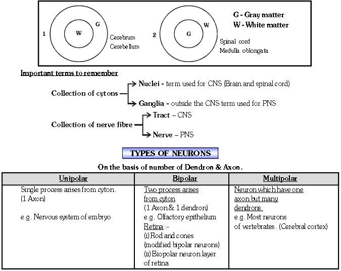
Gray matter : - It is composed of nerve cells. It consists of cytons & nonmedullated nerve fibres (Gray fibers).
White matter : - It contains myelinated nerve fibres (White fibres).
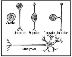
Apolar/Nonpolar Neuron :- No definite dendron/axon. Cell process are either absent or if present are not differentiated in axon and dendrons.Nerve impulse radiates in all directions. e.g. Hydra,amacrine cell of retina.
Pseudounipolar :- In this type, nerve cell has only axon but a small process develop from axon which act as dendron. eg. Dorsal root ganglia of spinal cord.
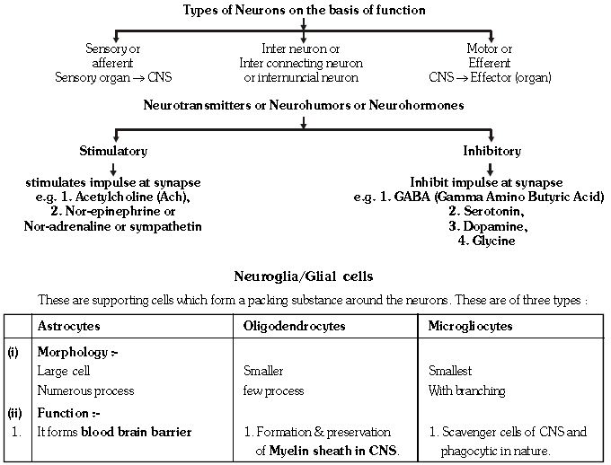
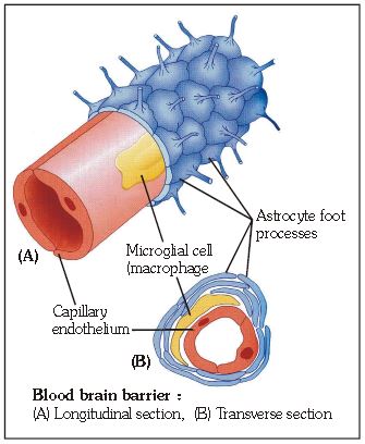
Blood Brain Barrier (BBB) :-
The Blood-Brain Barrier is formed by Astrocyte cells, which are coupled by tight junctions. The barrier prevents the entry of neurotoxins.
GENERATION AND CONDUCTION OF NERVE IMPULSE
Excitable cells - Neurons are excitable cells because their membranes are in a polarized state due to differential concentration gradient of ions across membrane. This axolemma is selectively permeable in nature.
1. RESTING PHASE :
☞ The potential difference (a charge) which exists across the cell surface membrane of nerve cells, negative inside the cell with respect to the outside. The membrane is said to be polarised.
☞ The potential difference across the membrane at rest is called the resting membrane potential and this is about - 70 mV (the negative sign indicates that inside the cell is negative with respect to the outside).
(Range→ - 60 to - 85 mV)
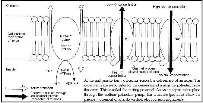
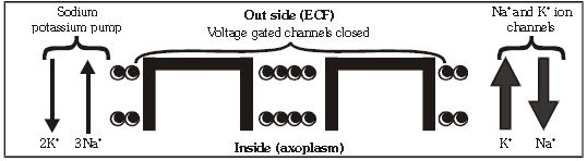
☞ The resting potential is maintained by active transport and passive diffusion of ions.
☞ Resting membrane potential is maintained by the active transport of ions against their electrochemical gradient by sodium potassium pump. These are carrier proteins located in the cell surface membrane. They are driven by energy supplied by ATP and couple the removal of three sodium ions from the axon with the up take of two potassium ions.
☞ The active movement of these ions is opposed by the passive diffusion of the ions. The rate of diffusion is determined by the permeability of the axon membrane to the ion.
☞ Potassium ions have a membrane permeability greater than that of sodium ions.
☞ Therefore potassium ions loss from the axon is greater than sodium ion gain.
☞ This leads to a net loss of positive ions from the axon, and the production of negative charge within the axon (Further there are many organic anions within the axoplasm, which also contribute to axoplasm negativity).
☞ Due to active transport (mainly) and diffusion process, positive charge is more outside and negative charge is more inside.
2. EXCITING STAGE :
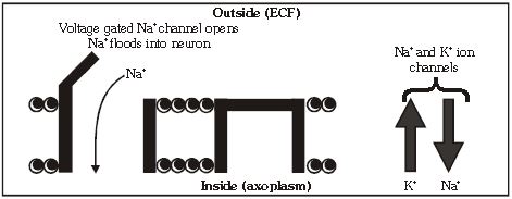
☞ Once the event of depolarization occurs, a nerve impulse or spike is initiated. Action potential is another name of nerve impulse. This is generated by a change in the sodium ion channels. These channels, and some of the potassium ion channels, are known as voltage gated channel, meaning they can be opened or closed with change in voltage. In resting state these channels are closed due to binding of Ca++.
☞ A potential is generated and it cause sudden opening of the sodium gates. Opening of gates increases the permeability of the axon membrane to sodium ions which enter by diffusion. This increases the number of positive ions inside the axon.
☞ A change of +10mV in potential difference from RMP through influx is sufficiently significant to trigger a rapid influx of Na+ ions leading to generation of action potential. This change of +10 mV is called as threshold stimulus.
☞ At the point where membrane (Axolemma) is completely depolarised due to rapid influx of Na+ ions, the negative potential is first cancelled out and becomes "0".
☞ Due to further entry of Na+, the membrane potential "over shoots" beyond the zero and becomes positive upto +30 to +45mV.
☞ This potential is called as action potential. In this state, the inner surface of axolemma becomes positively charged and outer surface becomes negatively charged. The rise in the stimulus-induced permeability to Na+ is extremely shortlived.It is quickly followed by a rise in permeability to K+.
3. REPOLARISATION :-
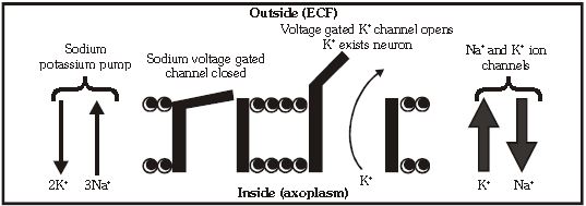
☞ After a fraction of second, the sodium gates get closed. Depolarisation of the axon membrane causes potasium gates to open.
☞ Within a fraction of a second, K+ diffuses outside the membrane and restores the resting potential of the membrane at the site of excitation and the fibre becomes once more responsive to further stimulation.
☞ Since potassium is positively charged, its exit makes the inside of cell less positive, or more negative and the process of repolarization or return to the original resting potential begins.
☞ The repolarization period returns the cell to its resting potential (70 mV). The neuron is now prepared to receive another stimulus and conduct it in the same manner.
☞ The time taken for restoration of resting potential is called refractory period, because during this periods the membrane is incapable of receiving another impulse.
☞ Nerve impulse travels as action potential which passes along the axon as a wave of depolarization.
☞ The whole process of depolarisation and repolarisation is very fast. It takes only about 1 to 5 milli second (ms).
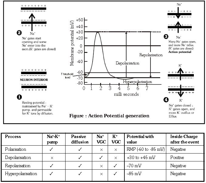
☞ Open/Operating →✓ , Closed → ×
SALTATORY CONDUCTION OF NERVE IMPULSE
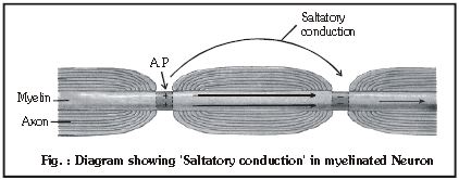
☞ This type of conduction occurs in myelinated fibre.
☞ This means, in effect that the action potential jumps from node to node and passes along the myelinated axon faster as compared to the series of small local circuits in a non-myelinated axon. This type of conduction is called saltatory conduction. Leakage of ions takes place only in nodes of Ranvier and less energy is required for saltatory conduction
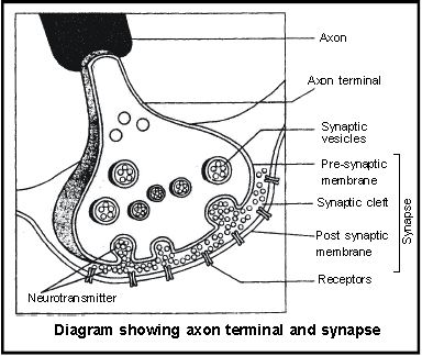
SYNAPSE
☞ It is the junctional region between two neurons where information is transferred from one neuron to another.
☞ Name synapse was proposed by Charles Sherrington
☞ Telodendria of one neuron form synapse with dendron of next neuron.
☞ It transmit stimulus in the form of electrochemical wave. Synapse = Presynaptic knob + synaptic cleft + postsynaptic membrane
☞ Telodendria membrane is called pre synaptic membrane & membrane of dendron of other neuron called as postsynaptic membrane. Space between pre and post synaptic membranes is called synaptic cleft. It may or may not be seperate.
☞ A nerve impulse is transmitted from the neuron to another through junction called synapse.
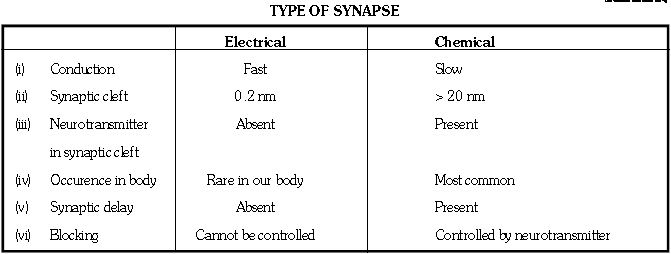
☞ There are two types of synapses, namely, electrical synapses and chemical synapses.
☞ At electrical synapses, the membranes of pre- and post-synaptic neurons are in very close proximity.
☞ Electrical current can flow directly from one neuron into the other across these synapses.
☞ Transmission of an impulse across electrical synapses is very similar to impulse conduction along a single axon.
☞ Impulse transmission across an electrical synapse is always faster than that across a chemical synapse. Electrical synapses are rare in our system. On the other hand, chemical synapses are characterised by a synaptic cleft. At these synapse, impulse transmission occurs with the help of a chemical, called neurotransmitter.
☞ Mechanism :- When the AP develops in presynaptic membrane, it becomes permeable for Ca++. Ca++ enter presynaptic membrane & neurotransmitter vesicles burst due to the stimulation by Ca++ and they release neurotransmitters in synaptic cleft.
Neurotransmitter reaches the postsynaptic membrane via synaptic cleft & binds to specific receptors. This binding opens up ion channels, allowing the entry of ions which can generate a new potential on post synaptic membrane. The potential may be excitatory (EPSP) or inhibitory (IPSP). In Excitatory postsynaptic potential (EPSP) Ach is main neuro transmitter, which develop due to opening of Na+ gatted channels.
☞ On the rest of the Ach, cholinestrase enzyme functions, which is found in synaptic cleft. This enzyme decomposes the Ach into choline & Acetate.
☞ If neuro inhibitory transmitter (GABA) binds with post synaptic membrane to open the Cl- gatted channels and hyperpolarization of neuron occurs. Now the potential is called inhibitory postsynaptic potential (IPSP) & further nerve conduction is blocked.
FUNCTIONAL PROPERTIES OF NERVE FIBRE
☞ Conduction of nerve impulse is unidirectional.
☞ It follow all or none law. Magnitude of response will always be same irrespective of strength of stimulus above threshold stimulus.
☞ Velocity of nerve impulse μ Diameter of neuron.
☞ This velocity is affected by physical & chemical factor, such as pressure, cold, heat, chloroform and ether etc.
GOLDEN KEY POINTS
☞ Depolarization : Na+ influx.
☞ Repolarization : K+ efflux.
☞ If myelin sheath is continuous there will be no nerve impulse conduction in nerve fibres.
☞ If question is informing for only about channel opening and closing then consider only VGC.
☞ Simple diffusion channels are always open (In every state).
BEGINNER'S BOX-2
1. Which statement is false regarding nerve impulse ?
(1) After applying a stimulus on polarised membrane, that site become freely permeable to Na+ and leads to rapid eflux of Na+.
(2) The rise in the stimulus induced permeability to Na+ is extremely short lived.
(3) After depolarization K+ diffuses outside the membrane and restores the resting potential.
(4) Ionic gradients across the resting membrane are maintainined by the Na+ - K+ ATPase pump.
2. Resting membrane potential is achievied by :-
(1) Passive diffusion by ion channels/Leaky channels
(2) Na+ - K+ ATPase pump.
(3) Negatively charged proteins in axoplasm.
(4) All of the above
CENTRAL NEURAL SYSTEM
☞ It includes the brain and the spinal-cord.
☞ These are formed from the neural-tube which develops from the ectoderm after the gastrula stage of embryo.
Development of CNS :-
Anterior part of neural tube develops into brain while caudal part of neural tube develops into spinal cord.
Brain's approximately 70-80% part develops in 2 year of age & complete development is achieved in 6 years of age & spinal cord develops completely in 4 to 5 years of age.

HUMAN BRAIN
1. The brain is the central information processing organ of our body and acts as the 'Command and controlsystem'.
2. It controls the voluntary movements, balance of the body, functioning of vital involuntary organs (e.g. lungs, heart, kidney etc.), thermoregulation, hunger and thirst, circadian (24 hours) rhythms of our body, activities of several endocrine glands and human behaviour.
3. It is also site for processing of vision, hearing, speech, memory, intelligence, emotions and thoughts.
4. It is situated in cranial box which is made up of 1 frontal bone, 2 parietal bone, 2 temporal bone, 1 spnenoid, 1 ethmoid 1 occipital bone. The weight of brain of an adult male is 1400 gm and of female is 1250 gm.
BRAIN MENINGES
Brain is covered by three membranes of connective tissue termed as meninges or menix
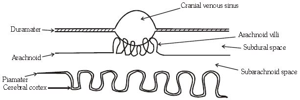
(1) Duramater :
☞ This is the first and the outermost membrane which is thick, strong and elastic layer.
☞ At several places it forms cranial venous sinuses containing blood.''
(2) Arachnoid :
☞ It is middle and delicate layer and found only in mammals. (Mammalian character)
☞ At several places it forms villi like foldings to absorb CSF called arachnoid villi.
☞ Space between duramater & arachnoid is called subdural space which is filled with serous fluid.
(3) Piamater :
☞ Inner most, thin and transparent membrane, which is firmly attached to the brain.
☞ At some places it merges deeply into sulci of brain to form telachoroidea.
☞ Space between arachnoid & piamater is called subarachnoid space, which is filled with CSF.
CEREBROSPINAL-FLUID (CSF)
☞ This fluid is clear and alkaline in nature just like lymph.
☞ CSF is present in ventricles of brain, subarachnoid space of brain & spinal cord.
☞ C.S.F. is formed in choroid plexus found in the ventricles of the brain.
Functions of C.S.F. :-
☞ Protection of Brain :- It acts as shock absorbing medium and works as cushion.
☞ It provides buoyancy to the brain, so net weight of the brain is reduced from about 1.4 kg to about 0.18 kg
FORE BRAIN
☞ The fore brain consists of Cerebrum, Diencephalon (containing epithalamus, thalamus, hypothalamus) and olfactory lobe.
1. CEREBRUM
☞ Cerebrum forms the major part of the brain which is most developed in human.
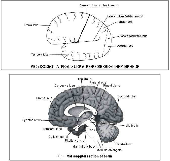
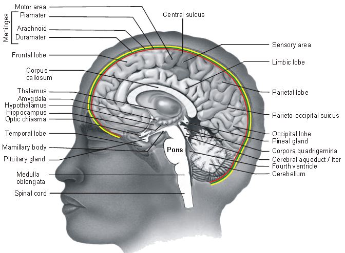
☞ Cerebrum consists of two cerebral hemispheres, on the dorsal surface a longitudinal groove is present between two cerebral hemispheres called as median fissure. Both the cerebral hemispheres partially connected with each other by curved white thick nerve fibre called corpus callosum (Largest commisure of brain) (Mammalian character).
☞ Each cerebral hemisphere is divided into 4 lobes-Anterior, Middle, Posterior and Lateral.
☞ Anterior lobe is also called frontal lobe (largest lobe) while middle lobe is called as parietal lobe. Frontal lobe is seprated by central sulcus or rolandic sulcus from parietal lobe.
☞ Posterior lobe is called as occipital lobe, it is separated from parietal lobe by a sulcus called parieto occipital sulcus.
☞ Lateral lobe or temporal lobe is separated from frontal lobe & parietal lobe by incomplete sulcus called lateral sulcus /sylvian sulcus.
☞ The outer part of cerebral hemisphere is called Cerebral cortex and thrown into prominent folds. These folds
are found as ridges and grooves on dorsal surface of cerebral hemisphere. Ridges are known as Gyri while grooves are called sulci. Gyri and sulci increase the surface area of cerebrum.
☞ The cerebral cortex referred to as the gray matter due to its greyish appearance. The neuron cell bodies are concentrated here giving the colour. This thick layer of gray matter is also known as Neopallium/Pallium.
☞ The cerebral cortex contains three types of functional areas :-
(i) Sensory area - Analysis of sensory impulses eg. Somesthetic area for general sensation (Touch, Pain, Temperature etc.)
(ii) Motor area - Generation of motor impulses eg. Broca's area for fine movement of tongue and speech. Motor area for voluntary movement of limb muscles.
(iii) Association area - These are large regions that are neither clearly sensory nor motor in function. They are responsible for certain complex functions like :-
• Intersensory associations : As you are aware that all sensory inputs like touch, sound, light, smell are sent to brain. These different sensation require association and inter connection with each other for their proper interpretation.
• Memory : Memory of past events is recorded by the association areas also with the different lobes of the cerebrum. Memory is basically of two types : Short term memory and long term memory.
• Communication : The ability of communication also controlled by the assoication areas of cerebral cortex.
Function of cerebrum : It is the most important part of brain because it controls and regulates different part of brain. This is the centre of conscious senses, will power. Voluntary movements, knowledge, etc.
☞ Different sense organs send impulse here and in this part of brain, analysis and coordination of impules is done then messages are transferred to organs.
2. DIENCEPHALON
☞ It is small chamber like posterior part of fore brain which is covered by cerebrum. It consists of 3 parts :
(i) Epithalamus (ii) Thalamus (iii) Hypothalamus
(i) Epithalamus : It form the roof of diencephalon. Pineal body (Epiphysis cerebri) is found on epithalamus & control sexual maturity.
(ii) Thalamus : It forms upper lateral wall of Diencephalon. It form 80% of diencephalon. It acts as a relay centre. It receives all sensory inputs from all part of body & these impulses are send to the cerebral cortex. Cerebrum wraps around the thalamus. It is a major coordinating centre for sensory & motor signalling.
(iii) Hypothalamus : It forms the lower or ventral part of diencephalon. It lies at the base part thalamus.
☞ The hypothalamus contains a number of centre which control body temperature, urge for eating and drinking (Hunger and thirst).
☞ It also contains several group of neurosecretory cells, which secrete hormone called hypothalamic hormone.
☞ A cross like structure is found on anterior surface of hypothalamus called as optic chiasma, through Infundibulum pituitary body is attached to middle part of hypothalamus. Corpus mammillare or corpus albicans or mammillary body is found on the posterior part of hypothalamus. (Mammalian character)
Hypothalamus controls :-
(1) Thermoregulation
(2) Behaviour and emotion
(3) Endocrine control
(4) Biological clock system
(5) ANS control.
Limbic system :
The inner part of cerebral hemispheres and a group of associated deep structures like amygdala, hippocampus etc. form a complex structure called Limbic system. Along with the hypothalmus, it is involved in the regulation of sexual behaviour, expression of emotional reactions (e.g. excitement, pleasure, rage, fear) and motivation, olfaction and autonomic responses.

* One pair of broad bean size organ called olfactory lobe/bulb are found an ventral surface of frontal lobe of cerebral hemisphere. It is a small spherical & solid structure in human brain.
It is connected to olfactory centre (temporal lobe) through olfactory tract and are extentions of the brain's limbic system.
Functions : It is supposed to be centre of smell intensity. Some animal like sharks and dogs have well developed olfactory lobes.

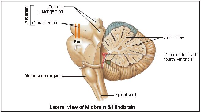
☞ It is a small part of brain. The midbrain is located between diencephalon of the fore brain and pons of the hind brain. A canal called Cerebral aqueduct/Aqueduct of sylvius passes through the midbrain.
☞ Anterior part of midbrain contains two longitudinal myelinated nerve fibre called cerebral peduncles or crus cerebri or crura cerebri.
☞ On posterior part of mid brain four spherical projections are found called colliculus or optic lobe. Four colliculus are collectively called as corpora quadrigemina. (2 upper & 2 lower) (Mammalian character)
☞ Mid brain and hind brain (except cerebellum) form the brain stem.
Function :
☞ The mid brain receives and integrates visual, tactile and auditory inputs.
☞ Crura cerebrai controls the muscle of limb while superior and inferior colliculus are related with pupillary (light) reflex and acoustic (Sound) reflex action respectively.
HIND BRAIN
☞ The hind brain comprises pons, cerebellum and medulla (also called the medulla oblongata).
1. PONS
☞ It is a small spherical projection. Which is situated below the midbrain and upper side of medulla oblongata. It consists of many transverse and longitudinal nerve fibre. Transverse nerve fibre connect with cerebellum while longitudinal nerve fibre connect cerebrum to medulla oblongata.
Function :- It regulates the breathing reaction through pneumotaxic centre.
2. CEREBELLUM
☞ It is made up of 3 lobe (2 lateral lobe and 1 vermis). Both lateral lobes become enlarged and spherical in shape, so lateral lobe of cerebellum are also called as cerebellar hemisphere. Cerebellum has very convoluted surface in order to provide the additional space of for many more neurons.
☞ Outer part of cerebellum is made up of gray matter while inner part is of white matter. White matter projects outside & forms a branched tree like structure known as Arbor Vitae.
Functions : The cerebellum integrates information received from the semicircular canals of the ear and the auditory system and also by this portion of hind brain impulses are received from different voluntary muscles and joints.
☞ Due to this region, coordination of voluntary muscle through involuntary regulation is more developed in human compared to other animals i.e. Body balance.
☞ The person who take alcohol in excess their cerebellum gets affected as a result that person can not maintain his balance and walking is disturbed.
☞ Thus it is related with fine and skill full voluntary movements.
3. MEDULLA OBLONGATA
☞ Posterior part of hind brain is tubular and cylindrical in shape. Medulla of brain is connected to spinal cord.
☞ Mid brain, pons and medulla are situated in one axis and it is called as Brain stem.
Functions : It is the most important part of brain which controls all the involuntary activities of the body e.g. cardiovascular reflex, respiration, metabolism, gastric secretion etc. As well as this act as conduction path for all impulses between spinal cord and remaining portions of brain.
☞ It is also concerned with cranial reflex action like sneezing reflex, salivation reflex, coughing reflex, swallowing reflex, vomiting reflex, yawning reflex.
Neural Control and Coordination
POINTS TO REMEMBER
HUMAN NEURAL SYSTEM :
• The human neural system divided into two parts –
o The central nervous system (CNS)
o The peripheral nervous system (PNS)
• The CNS includes the brain and spinal cord and is the site of information processing and control.
• The PNS comprises all nerves of the body associated with CNS.
o Cranial nerves: nerves arises from the brain (12 pairs)
o Spinal nerves: nerves arises from the spinal cord (33 pairs)
• The nerve fibres (Cranial and spinal nerves) are of two types –
o Afferent fibres: transmits impulses from the tissues to the CNS
o Efferent fibres: transmits impulses from the CNS to the tissues.
• The PNS is divided into two divisions –
o Somatic neural system.
o Autonomic neural system.
• Sympathetic neural system.
• Parasympathetic neural system.
• The somatic neural system relays impulses from the CNS to skeletal muscles.
• The autonomic neural system transmits impulses from the CNS to the involuntary organs and smooth muscles of the body.
NEURON AS STRUCTURAL AND FUNCTUIONAL UNIT OF NERVOUS SYSTEM :
• A neuron composed of three major parts –
o Cell body
o Dendrites
o Axon
• The cell body contains cytoplasm with typical cell organelles and specific granular body called Nissl’s granules.
• Short fibres which profusely branched projects out of cell body called dendrites.
• The axon is a long fibre, branched at the end.
• Each branch terminates as a bulb-like structure called synaptic knob.
• Based on the number of axon and dendrites the neurons are of following types –
o Multipolar: one axon and several dendrites - found in cerebral cortex.
o Bipolar: one axon and one dendrite - found in retina of eye.
o Unipolar: cell body with one axon only – found in embryonic stage.
• The axon may be myelinated or non-myelinated.
• The myelinated nerve fibres are enveloped with Schwann cells, which form myelin sheath around the axon. The gaps between two adjacent myelin sheath are called Nodes of Ranvier.
• Cranial and spinal nerves are myelinated.
• Autonomic and somatic neural fibres are non-myelinated.
GENERATIONA ND CONDUCTION OF NERVE IMPULSE :
Polarized membrane/Resting Potential :
• In resting phase when neuron is not conducting an impulse, the axonal membrane is called polarized. This is due to difference in concentration of ions across the axonal membrane.
• At Rest :
o Axoplasm inside the axon contains high conc. of K+ and low conc. of Na+.
o The fluid outside the axon contains low conc. of K+ and high conc. of Na+.
• As a result the outer surface of axonal membrane is positively charged and inner surface is negatively charged. The electric potential difference across the resting plasma membrane is called resting potential.
Action Potential :
• When a nerve fibre is stimulated, the permeability of membrane to Na+ is greatly increased at the point of stimulus (rapid influx of Na+) and hence polarity of membrane is reversed and now membrane is said to be depolarized.
• The electric potential difference across the plasma membrane at that site is called action potential, which in fact termed as nerve impulse.
• Depolarization is very rapid, so that conduction of nerve impulse along the entire length of axon occurs in fractions of second.
• Depolarization is followed by the increase in permeability of K+ to the membrane leads to change in polarization i.e. +ve charge outside and –ve charge inside. It is called repolarization.
• Regain of resting potential takes place due to action of Na+/K+ ATPase enzyme which transports three Na+ inside and two K+ inside with expense of one ATP. It continues till the resting potential becomes -70 mv.
Transmission of impulses through synapse :
• The functional junction between two neurons is called synapse.
• A synapse is formed by the membranes of a pre-synaptic neuron and a post-synaptic neuron, which may or may not be separated by a gap called synaptic cleft.
• There are two types of synapses:
o Electrical synapse: pre and post synaptic
Conduction of impulse in chemical synapse :
• The axon terminals contains vesicles filled with chemicals calledneurotransmitters.
• When the action potential arrives at the axon terminals, it stimulates the movement of synaptic vesicles towards the membrane.
• Synaptic vesicle fused with the pre-synaptic membrane and releases the neurotransmitter into the synaptic cleft.
• The neurotransmitter binds with the receptors located on the post-synaptic membrane.
• Activation of receptors on post-synaptic membrane makes it permeable to Na+ and generates action potential as it done by stimulus.
• The new potential developed may be either excitatory or inhibitory depends on the nature of the neurotransmitter.
CENTRAL NERVOUS SYSTEM :
• Brain is the central control and command system in neural coordination.
• The human brain is well protected by the skull.
• Inside the skull the brain is covered by cranial meninges.
• Meninges consists of following layers –
o Outer layer – dura mater.
o Middle layer – thin arachnoid.
o Inner layer – pia mater remain close contact with the brain.
• The human brain is divided into three major parts –
o Fore brain.
• Cerebrum.
• Thalamus.
• Hypothalamus.
o Mid brain.
o Hind brain.
• Pons.
• Cerebellum
• Medulla oblongata.
Fore brain :
• Cerebrum is the major part of the fore brain.
• Deep median fissure divides the cerebrum into two equal cerebral hemisphere.
• The hemispheres are connected by tract of nerve fibres called corpus callosum.
• The thin layers of cells covers the cerebral hemispheres called cerebral cortex and are thrown into prominent folds.
• The cerebral cortex is referred as the grey matter.
• The cerebral cortex differentiated into –
o Motor areas – sends information to the body
o Sensory areas – receives information from the body
o Association area-neither sensory nor motor (co-ordinates the information)
• Interior of the brain is called white matter due to myelin sheath of tract of nerve fibres.
• The cerebrum is wraps around a structure called thalamus, which is a major coordinating centre for sensory and motor signaling.
• At the base of the thalamus is the hypothalamus.
• The hypothalamus have following functions –
o Control body temperature.
o Urge for eating and drinking.
o Neurosensory cells secrete different hormones.
• The inner part of the cerebral hemispheres and a group of associated deep structures like amygdala, hippocampusetc. forms complex structure called the limbic lobe or limbic system.
• Along with the hypothalamus it is involved in the regulation of sexual behaviour, expression of emotional reactions (excitement, pleasure, rage and fear) and motivation.
Mid brain:
• The mid brain is located between the thalamus and pons of the hind brain.
• A canal called cerebral aqueduct passes through the mid brain.
• The dorsal part of the mid brain consists of four swelling called corpora quadrigemina.
Hind brain:
• Comprises pons, cerebellum and medulla oblongata.
• Pons consists of fibre tracts that interconnect different regions of the brain.
• Cerebellum has very convoluted surface in order to provide the additional space for many more neuron.
• Medulla of the brain is continued as spinal cord.
• Medulla contains centers which control respiration, cardiovascular reflexes and gastric secretion.
REFLEX ACTION AND REFLEX ARC :
• Sudden spontaneous, involuntary reaction to a stimulus without involvement of brain is called reflex action.
• Some examples of such actions are –
o Sudden withdrawal of the body part which comes in contact with objects that are extremely hot, cold, pointed.
• Reflex arc: sensory organ → sensory neuron → spinal cord → motor neuron → effector organ.
SENSORY RECEPTION AND PROCESSING :
Eye :
• Eye is the sensory organ of vision.
• Our paired eyes are located in sockets of the skull called orbit.
• Eye consists of three layer –
• Sclera: tunica fibrosa.
o External layer composed of dense connective tissue.
o It is the only complete layer of the eye.
o The anterior portion of this layer is transparent and called cornea.
• Choroid: tunica vascularis.
o It is the middle layer of the eye.
o It is well vascularized and looks bluish color.
o Posterior two third parts is thin.
o Anterior part is thick and form ciliary body.
o The ciliary body itself continues forward to form a pigmented and opaque structure called iris (the visible coloured portion of the eye).
o Iris contains a central aperture called pupil.
o The diameter of pupil is regulated by the muscle of iris.
• Retina or tunica nervosa.
o It is the innermost layer of the eye.
o It consists of three layer of cells – from inside to outside
• Ganglion cells
• Bipolar cells
• Photoreceptor cells.
o There are two types of photoreceptor cells namely rods and cones.
o Cones contain photopigment called iodopsin.
o Cones responsible for daylight (photopic) vision and color vision.
o Rods contain photopigment called rhodopsin or visual purple, which contain a derivative of Vitamin-A.
o Rods responsible for twilight (scotopic) vision.
o The optic nerves leave the eye and the retinal blood vessel enters it at a point where rods and cones are absent hence called blind spot.
o At the posterior pole of the eye lateral to blind spot there is a yellowish pigmented spot called macula lutea.
o Macula lutea with highly concentrated cones, where the vision is sharpest ( high resolution vision)
o In the centre of macula lutea there is a central pit called fovea centralis, a tightly packed array of specializedphotosensor-receptor cells. It prevents the entry of high intensity light by closing the eye by reflex action.
• The lens composed of crystalline protein, is suspended behind the pupil by a suspensory ligament attached to the ciliary body.
• The lens and suspensory ligament divide the cavity of the eye ball into two chambers.
• Chamber in front of lens called aqueous chamber filled with aqueous humor.
• Chamber behind the lens is called vitreous chamber filled with transparent gel called vitreous humor.
Mechanism of vision :
• The light rays in visible spectrum focused on the retina through the cornea and lens generate potentials (impulses) in rods and cones.
• Photosensitive pigments composed of opsin (a protein) and retinal (an aldehyde of vitamin-A).
• Light induces dissociation of the retinal from opsin resulting changes in structure of opsin.
• This causes change in membrane permeability. As a result, potential differences are generated in the photoreceptor cells.
• This produces a signal that generates action potential in the ganglion cells through bipolar cells.
• These action potentials transmitted by optic nerves to the visual cortex area of brain where the neural impulses are analyzed and the image formed on the retina is recognized.
THE EAR :
• The ear performs two sensory function, hearing and maintenance of body balance.
• Anatomically, the ear can be divided into three major section –
o Outer ear or external ear.
o Middle ear.
o Internal ear or inner ear
External ear :
• Outer ear consists of the pinna and external auditory meatus (canal).
• Pinna collects the vibration in the air which produces sound.
• Auditory meatus extends upto the tympanic membrane (the ear drum).
• Tympanic membrane is made of connective tissue covered with skin.
Middle ear :
• Middle ear contains three ear ossicles called Malleus (hammer),Incus (anvil) and stapes (stirrup).
• The Malleus is attached to the tympanic membrane and the stapes is attached to the oval window of the cochlea.
• The ear ossicles amplify the sound waves comes from the tympanic membrane.
• A Eustachian tube connects the middle ear cavity with the pharynx.
• Eustachian tube helps in equalizing the pressures on either sides of the ear drum.
Internal ear :
• The fluid filled internal ear is called labyrinth consists of two parts, the bony and membranous labyrinth.
• The bony labyrinth is a series of channels, inside these channels lies the membranous labyrinth, which is surrounded by a fluid called perilymph.
• The membranous labyrinth is filled by a fluid called endolymph.
• The labyrinth consists of two portions –
o The coiled portion called cochlea.
o The complex above the cochlea called vestibular apparatus.
Cochlea:
• The coiled portion of the labyrinth is called cochlea.
• The membrane constituting cochlea are-
o The reissner’s membrane
o The basilar membrane.
• Reissner’s and basilar membrane divide the surrounding perilymph into an upper scala vestibuli and lower scala tympani.
• The space within cochlea called scala media is filled with endolymph.
• At the base of the cochlea, the scala vestibule ends at the oval window (fenestra ovalis), while scala tympani terminate at the round window (fenestra rotundus) which opens into the middle ear.
• The organ of corti is a structure located on the basilar membrane which contains hair cells that act as auditory receptors.
• The basal end of hair cells is in close contact with the afferent nerve fibres.
• Hair cells contain stereo cilia projected from the apical part of each hair cell.
• Hair cells covered by a thin elastic membrane called tectorial membrane.
Vestibular apparatus:
• Vestibular apparatus located above the cochlea.
• Vestibular apparatus consists of –
o Three semi-circular canals
o Otolith organ consisting saccule and utricle.
• Each semicircular canal lies in a different plane at right angles to each other.
• Membranous semi-circular canals are suspended in the perilymph of bony canal.
• The base of canals is swollen and is called ampulla, which contain a projecting ridge called crista ampullaris with hair cells.
• The saccule and utricle contain a projecting ridge called macula.
• Crista and macula are the specific receptors of the vestibular apparatus responsible for maintenance of balance of the body and posture.
Mechanism of hearing:
• The external ear receives sound waves and directs them to the ear drum.
• Sound waves are amplified by the ear ossicles and send it to the oval window in the middle ear.
• The vibration of the oval window creates waves in the perilymph of scala vestibuli.
• The waves in perilymph induce a ripple in the basilar membrane.
• Movements of the basilar membrane bend the hair cells, pressing them against the tectorial membrane.
• As a result nerve impulses are generated in the associated afferent neuron.
• These impulses are transported to the auditory cortex of the brain where the impulses are analysed and the sound is recognized.
Sense organs
We humans are responsive organisms. Aroma of a freshly cooked dish makes our mouth water, loud thunder makes us jump in our seat and stepping on a nail causes intense pain. We sense the changes in our environment (both internal and external) with the help of special sensory receptors. These environmental changes, called stimuli, once detected by the special sensory cells, are conveyed to the brain in the form of nerve impulses. The meaning of each stimulus is interpreted in the brain and appropriate order is sent to the body parts for its appropriate response to ensure well being.
Traditionally, there are five senses: touch, vision, hearing, smell and taste. While touch is a complex general sense, the other four are special senses. The general sensory receptors are simple receptors that are mostly modified dendritic ends of sensory neurons. Such receptors are present throughout the body __ in the skin, mucous membranes, connective tissues and muscles. These monitor most of the types of general sensory information such as tactile sensation (a mix of touch, pressure, stretch and vibration), heat, cold, pain and muscle sense (proception).
In contrast, special sensory receptors are distinct receptor cells that are actually confined to the head region and are highly localized within complex sensory organs like eyes and ears and tissues of the taste buds and olfactory epithelium .These sensory organs and tissues are collection of cells of many different types (receptor and non receptor cells}, working together to accomplish a specific receptive process. Recall the structure of eye and ear that you have studied. Which type of sensory receptor are these made of? Yes, the special sensory receptors called the photoreceptors and the auditory receptors respectively.
Though the complex sense organs are more familiar to us, the simple sensory receptors associated with general senses are no less important. These keep the central nervous system well informed about what is happening, both deep within the body and on its surface. In this lesson you will learn about a few of these simple receptors present in the skin. You will also learn about the special senses of taste and smell.
The chemical senses: the taste and smell
The receptors for taste and smell are classified as chemoreceptors as these respond to special chemicals in aqueous solution. In each case, the chemicals must go into solution in the film of liquid coating the membranes of the receptor cells before these can be detected. The taste receptors are specialized cells that detect chemicals present in quantity in the mouth itself, while smell receptors are modified sensory neurons in the nasal passage which detect the volatile chemicals that get wafted up the nostrils from distant sources. These two types of receptors complement each other and often respond to the same stimulus. You can now guess why a very strong perfume leaves a peculiar taste in your mouth. The smell receptors can be as much as 3,400 times more sensitive than the taste receptors.
Sense of smell (olfaction): Nose contains the receptors of smell, in the mucous coated thin, yellowish patch (about 5 cm2) of modified pseudo stratified epithelium called olfactory epithelium. It is located way up at the roof of the nasal cavity on either sides of the nasal septum.
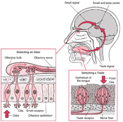
Figure 1 Human nose showing olfactory bulb and magnified view of olfactory epithelium
The olfactory epithelium contains three types of cells: (a) millions of olfactory receptor cells; (b) columnar supportive cells; (c) short basal cells. Olfactory receptors are unusual bipolar sensory neurons. The thin dendrites of each of these neurons run to the surface of the epithelium where these bear a cluster of about 20 modified cilia which function as receptor sites. These cilia extend from the olfactory epithelium into the thin coat of nasal mucous secreted by the supportive cells and olfactory glands. This mucous is a solvent that captures and dissolves the air borne odour molecules. Once dissolved, the chemicals bind to the specific receptors on the cilia stimulating the receptor cells. This causes depolarization and ultimately action potential in the receptor cell. The axons of the olfactory receptors unite to form the olfactory nerve which transmits the information directly to the overlying olfactory bulb, a relay station in the brain. Unlike receptor ends of other senses, the axons of the olfactory receptors directly extend from the outside environment (the nasal cavity) into the olfactory bulb, a part of the brain. The number of receptors stimulated indicates the strength of the stimulus. As with taste, some of what we call smell, can be really painful. The nasal cavity contains pain receptors that respond to irritants such as ammonia, vinegar or hot chilly pepper. Impulses from these pain receptors reach the brain. The brain combines these sensations with those of smell to identify the odours. Although humans do have a good sense of smell - we can detect about 10,000 different odours - our olfactory capability is not as good as those of many vertebrates, especially fish and mammals such as a dog.
Sense of taste (gustation)
The sense of taste and smell work closely together. If we cannot smell some thing we cannot taste it either. When we speak of taste sensations we are often referring to the combined sensation produced by both taste and smell receptors. One reason why we cannot taste (or smell) food well with a common cold is that with the nasal passages inflamed and coated with thick mucus layer the smell receptors are practically non functional. The receptor cells for taste are located in taste buds. Humans have about 10,000 taste buds. The majority of taste buds are located in pockets around the papillae (peg-like projections of the mucous membrane) on the surface and sides of the tongue, but there are some on the surface of the pharynx and the larynx. Each taste bud contains about 40 specialized receptor cells or gustatory cells, many more supporting cells and some basal cells that replace the worn out cells of the taste buds. Unlike the receptors for smell, that are modified sensory neurons, the receptor cells for taste are not neurons, but rather specialized cells with slender microvilli on their outer ends. The microvilli protrude into the surrounding fluids through a narrow opening called the taste pore. Dissolved chemicals contacting the microvilli bind to specific receptor proteins on the microvilli, thereby depolarizing the cell. The dendrites of the associated sensory neurons coil intimately around the receptor cells and synapse with them so that, when a receptor cell is stimulated and depolarized, it releases neurotransmitter which leads to the generation of an action potential in the associated sensory neuron. Each dendrite receives signals from several receptor cells within the taste bud. Nerve fibers emerging from the taste buds pass to the brain stem. From here the nerve impulse is relayed to the taste centre in the cerebral cortex of the brain that perceives the taste sensation.
Normally our taste sensations are complicated mixture of qualities. In humans there are four basic taste senses: sweet, sour, salt, and bitter. The receptors for these four basic tastes have their areas of greatest concentration on different parts of the tongue _ sweet and salty on the front, bitter on the back, and sour on the sides. A few substances stimulate only one of the four types of receptors, but most stimulate two, three, or all four types to varying degrees. The sensation and flavour of the food we experience are thus produced by a combination of these four basic sensations, modified by accompanying sensations of smell, texture and temperature.
Sense of touch
Skin is the sensory organ for touch and is also the largest sense organ. Our sense of touch allows us to feel light sensation like the touch of a feather as well as a heavy sensation like a stone falling on the toe. These sensations come from millions of microscopic simple sensory receptors located all over the skin and associated with the general sensations of contact or pressure, heat, cold, and pain. The receptors are located at different levels within the skin and distributed unevenly. Some parts of the body have a large number of these such as the finger tips, making them more sensitive. Can you name the parts of your body that are less sensitive and why?
Structurally, these touch receptors are either free dendritic endings or encapsulated dendritic endings present in the skin (and other parts of the body). When stimulated, these transmit the sensation to the brain. Given below is a list of some of these receptors present in the skin.
Free or bare dendritic nerve endings are present throughout the epidermis taking an extensive branching or “zigzag” form .These respond chiefly to pain and temperature but some respond to pressure as well. The root hair plexuses, net work of free nerve endings that surround hair follicles, are light touch receptors that detect bending of hairs. These report on wind blowing through your hair.
Meissner’s corpuscles are small receptors in which a few spiraling dendrites are surrounded by specialized capsule (Schawann) cells. These are found just beneath the skin epidermis in dermal papillae and are especially abundant in finger tips and soles of the feet. These are light pressure receptors that allow us to become aware of a caress or feel of our shirt against our skin.
Pacinian corpuscles are the large egg shaped bodies .In each a single dendrite is surrounded by multilayers of capsule cells. These are scattered deep in the dermis and in the subcutaneous tissue of the skin .These are stimulated by deep pressure and respond only when pressure is firs applied. Thus, these receptors are best suited to monitor vibrations (on-off pressure stimulus the sense of touch allows us to detect different textures, temperatures, hardness and pain. Pain serves as a warning or alert system for the body. Whenever one or more of these sensory receptors are stimulated (by heat, cold, vibrations, pressure or pain) an impulse or action potential is generated. This impulse is then taken to the spinal cord and from there to the brain which analyses the stimulus and then generates appropriate response .The way brain interprets the sensation is our lives is also shaped by our personal experience in the past .Try to recollect your experience of touching a sharp object/ a hot plate by accident.
Important Questions for NCERT Class 11 Biology Neural Control and Coordination
Ques. Alzheimer’s disease in humans is associated with the deficiency of
(a) glutamic acid (b) acetylcholine
(c) gamma aminobutyric acid (GABA)
(d) dopamine.
Answer: B
Ques. During the propagation of a nerve impulse, the action potential results from the movement of
(a) K+ ions from intracellular fluid to extracellular fluid
(b) Na+ ions from extracellular fluid to intracellular fluid
(c) K+ ions from extracellular fluid to intracellular fluid
(d) Na+ ions from intracellular fluid to extracellular fluid.
Answer: B
Ques. During the transmission of nerve impulse through a nerve fibre, the potential on the inner side of the plasma membrane has which type of electric charge?
(a) First positive, then negative and continue to be negative
(b) First negative, then positive and continue to be positive
(c) First positive, then negative and again back to positive
(d) First negative, then positive and again back to negative.
Answer: D
Ques. Which one of the following does not act as a neurotransmitter?
(a) Cortisone (b) Acetylcholine
(c) Epinephrine (d) Norepinephrine
Answer: A
Ques. Parkinson’s disease (characterized by tremors and progressive rigidity of limbs) is caused by degeneration of brain neurons that are involved in movement control and make use of neurotransmitter
(a) acetylcholine (b) norepinephrine
(c) dopamine (d) GABA.
Answer: C
Ques. In the resting state of the neural membrane, diffusion due to concentration gradients, if allowed, would drive
(a) K+ into the cell
(b) K+ and Na+ out of the cell
(c) Na+ into the cell
(d) Na+ out of the cell.
Answer: C
Ques. What used to be described as Nissl’s granules in a nerve cell are now identified as
(a) cell metabolites (b) fat granules
(c) ribosomes (d) mitochondria.
Answer: C
Ques. Which of the following statement is correct for node of Ranvier of nerve?
(a) Neurilemma is discontinuous.
(b) Myelin sheath is discontinuous.
(c) Both neurilemma and myelin sheath are discontinuous.
(d) Covered by myelin sheath.
Answer: B
Ques. Depolarization of axolemma during nerve conduction takes place because of
(a) equal amount of Na+ and K+ move out across axolemma
(b) Na+ move inside and K+ move more outside
(c) more Na+ outside
(d) none of these.
Answer: B
Ques. The junction between the axon of one neuron and the dendrite of the next is called
(a) constant bridge (b) junction point
(c) a joint (d) a synapse.
Answer: D
Ques. Which of the following is regarded as a unit of nervous tissue?
(a) Neurons (b) Myelin sheath
(c) Axons (d) Dendrites
Answer: A
Ques. The Nissl’s granules of nerves cell are made up of
(a) DNA (b) RNA
(c) ribosome (d) protein.
Answer: C
Ques. Which part of the brain is responsible for thermoregulation?
(a) Medulla oblongata (b) Cerebrum
(c) Hypothalamus (d) Corpus callosum
Answer: C
Ques. Which of the following structures or regions is incorrectly paired with its functions?
(a) Medulla oblongata : Controls respiration and cardiovascular reflexes
(b) Limbic system : Consists of fibre tracts that interconnect different
regions of brain controls movement
(c) Hypothalamus : Production of releasing hormones and regulation
of temperature, hunger and thirst
(d) Corpus callosum : Band of fibers connecting left and right cerebral hemispheres
Answer: B
Ques. Which of the following regions of the brain is incorrectly paired with its function?
(a) Corpus callosum - communication between the left and right cerebral cortices
(b) Cerebrum - calculation and contemplation
(c) Medulla oblongata - homeostatic control
(d) Cerebellum - language comprehension
Answer: D
Ques. Injury localized to the hypothalamus would most likely disrupt
(a) short - term memory
(b) co-ordination during locomotion
(c) executive functions, such as decision making
(d) regulation of body temperature.
Answer: D
Ques. The human hind brain comprises three parts, one of which is
(a) spinal cord (b) corpus callosum
(c) cerebellum (d) hypothalamus.
Answer: C
Please click the link below to download pdf file for CBSE Class 11 Biology Neural Control and Coordination Notes.
Free study material for Biology
CBSE Class 11 Biology Chapter 18 Neural Control and Coordination Notes
Students can use these Revision Notes for Chapter 18 Neural Control and Coordination to quickly understand all the main concepts. This study material has been prepared as per the latest CBSE syllabus for Class 11. Our teachers always suggest that Class 11 students read these notes regularly as they are focused on the most important topics that usually appear in school tests and final exams.
NCERT Based Chapter 18 Neural Control and Coordination Summary
Our expert team has used the official NCERT book for Class 11 Biology to design these notes. These are the notes that definitely you for your current academic year. After reading the chapter summary, you should also refer to our NCERT solutions for Class 11. Always compare your understanding with our teacher prepared answers as they will help you build a very strong base in Biology.
Chapter 18 Neural Control and Coordination Complete Revision and Practice
To prepare very well for y our exams, students should also solve the MCQ questions and practice worksheets provided on this page. These extra solved questions will help you to check if you have understood all the concepts of Chapter 18 Neural Control and Coordination. All study material on studiestoday.com is free and updated according to the latest Biology exam patterns. Using these revision notes daily will help you feel more confident and get better marks in your exams.
FAQs
You can download the teacher prepared revision notes for CBSE Class 11 Biology Neural Control And Coordination Notes Set 01 from StudiesToday.com. These notes are designed as per 2026-27 academic session to help Class 11 students get the best study material for Biology.
Yes, our CBSE Class 11 Biology Neural Control And Coordination Notes Set 01 include 50% competency-based questions with focus on core logic, keyword definitions, and the practical application of Biology principles which is important for getting more marks in 2026 CBSE exams.
Yes, our CBSE Class 11 Biology Neural Control And Coordination Notes Set 01 provide a detailed, topic wise breakdown of the chapter. Fundamental definitions, complex numerical formulas and all topics of CBSE syllabus in Class 11 is covered.
These notes for Biology are organized into bullet points and easy-to-read charts. By using CBSE Class 11 Biology Neural Control And Coordination Notes Set 01, Class 11 students fast revise formulas, key definitions before the exams.
No, all study resources on StudiesToday, including CBSE Class 11 Biology Neural Control And Coordination Notes Set 01, are available for immediate free download. Class 11 Biology study material is available in PDF and can be downloaded on mobile.