
Get the most accurate NCERT Solutions for Class 11 Biology Chapter 18 Neural Control and Coordination here. Updated for the 2026-27 academic session, these solutions are based on the latest NCERT textbooks for Class 11 Biology. Our expert-created answers for Class 11 Biology are available for free download in PDF format.
Detailed Chapter 18 Neural Control and Coordination NCERT Solutions for Class 11 Biology
For Class 11 students, solving NCERT textbook questions is the most effective way to build a strong conceptual foundation. Our Class 11 Biology solutions follow a detailed, step-by-step approach to ensure you understand the logic behind every answer. Practicing these Chapter 18 Neural Control and Coordination solutions will improve your exam performance.
Class 11 Biology Chapter 18 Neural Control and Coordination NCERT Solutions PDF
Question. Briefly describe the structure of the following:
(a) Brain
(b) Eye
(c) Ear
Answer
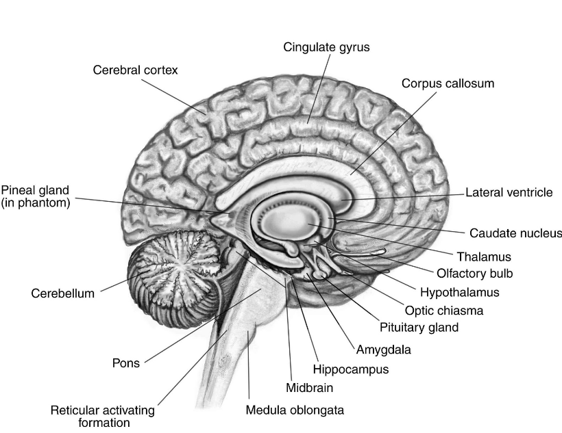
(a) Brain: The human brain is well protected by the skull. Inside the skull, the brain is covered by cranial meninges consisting of an outer layer called dura mater, a very thin middle layer called arachnoid and an inner layer (which is in contact with the brain tissue) called pia mater. The brain can be divided into three major parts:
(i) Forebrain: The forebrain consists of cerebrum, thalamus and hypothalamus.
(ii) Midbrain: It is located between the thalamus/hypothalamus of the forebrain and pons of the hindbrain.
(iii) Hindbrain: The hindbrain comprises pons, cerebellum and medulla.
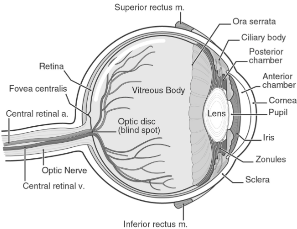
(b) Eye: The adult human eye ball is nearly a spherical structure. The wall of the eye ball is composed of three layers. The external layer is composed of a dense connective tissue and is called the sclera. The anterior portion of this layer is called the cornea. The middle layer, choroid, contains many blood vessels and looks bluish in colour. The choroid layer is thin over the posterior two-thirds of the eye ball, but it becomes thick in the anterior part to form the ciliary body. The ciliary body itself continues forward to form a pigmented and opaque structure called the iris which is the visible coloured portion of the eye. The eye ball contains a transparent crystalline lens which is held in place by ligaments attached to the ciliary body. In front of the lens, the aperture surrounded by the iris is called the pupil whose diameter is regulated by the muscle fibres of iris. The inner layer is the retina and it contains three layers of neural cells from inside to outside - ganglion cells, bipolar cells and photoreceptor cells. There are two types of photoreceptor cells, namely, rods and cones. The daylight (photopic) vision and colour vision are functions of cones and the twilight (scotopic) vision is the function of the rods. The innermost ganglionic cells give rise to optic nerve fibre that forms optic nerve in each eye and is connected with the brain.
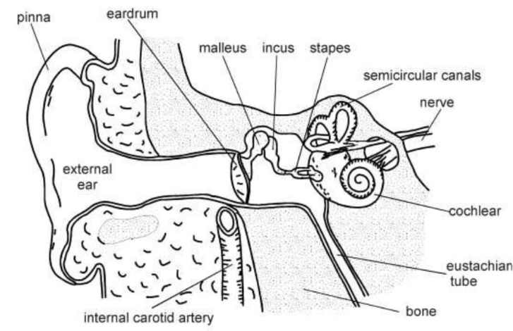
(c) Ear: It perform two sensory functions, hearing and maintenance of body balance. It can be divided into three major sections called the outer ear, the middle ear and the inner ear:
-- Outer ear: It consists of the pinna and external auditory meatus (canal). The pinna collects the vibrations in the air which produce sound. The external auditory meatus leads inwards and extends up to the tympanic membrane (the ear drum). There are very fine hairs and wax-secreting glands in the skin of the pinna and the meatus. The tympanic membrane is composed of connective tissues covered with skin outside and with mucus membrane inside. No part of this publication may be reproduced, distributed, or transmitted in any form or by any means, including photocopying, recording, or other electronic or mechanical methods, without the prior written permission.
-- Middle ear: It contains three ossicles called malleus, incus and stapes which are attached to one another in a chain-like fashion. The malleus is attached to the tympanic membrane and the stapes is attached to the oval window of the cochlea. The ear ossicles increase the efficiency of transmission of sound waves to the inner ear. An Eustachian tube connects the middle ear cavity with the pharynx. The Eustachian tube helps in equalising the pressures on either sides of the ear drum.
-- Inner ear: It is also known as labyrinth. Labyrinth is divided into bony labyrinth and a membranous labyrinth. Bony labyrinth is filled with perilymph while membranous labyrinth is filled with endolymph. Membranous labyrinth is divided into two parts - Vestibular apparatus and Cochlea. The vestibular apparatus is composed of three semi-circular canals and the otolith (macula is the sensory part of saccule and utricle). Each semi-circular canal lies in a different plane at right angles to each other. The membranous canals are suspended in the perilymph of the bony canals. The base of canals is swollen and is called ampulla, which contains a projecting ridge called crista ampullaris which has hair cells. The saccule and utricle contain a projecting ridge called macula. The crista and macula are the specific receptors of the vestibular apparatus responsible for maintenance of balance of the body and posture. Cochlea is a long and coiled outgrowth of sacculus. It is the main hearing organ. Cochlea consists of three membranes. The organ of corti, a hearing organ, is located on the basilar membrane that has hair cells.
Question. Compare the following:
(a) Central neural system (CNS) and Peripheral neural system (PNS)
(b) Resting potential and action potential
(c) Choroid and retina
Answer
(a) Central neural system (CNS) and Peripheral neural system (PNS)
| Central neural system (CNS) | Peripheral neural system (PNS) |
| It is the main coordinating centre of the body. |
It is not the main coordinating centre of the body. |
| It lies inside the skull. | It does not lie inside the skull. |
| This includes brain and spinal cord. | This includes all the nerves of the body associated with the CNS (brain and spinal cord). |
(b) Resting potential and action potential
| Resting potential | Action potential |
| It is the potential difference across the nerve fibre when there is no conduction of nerve impulse. | It is the potential difference across nerve fibre when there is conduction of nerve impulse. |
| The interior of the neuron is electronegative and the exterior is electropositive. | The interior of the neuron is electropositive and the exterior is electronegative. |
| An active sodium pump operates. | No sodium pump operates. |
(c) Choroid and retina
| Choroid | Retina |
| Choroid is the middle vascular layer of eye. | Retina is the innermost nervous coat of eye. |
| It is rich in blood cells | It is rich in neurons |
| Its function is to supply nutrients and oxygen to other parts of eye like retina | Its function is to form an image of an object over it. |
Question. Explain the following processes:
(a) Polarisation of the membrane of a nerve fibre
(b) Depolarisation of the membrane of a nerve fibre
(c) Conduction of a nerve impulse along a nerve fibre
(d) Transmission of a nerve impulse across a chemical synapse
Answer
(a) When the resting potential of the membrane changes it becomes polarized. During resting condition, the axoplasm inside the axon contains high concentration of K+ and negatively charged proteins and low concentration of Na+. As a result, the potassium ions move faster from inside to outside as compared to sodium ions. Therefore, the membrane becomes positively charged outside and negatively charged inside. This is known as polarization of membrane or polarized nerve.
(b) When an electrical stimulus is given to a nerve fibre, an action potential is generated. The membrane becomes permeable to sodium ions than to potassium ions. This results into positive charge inside and negative charge outside the nerve fibre. Hence, the membrane is said to be depolarized.
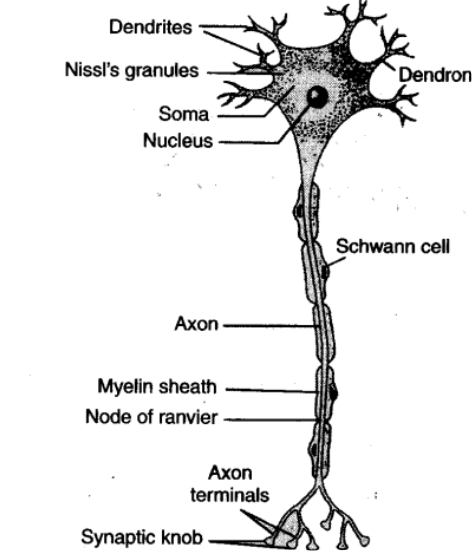
(c) There are two types of nerve fibres - myelinated and non-myelinated. In myelinated nerve fibre, the impulse is conducted from node to node in jumping manner as myelinated nerve fibre is are enveloped with Schwann cells, which form a myelin sheath around the axon. The myelin sheath is impermeable to ions. As a result, the ionic exchange and depolarisation of nerve fibre is not possible along the whole length of nerve fibre. It takes place only at some point, known as nodes of Ranvier. In non-myelinated nerve fibre, the ionic exchange and depolarization of nerve fibre takes place along the whole length of the nerve fibre because of this ionic exchange, the depolarized area becomes repolarised and the next polarized area becomes depolarized.
(d) At a chemical synapse, the membranes of the pre- and post-synaptic neurons are separated by a fluid-filled space called synaptic cleft. When an impulse arrives at the axon terminal, it stimulates the movement of the synaptic vesicles towards the membrane where they fuse with the plasma membrane and release their neurotransmitters in the synaptic cleft. The released neurotransmitters bind to their specific receptors, present on the post-synaptic membrane. This binding opens ion channels allowing the entry of ions which can generate a new potential in the post-synaptic neuron.
The new potential developed may be either excitatory or inhibitory.
Question. Draw labelled diagrams of the following:
(a) Neuron (b) Brain (c) Eye (d) Ear
Answer
(a) Neuron
(b) Brain
(c) Eye
(d) Ear
Question. Write short notes on the following:
(a) Neural coordination (b) Forebrain (c) Midbrain (d) Hindbrain (e) Retina (f) Ear ossicles
(g) Cochlea (h) Organ of Corti (i) Synapse
Answer
(a) The process through which two or more organs interact and complement the functions of one another through the neural system, it is called neural coordination. All the physiological processes in the body are closed linked and dependent upon each other. The neural system and the endocrine system jointly coordinate and integrate all the activities of the organs so that they function in a synchronised fashion. The neural system provides an organised network of point-to-point connections for a quick coordination. The endocrine system provides chemical integration through hormones.
(b) The forebrain consists of cerebrum, thalamus and hypothalamus.
-- Cerebrum forms the major part of the human brain. A deep cleft divides the cerebrum longitudinally into two halves, which are termed as the left and right cerebral hemispheres. The hemispheres are connected by a tract of nerve fibres called corpus callosum. The layer of cells which covers the cerebral hemisphere is called cerebral cortex and is thrown into prominent folds. The cerebral cortex is referred to as the grey matter due to its greyish appearance. The cerebral cortex contains motor areas, sensory areas and large regions that are neither clearly sensory nor motor in function. These regions called as the association areas are responsible for complex functions like intersensory associations, memory and communication.Fibres of the tracts are covered with the myelin sheath, which constitute the inner part of cerebral hemisphere. They give an opaque white appearance to the layer and, hence, is called the white matter.
-- Thalamus: It is a region present at the centre of the forebrain and wrapped by cerebrum. It is coordination center for sensory and motor signalling.
-- Hypothalamus: It lies at the base of the thalamus which contains a number of centres which control body temperature, urge for eating and drinking. It also contains the nerve centres for temperature regulation, hunger, thirst, heart beat and respiration regulation and emotions such as anger, love, cool, etc. It has connection with pituitary gland hence also controls growth and sexual behaviour.
(c) The midbrain is located between the thalamus/hypothalamus of the forebrain and pons of the hindbrain. A canal called the cerebral aqueduct passess through the midbrain. The dorsal portion of the midbrain consists mainly of four round swellings (lobes) called corpora quadrigemina. Midbrain and hindbrain form the brain stem.
(d) The hindbrain comprises pons, cerebellum and medulla.
-- Pons consists of fibre tracts that interconnect different regions of the brain.
-- Cerebellum has very convoluted surface in order to provide the additional space for many more neurons.
-- The medulla of the brain is connected to the spinal cord. The medulla contains centres which control respiration, cardiovascular reflexes and gastric secretions.
(e) Retina is the innermost layer which contains three layers of neural cells – from inside to outside – ganglion cells, bipolar cells and photoreceptor cells. There are two types of photoreceptor cells, namely, rods and cones. The daylight vision and colour vision are functions of cones and twilight vision is the function of the rods. The light enters through cornea, the lens and the images of objects are formed on the retina.
(f) The middle ear contains three ear ossicles called malleus, incus and stapes which are attached to one another in a chain-like fashion. The malleus is attached to the tympanic membrane, incus is connected with stapes. and the stapes is attached to the oval window of the cochlea.The ear ossicles increase the efficiency of transmission of sound waves to the inner ear.
(g) The coiled portion of the labyrinth is called cochlea. The membranes constituting cochlea, the reissner’s and basilar, divide the surounding perilymph filled bony labyrinth into an upper scala vestibuli and a lower scala tympani. The space within cochlea called scala media is filled with endolymph. At the base of the cochlea, the scala vestibuli ends at the oval window, while the scala tympani terminates at the round window which opens to the middle ear.
(h) The organ of corti is a structure located on the basilar membrane which contains hair cells that act as auditory receptors. The hair cells are present in rows on the internal side of the organ of corti. (i) A synapse is formed by the membranes of a pre-synaptic neuron and a post-synaptic neuron, which may or may not be separated by a gap called synaptic cleft. These are of two types electrical synapses and chemical synapses.
Question. Give a brief account of:
(a) Mechanism of synaptic transmission
(b) Mechanism of vision
(c) Mechanism of hearing
Answer
(a) A nerve impulse is transmitted from one neuron to another through junctions called synapses which is formed by the membranes of a pre-synaptic neuron and a post-synaptic neuron may or may not be separated by synaptic cleft. There are two types of synapses, namely, electrical synapses and chemical synapses.
At electrical synapses, the membranes of pre- and post-synaptic neurons are in very close proximity so electrical current can flow directly from one neuron into the other across these synapses.
Transmission of an impulse across electrical synapses is very similar to impulse conduction along a single axon and transmission is always faster than that across a chemical synapse however it is not common in human body.
At a chemical synapse, the membranes of the pre- and post-synaptic neurons are separated by a fluid-filled space called synaptic cleft. Chemicals called neurotransmitters are involved in the transmission of impulses at these synapses.
(b) The light rays in visible wavelength focussed on the retina through the cornea and lens generate impulses in rods and cones. The photosensitive compounds in the human eyes is composed of opsin and retinal. Light induces dissociation of the retinal from opsin resulting in changes in the structure of the opsin. This causes membrane permeability changes therefore potential differences are generated in the photoreceptor cells. This produces a signal that generates action potentials in the ganglion cells through the bipolar cells. These impulses are transmitted by the optic nerves to the visual cortex area of the brain, where the neural impulses are analysed and the image formed on the retina is recognised based on earlier memory and experience.
(c) The external ear receives sound waves and directs them to the ear drum.The ear drum vibrates in response to the sound waves and these vibrations are transmitted through the ear ossicles to the oval window. The vibrations are passed through the oval window on to the fluid of the cochlea, where they generate waves in the lymphs. The waves in the lymphs induce a ripple in the basilar membrane. These movements of the basilar membrane bend the hair cells, pressing them against the tectorial membrane therefore nerve impulses are generated in the associated afferent neurons. These impulses are transmitted by the afferent fibres via auditory nerves to the auditory cortex of the brain, where the impulses are analysed and the sound is recognised.
Question. Answer briefly:
(a) How do you perceive the colour of an object?
(b) Which part of our body helps us in maintaining the body balance?
(c) How does the eye regulate the amount of light that falls on the retina?
Answer
(a) The cones are responsible for color vision. There are three types of cones cells that respond to green light, blue light and red light according to their characteristics. These cells are stimulated by different lights, from different sources. The combinations of the signals generated help us see the different colours.
(b) The Inner ear has three semi-circular canals forming cochlea. Cochlea is responsible for maintaining the body balance.
(c) Pupil is the small aperture surrounded by the the iris that regulates the amount of light entering the eye. This expand in case of low light and contract in case of intense light thereby regulating the amount of light falling on the retina.
Question. Explain the following:
(a) Role of Na+ in the generation of action potential.
(b) Mechanism of generation of light-induced impulse in the retina.
(c) Mechanism through which a sound produces a nerve impulse in the inner ear.
Answer
(a) The action potential is determined by Na+ ions. The Na+ channels which are closed in the resting state, open and cause the inflow of Na+ ions by diffusion into the inside of axoplasm. The electrical potential of the membrane changes from 70 mV towards zero and then the membrane is said to be depolarised.
(b) The photopigments of the retina are photosensitive compounds in the eye that are composed of retinal and opsin. Light induces dissociation of retinal from opsin which changes the structure of the opsin. It generates action potential in the bipolar neurons. These impulses/action potential are transmitted by the optic nerves to the visual cortex of the brain where the neural impulses are analysed and the erect image is recognised.
(c) When sound falls over the ear drum, it is then transmitted to the inner ear by ear ossicles. The vibrations are passed through the oval window onto the fluid of the cochlea, where they generate waves in the lymphs. The waves induce a ripple in the basilar membrane that bend the hair cells, pressing them against the techtonial membrane. As a result nerve impulses are generated in the associated afferent neurons and transmitted to auditory cortex of brain via auditory nerves, where the impulses are analysed and the sound is recognised.
Question. Differentiate between:
(a) Myelinated and non-myelinated axons
(b) Dendrites and axons
(c) Rods and cones
(d) Thalamus and Hypothalamus
(e) Cerebrum and Cerebellum
Answer
(a) Myelinated and non-myelinated axons
| Myelinated axons | Non myelinated axons |
| These appear whitish in colour. | These appear greyish in colour. |
| Myelin sheath is present | Myelin sheath is absent |
| Nodes of Ranvier are present at intervals. | Nodes of Ranvier are absent. |
| Transmission of nerve impulse is faster. | Transmission of nerve impulse is slower |
| Ion exchange can occur only at the nodes. | Ion exchange occurs all over the surface. |
(b) Dendrites and axons
| Dendrites | Axons |
| These are extension of cyton present at anterior position. | These are extension of cyton present at posterior position. |
| These conduct impulses towards the cell body. | These conduct impulses away from the cell body. |
| Dendrites are always non-myelinated. | Axons can be myelinated or non-myelinated. |
| The terminals of dendrites become receptors. |
The terminals of axon ends in a group of branches called terminal arborisations. |
(c) Rods and cones
| Rods | Cones |
| Rods are sensitive to dim light. | Cones are sensitive to bright light only. |
| They have visual purple pigment called rhodopsin. | They have visual violet pigment called iodopsin. |
| Colours are not visible with rod cells. | Cones help to see colours. |
(d) Thalamus and Hypothalamus
| Thalamus | Hypothalamus |
| It represent the side of diencephalon. | It represents the lower part of diencephalon. |
| It is the major coordinating centre for sensory and motor signalling. |
It is the major centre for regulation of body temperature, thirst, hunger etc. |
| It does not secrete any hormone. | It secretes several hormones. |
(e) Cerebrum and Cerebellum
| Cerebrum | Cerebellum |
| It is part of fore brain | It is part of hind brain |
| It consists of two cerebral hemispheres. | It consists of two cerebellar hemispheres and a median vermis. |
| It initiates voluntary movements. | It maintains posture and equilibrium. |
Question. Answer the following:
(a) Which part of the ear determines the pitch of a sound?
Answer Cochlea
(b) Which part of the human brain is the most developed?
Answer Cerebrum
(c) Which part of our central neural system acts as a master clock?
Answer Hypothalamus
Question. The region of the vertebrate eye, where the optic nerve passes out of the retina, is called the
(a) fovea
(b) iris
(c) blind spot
(d) optic chaisma
Answer (c) blind spot
Question. Distinguish between
a) Afferent neurons and Efferent neurons
b) Impulse conduction in a myelinated nerve fibre and an unmyelinated nerve fibre
c) Aqueous humour and Vitreous humour
d) Blind spot and Yellow spot
e) Cranial nerves and Spinal nerves
Answer
a) Afferent neurons and Efferent neurons
| Afferent neurons | Efferent neurons |
| They conduct impulses towards the central neural system. |
They conduct impulses away from the central neural system |
| It provides stimulus and evoke senses. | It results in the response by the effectors. |
| They are sensory in nature. | They are motor in nature. |
| These take information from the receptors. | These take information to effectors. |
b) Impulse conduction in a myelinated nerve fibre and an unmyelinated nerve fibre
| Impulse conduction in a myelinated nerve fibre | Impulse conduction in a unmyelinated nerve fibre |
| The depolarisation occurs only in the nodes of Ranvier where the myelin sheath is absent. |
The depolarisation occurs all along the length of the nerve fibre. |
| Action potential jumps from one node of Ranvier to another. | Action potential travels along the entire length of the fibre. |
| Conduction is faster. | Conduction is slower. |
| Less amount of energy is required. | More amount of energy is required. |
c) Aqueous humour and Vitreous humour
| Aqueous humour | Vitreous humour |
| It is a watery and transparent fluid. | It is thick gelly-like transparent fluid |
| It is present between lens and cornea. | It is present between lens and retina. |
| It is continuously secreted by ciliary processes and drained out of the eyes. |
It is not replaced. |
| It provides nourishment to lens, cornea and other parts of anterior chamber. |
It does not have nutritive value. |
d) Blind spot and Yellow spot
| Blind spot | Yellow spot |
| Blind spot is a spot on the retina present at the point of origin of the optic nerve. |
Yellow spot is a small area on the retina present at the posterior pole of the eye, lateral to the blind spot. |
| It does not have a shallow depression. | It has a shallow depression called fovea centralis. |
| Photoreceptor cells are absent from this region. |
Only cones are present in this region. |
| It is insensitive to light. | It is sensitive to bright light. |
| Photoreceptor cells are absent from this region. |
Only cones are presentAn image is formed at the yellow spot. in this region. |
| No image is formed at the blind spot. | An image is formed at the yellow spot. |
e) Cranial nerves and Spinal nerves
| Cranial nerves | Spinal nerves |
| They originate from the brain. | They originate from the spinal cord. |
| There are 12 pairs of cranial nerves. | There are 31 pairs of spinal nerves. |
| These are sensory, motor or mixed in nature. | They are mixed in nature. |
| They extend between the brain and body parts. | They extend between the spinal cord and body parts. |
Free study material for Biology
NCERT Solutions Class 11 Biology Chapter 18 Neural Control and Coordination
Students can now access the NCERT Solutions for Chapter 18 Neural Control and Coordination prepared by teachers on our website. These solutions cover all questions in exercise in your Class 11 Biology textbook. Each answer is updated based on the current academic session as per the latest NCERT syllabus.
Detailed Explanations for Chapter 18 Neural Control and Coordination
Our expert teachers have provided step-by-step explanations for all the difficult questions in the Class 11 Biology chapter. Along with the final answers, we have also explained the concept behind it to help you build stronger understanding of each topic. This will be really helpful for Class 11 students who want to understand both theoretical and practical questions. By studying these NCERT Questions and Answers your basic concepts will improve a lot.
Benefits of using Biology Class 11 Solved Papers
Using our Biology solutions regularly students will be able to improve their logical thinking and problem-solving speed. These Class 11 solutions are a guide for self-study and homework assistance. Along with the chapter-wise solutions, you should also refer to our Revision Notes and Sample Papers for Chapter 18 Neural Control and Coordination to get a complete preparation experience.
FAQs
The complete and updated is available for free on StudiesToday.com. These solutions for Class 11 Biology are as per latest NCERT curriculum.
Yes, our experts have revised the as per 2026 exam pattern. All textbook exercises have been solved and have added explanation about how the Biology concepts are applied in case-study and assertion-reasoning questions.
Toppers recommend using NCERT language because NCERT marking schemes are strictly based on textbook definitions. Our will help students to get full marks in the theory paper.
Yes, we provide bilingual support for Class 11 Biology. You can access in both English and Hindi medium.
Yes, you can download the entire in printable PDF format for offline study on any device.