
Download CBSE Class 11 Biology Locomotion And Movement Notes Set A in PDF format. All Revision notes for Class 11 Biology have been designed as per the latest syllabus and updated chapters given in your textbook for Biology in Class 11. Our teachers have designed these concept notes for the benefit of Class 11 students. You should use these chapter wise notes for revision on daily basis. These study notes can also be used for learning each chapter and its important and difficult topics or revision just before your exams to help you get better scores in upcoming examinations, You can also use Printable notes for Class 11 Biology for faster revision of difficult topics and get higher rank. After reading these notes also refer to MCQ questions for Class 11 Biology given on studiestoday
Revision Notes for Class 11 Biology Chapter 17 Locomotion and Movement
Class 11 Biology students should refer to the following concepts and notes for Chapter 17 Locomotion and Movement in Class 11. These exam notes for Class 11 Biology will be very useful for upcoming class tests and examinations and help you to score good marks
Chapter 17 Locomotion and Movement Notes Class 11 Biology
Locomotion And Movement Class 11 Notes
POINTS TO REMEMBER
Types of Movement :
• Amoeboid movement: This movement takes place in phagocytes where leucocytes and macrophages migrate through tissue. It is affected by pseudopodia formed by the streaming of protoplasm (as in amoeba)
• Ciliary movement: These movements occur in internal organs which are lined by ciliary epithelium.
• Muscular Movement: This movement involves the muscle fibers, which have the ability to contract and relax.
MUSCLES :
Properties of Muscle :
• Excitability
• Contractility
• Extensibility
• Elasticity
Types of Muscles :
• Skeletal muscles or striated muscles –
o Closely associated with skeleton.
o They are striped appearance under the microscope and called Striated muscles.
o They are under voluntary control of nervous system, hence called voluntary muscles.
o These involved in locomotion and change of body postures.
o Unbranched and multinucleated.
• Visceral muscles or smooth muscles
o These are located in inner wall of hollow visceral organ.
o Spindle shaped and uni-nucleated.
o They do not exhibit any striation and are smooth in appearance.
o They are called smooth muscles or non-striated muscles.
o Their activities are not under voluntary control of nervous system hence called as involuntary muscles.
o They assist in transport of food through digestive tract and gametes through the genital tract.
• Cardiac muscles –
o The muscles of heart, involuntary in nature.
o Cardiac muscle cells assemble in a branching pattern to form a cardiac muscle.
o These are uni-nucleated with characteristic intercalated disc.
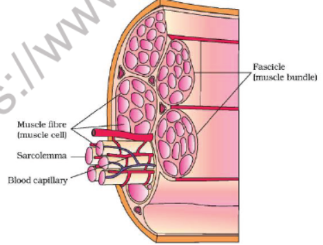
Structure of skeletal muscle :
• Each organized skeletal muscle in our body is made of a number of muscle bundles called fascicles held together by common fibrous covering called fascia.
• Each fascicle consists of a number of muscle fibres (cell) covered by a common fibrous perimysium.
• Each muscle fibre is lined by the plasma membrane called sarcolemma, enclosing cytoplasm called sarcoplasm.
• The sarcoplasm contain endoplasmic reticulum, called sarcoplasmic reticulum is the store house of calcium ion.
• Muscle fibre is a syncitium as the sarcoplasm contain many nuclei.
• Muscle fibres contain a large number of parallelly arranged filaments in the sarcoplasm called myofilaments ormyofibrils.
• There are two types of myofibrils are present in the sarcoplasm –
• The arrangement of thick and thin filament gives the characteristic striated appearance.
• The light bands contain only actin filaments and are called I-band or isotropic band.
• The dark band called ‘A’ or anisotropic band contains both actin and myosin.
• In the centre of each ‘I’ band is an elastic fibre called ‘Z’ line which bisects it.
• The thin filaments or actin are firmly attached with the ‘Z’ line.
• The thick filaments or myosin in the ‘A’ band are also held together in the middle by a thin fibrous membrane called‘M’ line.
• The portion between two successive ‘Z’ lines is considered as the functional unit of the muscle called sarcomere.
• Each ‘A’ band contains two overlap zone of thick and thin filament called ‘O’ band.
• The central part of thick filament, not overlapped by thin filament is called ‘H’ band.
• ‘A’ band = 2(O) + H.
Structure of Contractile proteins :
Thin filament or Actin :
• Each actin filament is made of two ‘F’ actins helically wound to each other.
• Each ‘F’ actin is made of polymer of monomeric ‘G’ (Globular) actin.
• Each ‘F’ actin associated with another protein, tropomyosin also run throughout its length.
• Another complex protein, Troponin is distributed at regular intervals on the tropomyosin.
• Each troponin has three component –
o Troponin-C binds with calcium.
o Troponin-M, binds with the tropomyosin.
o Troponin T, masks the active site on the ‘G’ actin (thin filament)
• In the resting state a sub-unit of Troponin (Tn-T), masks the active binding sites on the thin filaments for myosin.
Thick filament :
• Each myosin (thick) filament is consists of many monomeric protein called Meromyosins.
• Each meromyosin has two parts –
o Heavy meromyosin (HMM) - A globular head with a short arm.
o Light meromyosin (LMM) – a tail.
• The HMM component, i.e. the head and short arm projects outwards at regular distance and angle from each other from the surface of a polymerized myosin filament and is known as cross arm.
• The globular head is an active ATPase enzyme and has binding sites for ATP and active sites for actin.
Mechanism of muscle contraction :
• Mechanism of muscle contraction is explained by sliding filament theory which states that contraction of a muscle fibre takes place by the sliding of the thin filaments over the thick filaments.
• Muscle contraction is initiated by a signal sent by the central nervous system via a motor neuron.
• A motor neuron along with the muscle fibres connected to it constitutes a motor unit.
• The junction between a motor neuron and the sarcolemma of the muscle fibre is called neuromuscular junction ormotor-end plate.
• Neurotransmitter releases here which generates an action potential in sarcolemma.
• These causes release of Ca++ into sarcoplasm.
• These Ca++ binds with troponin, thereby remove masking of active site.
• Myosin head binds to exposed active site on actin to form a cross bridge, utilizing energy from ATP hydrolysis.
• This pulls the actin filament towards the centre of ‘A’ band.
• ‘Z’ lines also pulled inward thereby causing a shortening of sarcomere i.e. contraction.
• ‘I’ band get reduced, whereas the ‘A’ band retain the length.
• During relaxation, the cross bridge between the actin and myosin break.
• Ca++pumped back to sarcoplasmic cisternae.
• Actin filament slide out of ‘A’ band and length of ‘I’ band increases. This returns the muscle to its original state.
• Repeated muscle contraction causes accumulation of lactic acid, produced from anaerobic breakdown of glycogen leads to muscle fatigue.
• Muscle contains red coloured oxygen storing pigment called myoglobin.
• Muscle with myoglobin called red muscle fibres, they are also contain large number of mitochondria which can utilize large amount of oxygen stored in them for ATP production also called aerobic muscle.
• Some muscles possess very less quantity of myoglobin and less mitochondrion hence called white fibres. Amount of sarcoplasmic reticulum is high in these muscles. They depend on anaerobic process for energy.
SKELETAL SYSTEM :
• Human skeleton consists of 206 bones in adult.
o Axial skeleton – 80 bones
o Appendicular skeleton – 126 bones.
• Axial skeleton :
o Skull – 29 bones.
• Cranium – 8 bones forms the brain box.
• Facial – 14 bones forms the front part of the face.
• Hyoid – a single U-shaped bone at the base of the buccal cavity.
• Ear ossicles – 6 bones- 3 on either side (Malleus, Incus and stapes)
o The skull region articulates with the superior region of the vertebral column with the help of two occipital condyles hence called dicondylic skull.
o Vertebral column – 26 bones
• Cervical – 7 vertebrae.
• Thoracic – 12 vertebrae.
• Lumber – 5 vertebrae.
• Sacral – 1 vertebra. (fused five bone)
• Caudal – 1 vertebra (fused four bones)
o Sternum or breast bone – 1 bone in the middle line of the thorax.
• Ribs – 12 pairs – (24 bones)
o 1-7 are true ribs (connected to the sternum directly)
o 8th, 9th, 10th pairs are called false ribs they attached to the 7th ribs.
o 11th and 12th not connected ventrally hence called floating ribs.
o Ribs attaché dorsally to the vertebra and ventrally with the sternum by hyaline cartilage.
o Thoracic vertebrae, ribs and sternum together form the rib cage.
• Appendicular skeleton: 126 bones
o Fore limb – 60 ( 30 in each)
• Humerus – 1 bone
• Radius and ulna – 2 bones
• Carpals (wrist bones) – 8 in numbers.
• Metacarpals (palm bones) – 5 in numbers
• Phalanges (digits) – 14 in number.
o Hind limb – 60 (30 in each)
• Femur (thigh bone- the longest and heaviest bone) – 1 number.
• Tibia and fibula – 2 bones.
• Tarsals (ankle bone) – 7 bones.
• Metatarsals – 5 in numbers.
• Phalanges (digits) – 14 in numbers.
• Patella (knee cap) – 1 bone.
o Pectoral girdles: consists of 2 bones each = 4 bones.
• Helps in articulation of fore limb with the axial skeleton.
• Each pectoral girdle made of two half.
• Each half made of two bone the clavicle and scapula.
• Scapula is a large triangular flat bone situated in the dorsal part of the thorax between the second and the seventh ribs.
• Scapula is characterized by spine with acromion process.
• Below acromion, is glenoid cavity to which head of humerus fits.
• Clavicle is commonly called collar bone.
o Pelvic girdle: 2 bones.
• Pelvic girdle consists of two coxal bones.
• Each coxal bone is formed of fusion of three bones
• Ilium
• Ischium
• Pubis.
• At the point of fusion of the three bones is a cavity called acetabulum to which the femur articulates.
• Two halves of the pelvic girdle meets ventrally to form the pubic symphysis containing fibrous cartilage.
JOINTS :
• Joints are the points of contact between bones, or between bones and cartilages.
• Force generated by the muscles is used to carry out movement through joints, where joint acts as a fulcrum.
• Joints are classified into three types:
o Fibrous joint
o Cartilaginous joint
o Synovial joint
Fibrous joints :
• Do not allow any movements.
• Found in flat bones which fuse end-to-end with the help of dense fibrous connective tissues in the form of sutures.
• These types of joints are found in the bones of cranium.
Cartilaginous joints :
• The bones involved are joined together with the help of cartilages.
• Permits very little movements.
• Joint between the vertebral column are the example of such joints.
Synovial joints :
• Characterized by the presence of a fluid filled synovial cavity between the articulating surfaces of the two bones.
• Allow free movement between two bones.
• The fluid inside it called synovial fluid covered by synovial membrane.
o Ball and socket joint - between humerus and pectoral girdle
o Hinge joint – knee joint
o Pivot joint – between atlas and axis.
o Gliding joint – between carpals.
o Saddle joint – between carpals and metacarpals of thumb.
DISORDERS OF MUSCULAR AND SKELETAL SYSTEM :
Myasthenia gravis :
• It is an auto-immuno disorder.
• Affects the neuromuscular junction leads to fatigue.
• Caused weakening and paralysis of skeletal muscle.
Muscular dystrophy : Progressive degeneration of skeletal muscle mostly due to genetic disorder.
Tetany: rapid spasms (wild contractions) in muscle due to low Ca++ in body fluid.
Arthritis : inflammation of joints.
Osteoporosis : age related disorder characterized by decreased bone mass and increased chances of fractures. Decrease levels of oestrogen are a common cause.
Gout : inflammation of joints due to accumulation of uric acid crystals.

2.1 (iii) Structure of Contractile Proteins :
(a) Actin : Each actin filament is made up of the following components-
(A) F- actin : In each actin filament, two ‘F’ (filamentous) actins helically wound to each other. Each ‘F’ actin is a polymer of monomeric ‘G’ (Globular) actins.
(B) Tropomyosin : Two filaments of another protein, tropomyosin also run close to the ‘F’ actins throughout its length.
(C) Troponin : It is a complex protein which is distributed at regular intervals on the tropomyosin. In the resting state a subunit of troponin masks the active binding sites for myosin on the actin filaments.
(b) Myosin : Each myosin (thick) filament is also a polymerised protein. Many monomeric proteins called
Meromyosins constitute one thick filament. Each meromyosin has two important parts, a globular head with a short arm and a tail, the former being called the heavy meromyosin (HMM) and the latter, the light meromyosin (LMM). The HMM component, i.e.; the head and short arm projects outwards at regular distance and angle from each other from the surface of a polymerised myosin fllament and is known as cross arm. The globular head is an active ATPase enzyme and has binding sites for ATP and active sites for actin.

2.1 (iv) Working of striated muscles :
• H.E. Huxley and A.F. Huxley in 1954 proposed a theory to explain the process of muscular contraction. This theory is known as ‘sliding
filament theory’.
• It was observed that when a fibril contracts :
(a) Its ‘A’ bands remain intact,
(b) ‘I’ bands progressively shorten and eventually disappear when the fibril has shortened to about 65% of its resting length.
(c) At this stage, ‘H’ zones also disappear because the actin filaments of both sides in each sarcomere reach, and may even overlap each other at the “M” line, and the ‘Z’ lines now touch the ends of myosin filaments.
(d) Sarcomere shortens
Note: It was further observed that if a fibre is mechanically streched, the zones of overlap between thick and thin filaments are shorter than in resting condition, resulting in wider ‘H’ zones.
• It was observed that when a fibril relax : All the phenomenona occur in reverse way to relax the muscle i.e. the muscle comes in normal condition.
Note : These observations led Huxley to propose that shortening of the fibrils in contraction is brought about by sliding movement of actin filaments over myosin filaments towards “M” line by means of rapidly forming and breaking cross bridges or rachets at the spurs of myosin filaments. Thus, the sarcomere were recognised as the ‘ultimate units of contraction

2.1 (v) Mechanism of Muscle Contraction
• Mechanism of muscle contraction is best explained by the sliding filament theory which states that contraction of a muscle fibre takes place by the sliding of the thin filaments over the thick filaments.
• Muscle contraction is initiated by a signal sent by the central nervous system (CNS) via a motor neuron.
A motor neuron along with the muscle fibres connected to it constitute a motor unit. The junction between a motor neuron and the sarcolemma of the muscle fibre is called the neuromuscular junction or motorend plate.
• A neural signal reaching this junction releases a neurotransmitter (Acetyl choline) which generates an action potential in the sarcolemma. This spreads through the muscle fibre and causes the release of calcium ions into the sarcoplasm.
• Increase in Ca2+ level leads to the binding of calcium with a subunit of troponin on actin filaments and thereby remove the masking of active sites for myosin.
• Utilising the energy from ATP hydrolysis, the myosin head now binds to the exposed active sites on actin to form a cross bridge.

• This pulls the attached actin filaments towards the centre of ‘A’ band. The ‘Z’ line attached to these actins are also pulled inwards thereby causing a shortening of the sarcomere, i.e., contraction. It is clear from the above steps, that during shortening of the muscle, i.e., contraction, the I bands get reduced, whereas the ‘A’ bands retain the length.
• The myosin, releasing the ADP and Pi goes back to its relaxed state. A new ATP binds and the crossbridge is broken. The ATP is again hydrolysed by the myosin head and the cycle of cross bridge formation and breakage is repeated causing further sliding.
• The process continues till the Ca2+ ions are pumped back to the sarcoplasmic cisternae resulting in the masking of actin filaments. This causes the return of’Z’ lines back to their original position, i.e., relaxation.
• The reaction time of the fibres can vary in different muscles.
Note : Repeated activation of the muscles can lead to the accumulation of lactic acid due to anaerobic breakdown of glycogen in them, causing fatigue.
Note : On the basis of quantity of myoglobin pigment muscles are categories as
(A) Red muscle fibre : Muscle contains a red coloured oxygen storing pigment called myoglobin. Myoglobin rich muscles gives a reddish appearance. Such muscles are called the Red fibres. These muscles also contain plenty of mitochondria which can utilise the large amount of oxygen stored in them for ATP production. These muscles, therefore, can also be called aerobic muscles.
(B) White muscle fibre : Muscles possess very less quantity of myoglobin, appear pale or whitish. These are the White fibres. Number of mitochondria are also few in them, but the amount of sarcoplasmic reticulum is high. They depend on anaerobic process for energy.

2.2 VISCERAL OR SMOOTH MUSCLES OR NON STRIATED
• These are called smooth, plain nonstriated involuntary or unstriped muscles due to absence of striations.
• These occur in the walls of hollow internal organs (alimentary canal, gall bladder, bile ducts, respiratory tracts, uterus, urinogenital ducts, urinary bladder, blood vessels, etc.), in capsules of lymph glands, spleen etc., in iris and ciliary body of eyes, skin dermis, penis and other accessory genitalia, etc.
• Smooth muscles of skin dermis, called errector pilli muscles, are associated with hair roots, and are responsible for flesh (erection of hairs). Those of penis form a muscular network which helps in its erection and limping.
• Structure : Smooth muscle fibre is unbranched goose-spindle shaped, uninucleated and has no sarcolemma. Contraction is slow, involuntary under the control of ANS.

• Functionally smooth muscles are of two types –
(1) Single-unit smooth muscle : Single unit smooth muscle fibres are composed of muscle fibres closely joined together, contract as a single unit. e.g., urinary bladder, gastrointestinal tract, small arteries and small veins.
(2) Multi-unit smooth muscles : are composed of more independent muscle fibres, contract as separate units e.g. – hair root muscle, muscles on the wall of large blood vessels, ciliary muscles, muscles of iris and bronchi.
2.3 CARDIAC MUSCLES
• Heart wall (also the wall of large veins just where these enter into the heart) is made up of cardiac muscles and, hence, called myocardium.
• Structurally, these muscles resemble striated muscles but, functioning independently of the conscious control of brain, these are involuntary like the smooth muscles.
• Cardiac muscle cells of fibres are comparatively shorter and thicker, cylindrical, mostly uninucleate with a central nucleus, somewhat branched and covered by a sarcolemma.
3. classification of body muscles
The total number of muscles in the body of adult man is 639. The muscles that act together to produce a movement are called synergists and the muscle that act in opposition to each other are antagonists. According to the type of motion they cause, the muscles are divided into the following types.
(i) Flexor and Extensor : Muscles that bend one part over another joint is called flexor. Extensor muscle is antagonist of flexor muscle. The contraction of an extensor extends a joint by pulling one of the articulating bone apart from another.

(ii) Pronator and Supinator : The contraction of a pronator rotates the forearm to turn the palm downward or backward Supinator is antagonist of pronator. A supinator contracts to rotate the forearm and thus to make palm face upward or forward.

(iii) Abductor and Adductor : An abductor contracts to draw a bone away from the body midline. Muscle that brings the limb away from midline is called abductor. An adductor draws a bone towards the body midline. Muscles that brings the limb towards midline is called adductor. Abductor muscle is antagonist of adductor muscle.

(iv) Protractor and Retractor : Protractor muscle pulls the lower jaw, tongue and the head forward. Retraction is opposite to protaction. Retractor muscle draws the lower jaw, tongue and the head backward

(v) Inversion and Eversion : Turning of feet so that the soles face one another in inversion. Eversion is the opposite of inversion. In this movement, the soles of the feet face laterally

(vi) Rotation : Rotation is term that indicates the partial revolving of a body part on the part’s long axis.

(vii) Errector : Raises hairs of skin.
(viii) Levator : Elevates a part of body.
(ix) Depressor : Lowers a part of body.
(x) Sphincter : Closes a natural orifice or passage.
(xi) Constrictor : Causes constriction or squeezing.
4 Skeleta Systeml
• Skeletal system consists of a framework of bones and a few cartilages. This system has a significant role in movement shown by the body.
• Bone and cartilage are specialised connective tissues. The former has a very hard matrix due to calcium salts in it and the latter has slightly pliable matrix due to chondroitin salts.
• The study of bone structure and treatment of bone disorder called osteology.
• The specialized branch of medicine that deals with preservation and restoration of skeletal system, joints is called orthopedics.
• Bones are made up of a protein called ossein and cartilage are made of a protein called chondrin.
Hence study of bones is called osteology and study of cartilage is called chondrology.
4.1 SKELETON
The hardened tissues of the body together form the skeleton (sclero = hard). Skeleton of invertebrates is most often secreted on the surface, forming a lifeless or dead exoskeleton. Whereas skeleton of vertebrates develops most often underneath the surface forming a living or growing endoskeleton.
• Three types of skeletons develop in vertebrates :
(1) Epidermal/Horny exoskeleton : These include hard and horny of keratinized derivatives of epidermal layer of skin, such as claws, most reptilian’s scales, bird feathers and mammalian hairs, horns, nails and hoofs, etc.
(2) Dermal/Bony skeleton : Dermal bony skeleton is derived from the dermis of skin. It includes bony scales and plates. In fishes, dermal scales become exposed due to wearing out of epidermis, and form exoskeleton.
(3) Endoskeleton : Greater part of vertebrate skeleton lies more deeply, forming the endoskeleton. Endoskeleton is formed by bones in vertebrates.
4.2 SKELETON IN DIFFERENT ANIMALS
(a) Invertebrate –
(i) Protozoa – No skeleton.
(ii) Porifera – Calcarius spicules + silicious spicules
(iii) Coelentrata – Calcareous (corals) and chitinous
(iv) Helminth – No skeleton, cuticle present.
(v) Annelida – No skeleton, cuticle present.
(vi) Arthropoda – Dead Chitinus exoskeleton.
(vii) Mollusca – Calcarius shell
(viii) Echinodermata – Dermal calcareous plates are present.
(b) Vertebrates : In vertebrates dermal skeleton is formed by bones. Bone is the connective tissue with intercellular spaces filled with ossein matrix composed of 25% water, 25% protein fibers, 50% mineral salts. The inner most region is full of bone marrow having various types of cells.
(i) Cartilage bones : The bones which are formed by the ossification of preexisting cartilage are called cartilage bones or replacing bones. e.g., vertebra, Girdles, limbs bones, Incus, malleus, stapes.
(ii) Membrane or dermal bones : The bones which are formed by ossification in connective tissue are called dermal, bones. e.g., Ribs, sternum, clavicle, Nasal, palatine, maxilla.
(iii) Pneumatic bones : Bones with hollow spaces containing air e.g., bones of bird, Frontal, ethmoid, maxilla of human.
(iv) Irregular bones : Vertebrae are irregular bone.
(v) Flat bones : Cranial bone, scapula, Ribs.
(vi) Short bones : Carpals and tarsals.
Functions of bony skeleton : Chief function of vertebrate bony skeleton can be enumerated as follows –
(i) To provide physical support to body by forming a firm and rigid internal framework.
(ii) To give definite body shape and form.
(iii) To protect by surrounding delicate internal organs like brain, heart, lungs etc.
(v) To provide surface for attachment of muscles.
(vi) To serve as levers on which muscles can act.
(vii) To manufacture blood corpuscles in bone marrow.
(viii) To help in breathing (tracheal rings, ribs).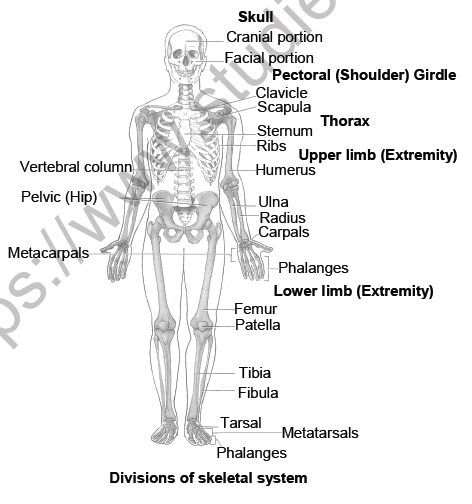
5.Human Skeletal System
In human beings, this system is made up of 206 bones and a few cartilages. It is grouped into two principal divisions - the axial and the appendicular skeleton.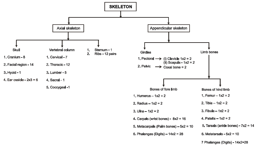
5.1 AXIAL SKELETON :
• Axial skeleton comprises 80 bones distributed along the main axis of the body. The skull, vertebral column, sternum and ribs constitute axial skeleton.
(a) Skull : The skull is composed of two sets of bones cranial and facial, that totals to 22 bones. Cranial bones are 8 in number. They form the hard protective outer covering, cranium for the brain.
• The facial region is made up of 14 skeletal elements which form the front part of the skull. A single Ushaped bone called hyoid is present at the base of the buccal cavity and it is also included in the skull.Each middle ear contains three tiny bones - Malleus, Incus and Stapes, collectively called Ear Ossicles.
• The skull region articulates with the superior region of the vertebral column with the help of two occipital condyles (dicondylic skull).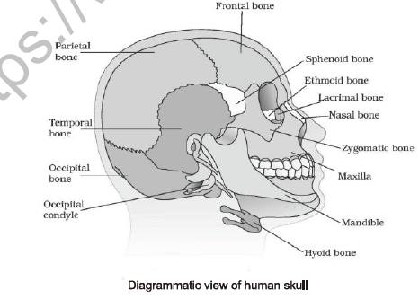
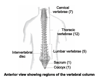
(b) Vertebral column :
• Our vertebral column is formed by 26 serially arranged units called vertebrae and is dorsally placed. It extends from the base of the skull and constitutes the main framework of the trunk.
• Each vertebra has a central hollow portion (neural canal) through which the spinal cord passes.
• First vertebra is the atlas and it articulates with the occipital condyles.
• The vertebral column is differentiated into cervical (7), thoracic (12), lumbar (5), sacral (1-fused) and coccygeal (1-fused) regions starting from the skull. The number of cervical vertebrae are seven in almost all mammals including human beings.
• The vertebral column protects the spinal cord, supports the head and serves as the point of attachment for the ribs and musculature of the back.
• Sternum is a flat bone on the ventral midline of thorax.
•Curvatures of vertebral column : In a foetus, there is only a single anteriorly concave curve, in adult there are 4 curves like, cervical, thoracic, lumber, and sacral. Cervical and lumber are anteriorly convex, while thoracic and sacral are anteriorly concave.
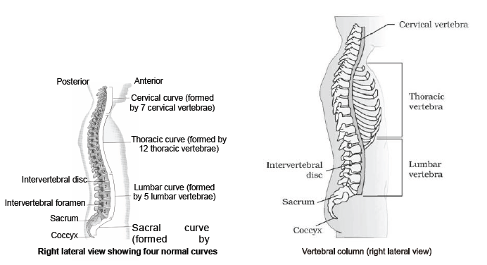
•The curves of vertebral column are important because they increases its strength, help maintain balance in upright position absorb shock during walking and running and help protect the column from fracture.
Certain abnormalities of curvature are :
i) Kyphosis : Exaggeration of thoracic curve, resulting in “round-shouldered” appearance, also called hunch back.
(ii) Lordosis : An exaggeration of lumber curve, also called sway back.
(iii) Scoliosis : An abnormal lateral curvature in any region of spine.
(c) Ribs & Sternum:
•There are 12 pairs of ribs. Each rib is a thin flat bone connected dorsally to the vertebral column and ventrally to the sternum. It has two articulation surfaces on its dorsal end and is hence called bicephalic.
•First seven pairs of ribs are called true ribs. Dorsally, they are attached to the thoracic vertebrae and ventrally connected to the sternum with the help of hyaline cartilage. The 8th, 9th and 10th pairs of ribs do not articulate directly with the sternum but join the seventh rib with the help of hyaline cartilage. These are called vertebrochondral (false) ribs.
•Last 2 pairs (11th and 12th) of ribs are not connected ventrally and are therefore, called floating ribs.
Thoracic vertebrae, ribs and sternum together form the rib cage or thoracic cage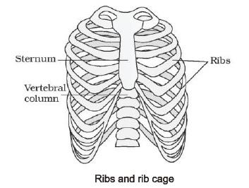
5.2 APPENDICULAR SKELETON
•The bones of the limbs along with their girdles constitute the appendicular skeleton which are followings
(a) Limb bone : Each limb is made of 30 bones.
(A) Fore limb bones : The bones of the hand (fore limb) are humerus, radius and ulna, carpals (wrist bones - 8 in number), metacarpals (palm bones - 5 in number) and phalanges (digits - 14 in number).
(B) Hind limb bones : Femur (thigh bone - the longest bone), tibia and fibula, tarsals (ankle bones - 7 in number), metatarsals (5 in number) and phalanges (digits - 14 in number) are the bones of the legs (hind limb). A cup shaped bone called patella cover the knee ventrally (knee cap).
(b) Girdle bones : Pectoral and Pelvic girdle bones help in the articulation of the upper and the lower limbs respectively with the axial skeleton. Each girdle is formed of two halves.
(A) Pectoral girdle : Each half of pectoral girdle consists of a clavicle and a scapula. Scapula is a large triangular flat bone situated in the dorsal part of the thorax between the second and the seventh ribs. The dorsal, flat, triangular body of scapula has a slightly elevated ridge called the spine which projects as a flat, expanded process called the acromion. The clavicle articulates with this. Below the acromion is a depression called the glenoid cavity which articulates with the head of the humerus to form the shoulder joint. Each clavicle is a long slender bone with two curvatures. This bone is commonly called the collar bone.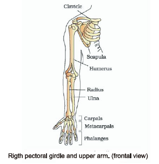
(B) Pelvic girdle : It consists of two coxal bones. Each coxal bone is formed by the fusion of three bones - ilium, ischium and pubis. At the point of fusion of the above bones is a cavity called acetabulum to which the thigh bone articulates. The two halves of the pelvic girdle meet ventrally to form the pubic symphysis containing fibrous cartilage.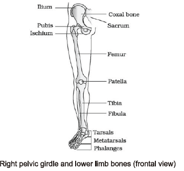
6.Jolnts
•Joints are essential for all types of movements involving the bony parts of the body. Locomotory movements are no exception to this. Joints are points of contact between bones, or between bones and cartilages.
•Force generated by the muscles is used to carry out movement through joints, where the joint acts as a fulcrum. The movability at these joints vary depending on different factors.
•Joints have been classified into three major structural forms, namely, fibrous, cartilaginous and synovial.
(a) Fibrous joints : These do not allow any movement. This type of joint is shown by the flat skull bones which fuse end-to-end with the help of dense fibrous connective tissues in the form of sutures, to form the cranium.
(b) Cartilaginous joints : In this, the bones involved are joined together with the help of cartilages. The joint between the adjacent vertebrae in the vertebral column is of this pattern and it permits limited movements.
(c) Synovial joints : These are characterised by the presence of a fluid filled synovial cavity between the articulating surfaces of the two bones. Such an arragement allows considerable movement. These joints help in locomotion and many other movements.
• Synovial joints are classified as :
(i) Ball and socket joint : Ball of one bone articulate in socket of another bone. e.g., head of humerus and glenoid cavity of pectoral girdle, femur and acetabulum of pelvic girdle.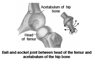
(ii) Hinge joint : Movement is possible in one direction only. e.g., Joint of malleus and incus, knee joint, elbow joint.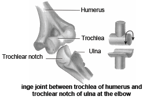
(iii) Pivot joint : Also known as rotatoria and helps in turning movement. e.g. between Atlas & Axis, Radius & Ulna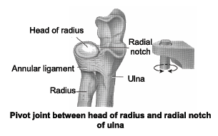
(iv) Gliding joint : Limited movement in all direction. e.g., Tarsals bones of ankle, between the carpals.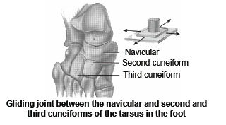
(v) Saddle joint : It is ball and socket like joint but not developed fully. e.g. between carpal & metacarpal of thumb.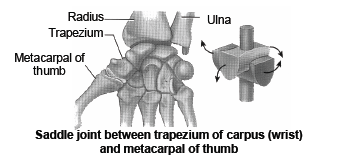
7.Disorders of Muscular & Skeletal System
1. MYASTHENIA GRAVIS : Auto immune disorder affecting neuromuscular junction leading to fatigue, weakening and paralysis of skeletal muscle.
•Auto antibodies against ach receptors.
2. MUSCULAR DYSTROPHY : Progressive degeneration of skeletal muscle mostly due to genetic disorder.
3. TETANY : Rapid spasms (wild contractions) in muscle due to low Ca2+ in body fluid.
4. SPRAIN : Sprain refers to injury to a joint capsule, typically involving a stretching or tearing of tendons or ligaments.
5. ARTHRITIS : Arthritis refers to inflammation of the joints. It is a common disease of the old age. Its common symptoms are pain and stiffness in the joints. It is differentiated in three given forms :-
(a) Osteoarthritis : Secretion of the lubricating synovial fluid between the bones at the joint stops. The smooth cartilage covering the ends of the bones at the joint wears out due to years of use and is replaced by uneven bony spurs. The joint becomes inflamed, its movement becomes painful, and its function is diminished. Such a stiffness or fixation of a joint is also called ankylosis. The condition of osteoarthritis is
more or less permanent. It is common in old persons, mainly affecting weight bearing joints.
(b) Rheumatoid arthritis : It is a chronic painful inflammation of the synovial membranes of many joints simultaneously. It usually starts in the small joints in the hand and progresses in centripetal and symmetrical manner. In severe cases, it eventually results in crippling deformities. There may be other manifestations such as fever, anaemia, loss of weight and morning stiffness. The rheumatoid arthritis involves erosion of joints. It usually starts at the age of 20 – 40 years, but may begin at any age. It affects the women more often than the men. Rest and exercise under medical advice may give relief.
(c) Gout : It is an inherited disorder of purine metabolism, occurring especially in men. Body forms excess amounts of uric acid and the crystals of sodium urate are deposited in the synovial joints, giving rise to a severe arthritis. It generally affects one or two joints only. It is very painful, particularly at night, and makes movement difficult. Redness and tenderness may be noticed in and about the affected joint. Gout generally affects the great toe. Occurrence of gout is related to diet. Persons suffering from gout should avoid meat.
There is no cure for arthritis. However, pain relieving (analgesic) drugs are available to give comfort.
6. OSTEOPOROSIS
(a) Meaning : Osteoporosis is reduction in bone tissue mass causing weakness of skeletal strength (G.osteon = bone, poros = pore, osis = condition). It results from excessive resorption of calcium and phosphorous from the bone.
(b) Causes : Osteoporosis occurs in postmenopausal women and elderly men. It may result from defective intestinal calcium absorption and menopause. Possible environment factors include smoking, excessive drinking, and decreased exercise. Osteoporosis is more common in women than in men, and in older than in middle-aged persons.
(c) Symptoms : Symptoms of osteoporosis are pain in the bone, particularly the back.
(d) Prevention : Preventive measures in high-risk patients include supplementary calcium and exercise, and, in postmenopausal women, estrogen replacement therapy. Supplementary calcium and sex hormones decrease bone resorption and may arrest or reduce disease progression.
7. DISLOCATION : Dislocation is displacement of bones from their normal positions at a joint, for instance, slipping out of the ball of one bone from the socket of another bone into which it is fitted. Dislocation is accompanied by pulling or even tearing of the ligaments. Dislocation also tends to become chronic.
8. SLIPPED DISC : Slipped disc is a displacement of vertebrae and the intervertebral fibrocartilage disc from their normal position. It may result from mechanical injury or defects of ligaments holding the vertebrae together.
9. FRACTURE : Fracture is a break of a bone. Fracture occurs rarely in children. The bones of children have a large quantity of organic matter and are, therefore, very flexible and less likely to break. With advancing age, mineral matter (calcium phosphate) is deposited in the bones. This decreases the organic matter, making the bones hard and brittle. Thus, old people are more liable to fracture of bones. Bones fractures are of many types –
(a) Green-stick fracture : It is merely a crack. The bones remains partly intact, occurs only in children.
(b) Simple or complete fracture : Bone breaks completely into two parts which remain close to each other.
(c) Comminuted fracture : Bone breaks into more than two pieces (smaller fragments between two main fragment,
(d) Compound fracture : Bone breaks completely but a fragment pierces out through the skin.
(e) Evulsive fracture : A small piece breaks off fully from the bone but remains attached to the ligament.
Fractures need surgical treatment for healing and should be promptly and properly attended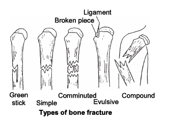
Key Concepts
• Antagonistic muscles : The striated muscles occur in antagonistic pairs; one pulls a bone in one direction, while the other pulls it back in reverse direction to its normal position. For example, the biceps muscle, extending from shoulder to radius, bends or flexes the arm at the elbow, whereas the triceps extending from ulna to the shoulder, straightens the arm. Thus, biceps is a flexor and triceps an extensor for bending the arm.
• Single twitch : When a muscle receives a single excitation impulse, it respond by a sudden partial contraction (twitch) lasting for about 0.5 second in man. Each twitch is followed by a refractory period during which the muscle does not respond to next stimulus. The refractory period is, however, so short 0.002 second) that the muscle can respond to the second stimulus while still in contraction phase in response to the first stimulus.
• Tetanus : Generally, whole muscles contract, not in a single twitch, but in sustained contractions evoked by a series of nerve impulses reaching them in rapid succession. Such a sustained contraction is called tetanus. Described above should not be confused with the disease of “tetanus” (lock jaw) caused by tetanus bacillus. This disease is characterised by abnormal muscular contractions.
• Muscle tone or “Tonus” : Even at rest the striated muscles normally remain in a state of mild sustained partial contraction to maintain the body posture. This is called muscle tone. It is a mild state of tetanus.
• Paralysis : When supply of motor impulses to a muscle is completely cut off due to destruction, either of the control centres in brain, or of the concerned motor nerves, or due to blocking of myoneural junctions by the use of certain drugs, the muscle function is completely impaired. This is called paralysis of the muscle.
• Muscle fatigue : A muscle that has contracted many times at short intervals, exhausts its store of ATP and glycogen and accumulates lactic acid. Hence its contractility gradually decreases and finally stops.
• Oxygen debt : During active work or exercise, the rate of oxygen supply by the lungs falls short of the requirement of the muscles. Hence, lactic acid accumulates in the muscles and the breathing gradually becomes hard to enhance O2 intake by the lungs. This is called oxygen debt.
• Involuntary action of skeletal muscles : Muscles are capable of utilizing, in their mechanical work, only about 20% to 40% of energy liberated from glucose. The unutilized energy is lost as “heat” dissipated into the environment. This heat helps in maintenance of body temperature. “Shivering with cold” in winter is caused by a quick involuntary reaction of striated muscles.
• Rigor mortis : Rigidity that develops in the muscles after death is known as rigor mortis. It is due to permanent irreversible contraction, establishment of permanent link between actin and myosin and also fall in the concentration of ATP molecules.
• Cori’s cycle : Lactic acid is transported by blood to liver and there it is converted to glycogen through Cori’s cycle.
• Contraction period : Time taken in sliding of filament is called contraction time. (10 to 100 milli second).
• Relaxation time : It is time taken in relaxation of fibre i.e. active transport of calcium from sarcoplasm to cisternae. (10 to 100 milli second)
• Refractory period : It is time in a muscle or nerve fibre when they are non responding to second stimulus. Infact in this period there is temporary loss of excitability. Refractory period for skeletal and cardiac muscle is 5 and 300 milli second respectively.
• Hypertrophy and Atrophy of muscles : Muscles which are put to excessive work become thick and strong. This is called their hypertrophy. Conversely, if certain muscles are not used for a long period, those become thin and weak. This is called their atrophy (disuse atrophy). Cardiac muscle have a poor regenrative power.
MCQ for NCERT Class 11 Biology Locomotion and Movement
Question. Consider the following figure and arrange them in correct sequence of muscle contraction.
(a) A-B-C-D
(b) D-B-A-C
(c) C-B-A-D
(d) A-D-B-C
Answer : B
Question. Ions that must be present for binding the cross bridge is
(a) Na+
(b) Ca2+
(c) K+
(d) Mg+
Answer : B
Question. One myosin filament in the myofibril of skeletal muscle fibre is surrounded by how many actin filaments?
(a) Two
(b) Four
(c) Six
(d) Three
Answer : C
Question. Select the incorrect pair.
(a) Amoeboid movement – Phagocytes
(b) Muscular movement – Jaws
(c) Flagellar movement – Ova
(d) Ciliary movement – Fallopian tubes
Answer : C
Question. Accumulation of which of the following in muscle causes fatigue?
(a) Acetic acid
(b) Carboxylic acid
(c) Hydrochloric acid
(d) Lactic acid
Answer : D
Question. Which of the following is true for the labelled parts in the figure below?
(a) A : Z-line – located at centre of I - band
(b) B : Thin filament – occurs in A-band only
(c) C : Thick filament – confined to I-band
(d) D : H-zone – located at centre of M-line
Answer : A
Question. Amoeboid movement involves
(a) cytoskeletal elements like microfilaments
(b) coordinated beats of cilia
(c) whip like action of flagella
(d) none of these.
Answer : A
Question. Label the correct part of the myosin monomer.
(a) (A) Actin binding site
(B) Head
(C) ATP binding site
(D) Cross arm
(b) (A) Cross arm (B) Actin binding site
(C) Head (D) ATP binding site
(c) (A) ATP binding site (B) Actin binding site
(C) Head (D) Cross arm
(d) (A) Head (B) Cross arm
(C) Actin binding site (D) ATP binding site
Answer : D
Question. I-band is bisected by
(a) Z-line
(b) H-zone
(c) M-line
(d) A-band.
Answer : A
Question. The junction between the motor neuron and the sarcolemma of the muscle fibre is called the
(a) motor unit
(b) motor end plate
(c) neuromuscular junction (NMJ)
(d) both (b) and (c).
Answer : D
Ques. Match the following joints with the bones involved:
(1) Gliding joint (i) Between carpal and metacarpal of thumb
(2) Hinge joint (ii) Between atlas and axis
(3) Pivot joint (iii) Between the carpals
(4) Saddle joint (iv) Between humerus and ulna.
Select the correct option from the following:
(a) (1)-(iii), (2)-(iv), (3)-(ii), (4)-(i)
(b) (1)-(iv), (2)-(i), (3)-(ii), (4)-(iii)
(c) (1)-(iv), (2)-(ii), (3)-(iii), (4)-(i)
(d) (1)-(i), (2)-(iii), (3)-(ii), (4)-(iv)
Answer: A
Ques. The pivot joint between atlas and axis is a type of
(a) cartilaginous joint
(b) synovial joint
(c) saddle joint
(d) fibrous joint.
Answer: B
Ques. Which of the following joints would allow no movements?
(a) Synovial joint
(b) Ball and socket joint
(c) Fibrous joint
(d) Cartilaginous joint
Answer: C
Ques. Select the correct matching of the type of the joint with the example in human skeletal system.
Type of joint Example
(a) Cartilaginousjoint – Between frontal and parietal
(b) Pivot joint – Between third and fourth cervical vertebrae
(c) Hinge joint – Between humerus and pectoral girdle
(d) Gliding joint – Between carpals
Answer: D
Ques. The characteristic and an example of a synovial joint in humans is
Characteristics Examples
(a) Fluid filled synovial cavity Joint betweenatlas and axis
between two bones
(b) Lymph filled between two Gliding joint between carpals
bones, limited movement
(c) Fluid cartilage between Knee joint
two bones, limited movements
(d) Fluid filled between two Skull bones
joints, provides cushion
Answer: A
Ques. Which one of the following is the correct description of a certain part of a normal human skeleton?
(a) Parietal bone and the temporal bone of the skull are joined fibrous joint.
(b) First vertebra is axis which articulates with the occipital condyles.
(c) The 9th and 10th pairs of ribs are called the floating ribs.
(d) Glenoid cavity is a depression to which the thigh bone articulates.
Answer: A
Ques. Elbow joint is an example of
(a) hinge joint
(b) gliding joint
(c) ball and socket joint
(d) pivot joint.
Answer: A
Ques. Which of the following pairs is correctly matched?
(a) Hinge joint – Between vertebrae
(b) Gliding joint – Between zygapophyses of the successive vertebrae
(c) Cartilaginous joint – Skull bones
(d) Fibrous joint – Between phalanges
Answer: B
Ques. What is the name of joint between ribs and sternum?
(a) Cartilaginous joint
(b) Angular joint
(c) Gliding joint
(d) Fibrous joint
Answer: A
Ques. The joint between atlas and axis is called
(a) angular joint
(b) hinge joint
(c) pivot joint
(d) saddle joint.
Answer: C
Ques. The type of joint between the human skull bones is called
(a) cartilaginous joint
(b) hinge joint
(c) fibrous joint
(d) synovial joint.
Answer:
Ques. Which of the following muscular disorders is inherited?
(a) Botulism
(b) Tetany
(c) Muscular dystrophy
(d) Myasthenia gravis
Answer: C
Ques. Osteoporosis, an age-related disease of skeletal system, may occur due to
(a) immune disorder affecting neuromuscular junction leading to fatigue
(b) high concentration of Ca++ and Na+
(c) decreased level of estrogen
(d) accumulation of uric acid leading to inflammation of joints.
Answer: C
Ques. Select the correct statement with respect to locomotion in humans.
(a) The vertebral column has 10 thoracic vertebrae.
(b) The joint between adjacent vertebrae is a fibrous joint.
(c) A decreased level of progesterone causes osteoporosis in old people.
(d) Accumulation of uric acid crystals in joints causes their inflammation.
Answer: D
Ques. Select the correct statement with respect to disorders of muscles in humans.
(a) Failure of neuromuscular transmission in myasthenia gravis can prevent normal swallowing.
(b) Accumulation of urea and creatine in the joints causes their inflammation.
(c) An overdose of vitamin D causes osteoporosis.
(d) Rapid contractions of skeletal muscles cause muscle dystrophy.
Answer: A
Ques. Select the correct statement regarding the specific disorder of muscular or skeletal system.
(a) Muscular dystrophy – Age related shortening of muscles
(b) Osteoporosis – Decrease in bone mass and higher chances of fractures with advancing age
(c) Myasthenia gravis – Autoimmune disorder which inhibits sliding of myosin filaments
(d) Gout – Inflammation of joints due to extra deposition of calcium
Answer: B
Refer to the given figures and answer the questions given below.
Question. What does Y represent?
(a) Thin filament
(b) Meromyosin
(c) Tropomyosin
(d) Both (a) and (c)
Answer : B
Question. Which of the following labelled part binds to Ca2+ to initiate the contraction process?
(a) A
(b) B
(c) C
(d) D
Answer : A
Question. The labelled part C is also called
(a) cross arm
(b) light meromyosin
(c) heavy meromyosin
(d) F actin
Answer : C
Question. Select the correct statements(s) regarding globular head of Y.
(a) It is an active ATPase enzyme
(b) It has binding site for ATP
(c) It has actin active sites
(d) All of these
Answer : D
Question. B is a
(a) single stranded a-helical rod
(b) fibrous molecule that attaches to F-actin
(c) is a double stranded a-helical rod
(d) both (b) and (c).
Answer : D
Assertion & Reasoning Based MCQs For question numbers , two statements are given-one labelled Assertion and the other labelled Reason. Select the correct answer to these questions from the codes (a), (b), (c) and (d) as given below.
(a) Both assertion and reason are true and reason is the correct explanation of assertion.
(b) Both assertion and reason are true but reason is not the correct explanation of assertion.
(c) Assertion is true but reason is false.
(d) Assertion is false but reason is true.
Question. Assertion : Athletic training enhances the capacity for aerobic contraction of muscles.
Reason : After strenous exercise, athletes are liable to much more oxygen debt than non-athlete.
Answer : C
Question. Assertion : The contraction and relaxation of muscle fibre is controlled by nerve impulses.
Reason : The threshold stimulus is the minimum stimulus required for the beginning of contraction.
Answer : B
Question. Assertion : Muscle contraction force increases with rise in strength of stimulus.
Reason : This is due to increased contraction of individual muscle fibres with increase in stimulus strength.
Answer : C
Short Answer Type Questions
Question. What are the different types of movements exhibited by the human cells? Explain each with the suitable examples?
Answer. Human cells exhibit three main types of movements, namely, amoeboid, ciliary and muscular. (i) Amoeboid movement : Certain specialised cells, e.g., macrophages and leucocytes exhibit amoeboid movement which occurs with the help of pseudopodia formed due to the streaming of protoplasm. Cytoskeletal elements, e.g., microfilaments are also involved in amoeboid movement. (ii) Ciliary movement : Large number of internal tubular organs in man are lined by ciliated epithelium. For instance, the cilia of the cells lining the trachea, oviducts and vasa efferentia propel dust particles, eggs and sperms respectively by their coordinated movements in specific directions in these organs. (iii) Muscular movement : Movement of limbs, jaws, tongue, etc., require the muscular movement. The universal property of alternate contraction and relaxation of muscles are effectively used for locomotion and movement in human beings.
Question. Elucidate the importance of organ level movements.
Answer. Movements of internal organs make many vital activities possible. For example : (i) Movement of muscle of heart circulate blood in the body. (ii) Peristalsis of alimentary canal propels food through it. (iii) Movements of diaphragm assist the chest in the flow of air through the respiratory tract. (iv) Movements of genital tract affect egg laying and delivery of the baby. (v) Visceral movements are also responsible for sound production, defecation and micturition.
Question. How muscle gets energy for its contraction?
Answer. The muscle gets its energy for contraction from ATP. During muscular contraction, ATP is hydrolysed to ADP and inorganic phosphate by myosin ATPase. ATP + H2O → ADP + Pi After that, ATP is soon replenished in the muscle fibres. For this, muscles contain another energy compound called creatine phosphate (CP). It helps in the conversion of ADP to ATP. This happens at the end of muscular contraction. ADP + CP ATP + Creatine.
Question. What is the purpose of locomotion in animals?
Answer. Locomotion occurs generally for the search of food, shelter, mate, suitable breeding locations and favourable climatic conditions. Animals also move to escape from predators.
Question. (a) How is amoeboid movement effective in our body?
(b) Why ciliary movement is important in respiratory tract and Fallopian tube of humans?
(c) How flagellar movement helps in the movement of sperms?
Answer. (a) Some specialised cells in our body like macrophages and leucocytes in blood exhibit amoeboid movement which occurs with the help of pseudopodia formed by the streaming of protoplasm. Cytoskeletal elements like microfilaments are also involved in amoeboid movement. (b) Cilia of the upper respiratory tract of humans keeps the invading microbes and dust particles out whereas, the cilia of the Fallopian tubes (oviducts) of human females transport ova. (c) Human sperms (a flagellated cell) exhibit the flagellar movement. The flagellum provides propulsion for the movement of sperm towards the ovum. This propulsion is brought about by the whip-like movement of the tail and the middle piece of the sperm.
Question. How do you distinguish between a skeletal muscle and a cardiac muscle?
Answer. Differences between skeletal and cardiac muscles are as follows :
| Skeletal muscles | Cardiac muscles | |
| (i) | They are associated with bones. | They are not associated with bones. |
| (ii) | They are voluntary in nature. | They are involuntary in nature. |
| (iii) | Skeletal muscle fibres are syncytial (multinucleated). | Cardiac muscle fibres are uninucleated. |
| (iv) | Fibres are unbranched. | Fibres are branched. |
| (v) | It soon gets fatigued. | It never gets fatigued. |
| (vi) | Oblique bridges and intercalated discs absent. | Oblique bridges and intercalated discs present. |
Question. Explain briefly :
(a) Oxygen debt
(b) Muscle twitch
(c) Rigor mortis
Answer. (a) Oxygen debt is the requirement of extra oxygen during recovery phase of muscle after vigorous exercise. During strenuous exercise, muscles do not get sufficient oxygen to meet its energy need immediately; so it contracts anaerobically and accumulates lactic acid produced by anaerobic gylcolysis. The increased oxygen consumption of muscle during recovery is called oxygen debt. The extra oxygen required during recovery phase is for regeneration of oxymyoglobin, restoration of depleted ATPs and creatine phosphate and oxidation of lactic acid. (b) Muscle twitch or muscle fasciculations are fine movements of small area of muscle, caused by minor muscle contractions in the area, or uncontrollable twitching of muscle group served by single motor nerve fibre, e.g., flittering eye lids. (c) Rigor mortis is a condition that sets few hours after the death. In this, muscles and joints become rigid, i.e., can neither contract nor stretch as cellular processes come to halt. Rigor mortis subsides about 15-24 hours after death.
Long Answer Type Questions
Question. (a) Draw a detailed labelled structure of a myofibril showing sarcomere.
(b) Differentiate between A-band and I-band.
Answer. (a) Structure of a myofibril showing sarcomere. 
(b) Differences between A-band and I-band are :
Question. Explain the electrical and biochemical events that occur during muscle contraction.
Answer. Electrical and biochemical events of muscle contraction are summarised as follows :
(i) As a nerve impulse reaches the terminal end of the axon, small sacs called synaptic vesicles fuse with the axon membrane and release a chemical transmitter, acetylcholine. Acetylcholine diffuses across the synaptic cleft (the space between the axon membrane and the motor end plate) and binds to receptor sites of the motor end plate. When depolarisation of the motor end plate reaches a certain level, it creates an action potential. After this, an enzyme cholinesterase present along with receptor sites for acetylcholine, breaks down acetylcholine into acetate and choline. A portion of the choline diffuses back to the axon and is reused to synthesise more acetylcholine for transmission of subsequent impulses. (ii) At the opening of each transverse tubule onto the muscle fibre surface, the action potential spreads inside the muscle fibre.
(iii) At each point where a transverse tubule touches part of the sarcoplasmic reticulum, it causes the sarcoplasmic reticulum to release Ca++ ions.
(iv) The calcium ions bind to troponin causing a change in its shape and position. This in turn alters shape and the position of tropomyosin, to which troponin binds. This shift exposes the active sites on the F - actin molecules.
(v) The heads of myosin molecules project laterally from thick myofilaments towards the surrounding thin myofilaments.
(vi) These heads are called crossbridges. The head of each myosin molecule contains an enzyme myosin ATPase. In the presence of myosin ATPase, Ca++ and Mg++ ions, ATP breaks down into ADP and inorganic phosphate, releasing energy in the head. (vii) Energy from ATP causes energised myosin crossbridges to bind to actin.
(viii) The energised cross-bridges move, causing thin myofilament to slide along the thick myofilaments.
Please click the link below to download pdf file for CBSE Class 11 Biology Locomotion And Movement Notes.
| CBSE Class 11 Biology The Living World Notes Set A |
| CBSE Class 11 Biology The Living World Notes Set B |
| CBSE Class 11 Biology Biological Classification Notes Set A |
| CBSE Class 11 Biology Biological Classification Notes Set B |
| CBSE Class 11 Biology Plant Kingdom Notes Set A |
| CBSE Class 11 Biology Plant Kingdom Notes Set B |
| CBSE Class 11 Biology Animal Kingdom Notes Set A |
| CBSE Class 11 Biology Animal Kingdom Notes Set B |
| CBSE Class 11 Biology Morphology Of Flowering Plants Notes Set A |
| CBSE Class 11 Biology Morphology Of Flowering Plants Notes Set B |
| CBSE Class 11 Biology Anatomy Of Flowering Plants Notes Set A |
| CBSE Class 11 Biology Anatomy Of Flowering Plants Notes Set B |
| CBSE Class 11 Biology Structural Organisation In Animals Notes Set A |
| CBSE Class 11 Biology Structural Organisation In Animals Notes Set B |
| CBSE Class 11 Biology Cell The Unit Of Life Notes Set A |
| CBSE Class 11 Biology Cell The Unit Of Life Notes Set B |
| CBSE Class 11 Biology Biomolecules Notes Set A |
| CBSE Class 11 Biology Biomolecules Notes Set B |
| CBSE Class 11 Biology Cell Cycle And Cell Division Notes Set A |
| CBSE Class 11 Biology Cell Cycle And Cell Division Notes Set B |
| CBSE Class 11 Biology Photosynthesis In Higher Plants Notes Set A |
| CBSE Class 11 Biology Photosynthesis In Higher Plants Notes Set B |
| CBSE Class 11 Biology Respiration In Plants Notes Set A |
| CBSE Class 11 Biology Respiration In Plants Notes Set B |
| CBSE Class 11 Biology Plant Growth And Development Notes Set A |
| CBSE Class 11 Biology Plant Growth And Development Notes Set B |
| CBSE Class 11 Biology Breathing And Exchange Of Gases Notes Set A |
| CBSE Class 11 Biology Breathing And Exchange Of Gases Notes Set B |
| CBSE Class 11 Biology Body Fluids And Circulation Notes Set A |
| CBSE Class 11 Biology Body Fluids And Circulation Notes Set B |
| CBSE Class 11 Biology Excretory Products And Their Elimination Notes Set A |
| CBSE Class 11 Biology Excretory Products And Their Elimination Notes Set B |
| CBSE Class 11 Biology Locomotion And Movement Notes Set A |
| CBSE Class 11 Biology Locomotion And Movement Notes Set B |
| CBSE Class 11 Biology Neural Control And Coordination Notes Set A |
| CBSE Class 11 Biology Neural Control And Coordination Notes Set B |
| CBSE Class 11 Biology Chemical Coordination and Integration Notes Set A |
| CBSE Class 11 Biology OTBA Guidance Document Set A |
| CBSE Class 11 Biology OTBA Guidance Document Set B |
CBSE Class 11 Biology Chapter 17 Locomotion and Movement Notes
We hope you liked the above notes for topic Chapter 17 Locomotion and Movement which has been designed as per the latest syllabus for Class 11 Biology released by CBSE. Students of Class 11 should download and practice the above notes for Class 11 Biology regularly. All revision notes have been designed for Biology by referring to the most important topics which the students should learn to get better marks in examinations. Our team of expert teachers have referred to the NCERT book for Class 11 Biology to design the Biology Class 11 notes. After reading the notes which have been developed as per the latest books also refer to the NCERT solutions for Class 11 Biology provided by our teachers. We have also provided a lot of MCQ questions for Class 11 Biology in the notes so that you can learn the concepts and also solve questions relating to the topics. We have also provided a lot of Worksheets for Class 11 Biology which you can use to further make yourself stronger in Biology.
You can download notes for Class 11 Biology Chapter 17 Locomotion and Movement for latest academic session from StudiesToday.com
Yes, the notes issued for Class 11 Biology Chapter 17 Locomotion and Movement have been made available here for latest CBSE session
There is no charge for the notes for CBSE Class 11 Biology Chapter 17 Locomotion and Movement, you can download everything free of charge
www.studiestoday.com is the best website from which you can download latest notes for Chapter 17 Locomotion and Movement Biology Class 11
Come to StudiesToday.com to get best quality topic wise notes for Class 11 Biology Chapter 17 Locomotion and Movement