
Read and download the NCERT Class 11 Biology Structural Organization In Animals Important Notes. Designed for 2026-27, this advanced study material provides Class 11 Biology students with detailed revision notes, sure-shot questions, and detailed answers. Prepared by expert teachers and they follow the latest CBSE, NCERT, and KVS guidelines to ensure you get best scores.
Advanced Study Material for Class 11 Biology Chapter 7 Structural Organisation in Animals
To achieve a high score in Biology, students must go beyond standard textbooks. This Class 11 Chapter 7 Structural Organisation in Animals study material includes conceptual summaries and solved practice questions to improve you understanding.
Class 11 Biology Chapter 7 Structural Organisation in Animals Notes and Questions
Tissue:
A group of cells which is formed to carry out specific task is called a tissue.
Formation of tissues is the first step towards division of labour in multicellular animals. Subsequently, a group of tissues forms an organ and a group of organs forms the organ system.
ANIMAL TISSUES
There are four major types of animal tissues, viz. epithelial, connective, muscular and neural tissues.
EPITHELIAL TISSUE
Tissues which provide covering to the inner and outer linings of various organs are called epithelial tissue. The cells in the epithelial tissue are compactly packed. There is little intercellular matrix in the epithelial tissue.
The epithelial tissues are further divided into two main types, viz. simple epithelium and compound epithelium.
Simple Epithelium: The simple epithelium is composed of a single layer of cells. It is present in the lining of body cavities, ducts and tubes.
Compound Epithelium: The compound epithelium is composed of more than one layer of cells. The compound epithelium serves the protective function. Compound epithelium is present in skin, in the lining of buccal cavity, pharynx, ducts of salivary glands and pancreatic ducts.
The simple epithelium can be further divided into three types, viz. squamous, cuboidal and columnar.
Squamous Epithelium: The squamous epithelium is made up of flattened cells with irregular boundaries. Squamous epithelium is present in the walls of blood vessels and air sacs of lungs. They form a diffusion boundary.
Cuboidal Epithelium: The cuboidal epithelium is composed of cube-like cells. The cuboidal epithelium is usually found in ducts of glands and tubular parts of nephrons. The main functions of cuboidal epithelium are secretion and absorption.
Columnar Epithelium: The columnar epithelium is composed of tall and slender cells. The nuclei of the columnar cells are located at the base. Microvilli may be present on the free surface. Columnar epithelium is present in the lining of stomach and intestine. They help in secretion and absorption.
Ciliated Epithelium: Sometimes, cilia may be present in columnar or cuboidal epithelium. In that case, they are called ciliated epithelium. The cilia move particles or mucus in a specific direction over the epithelium. They are present in the inner surface of hollow organs; like bronchioles and fallopian tubes.
CONNECTIVE TISSUE
The connective tissues are most abundant and widely distributed in the body of complex animals. The connective tissues support and link other tissues or organs of the body. The cells of the connective tissue secrete fibres of structural proteins; called collagen or elastin. But blood is an exception; in which no structural fibre is secreted. The structural fibres provide strength, elasticity and flexibility to the tissue. The cells of the connective tissue also secrete polysaccharides. These polysachharides accumulate between cells and fibres and act as matrix (ground substance).
There are three types of connective tissues, viz. loose connective, dense connective and specialized connective tissues.
Loose Connective Tissue: The cells and fibres are loosely arranged in a semi-fluid matrix; in loose connective tissue. Areolar tissue is an example of loose connective tissue. Areolar tissue is present beneath the skin. It usually makes the support framework for epithelium. It contains fibroblasts, macrophages and mast cells. Adipose tissue is another example of loose connective tissue. Adipose tissue is usually located beneath the skin. The cells of the adipose tissue are specialized to store fat.
Dense Connective Tissue: The cells and fibres are compactly packed in dense connective tissue. The orientation of fibres can be regular or irregular. On this basis, the dense connective tissue is called dense regular or dense irregular connective tissue. In dense regular connective tissue, the collagen fibres are present in rows between many parallel bundles of fibres, e.g. tendons and ligaments. Tendons attach muscles to bones, while ligaments attach one bone to another. In dense irregular tissue, fibroblasts and fibres are oriented in irregular pattern. Dense irregular tissue is present in skin.
Specialised Connective Tissue: Cartilage, bones and blood are the specialized connective tissues.
Cartilage: The matrix of cartilage is solid and pliable and resists compression. The cells of cartilage are called chondrocytes. Chondrocytes are enclosed in small cavities within the matrix. Most of the cartilages in vertebrae embryo are replaced by bones in adulthood.
Bones: The matrix of bones is hard and non-pliable. The matrix is rich in calcium salts and collagen fibres; which give strength to the bone. The bone cells are called osteocytes. Osteocytes are present in spaces; called lacunae. The bone marrow in some bones is the site of production of blood cells.
Blood: Blood is a fluid connective tissue. It contains plasma, blood cells and platelets. Blood is the main circulating fluid which helps in transport of various substances.
MUSCLE TISSUE
A muscle is made of many long, cylindrical fibres which are arranged in parallel rays. The muscle fibres are composed of very fine fibrils; called myofibril. The muscle fibres can contract and relax in a coordinated fashion. Thus, muscles play an important role in movement and locomotion.
There are three types of muscles, viz. skeletal, smooth and cardiac muscles.
Skeletal Muscle: The skeletal muscle is closely attached to skeletal bones. Skeletal muscle fibres are bundled together in a parallel fashion. Several bundles of muscle fibres are covered by a sheath of tough connective tissue. Striations are present in skeletal muscles and hence they are also called striated muscles. The voluntary movements are facilitated by skeletal muscles. Skeletal muscles quickly get tired.
Smooth Muscle: The smooth muscle fibres are spindle-shaped. Striations are absent on smooth muscle. Various smooth muscle fibres are held together by cell junctions. They are bundled together in a sheath of connective tissue. Smooth muscles are present in those organs which are not under our conscious control. Smooth muscles can carry on their activity for a longer duration, as compared to skeletal muscles.
Cardiac Muscle: The muscles in the heart are called cardiac muscles. Cardiac muscles are multinucleated and have striations. The muscle fibres are held together by cell junctions. Communication junctions (intercalated discs) are present at some fusion points. These communication junctions allow the cells to contract as a unit. Due to this, when one cell receives a signal to contract, its neighbours are also stimulated to contract. Cardiac muscles keep on contracting and relaxing continuously throughout the life.
Neural Tissue
Neural tissue is made of neurons. Neurons are excitable cells. The neuroglial cell protects and supports neurons. Neuroglia make up more than half of the volume of the neural tissue in human body.
When a neuron is stimulated, an electrical disturbance is generated. The electrical disturbance travels along its plasma membrane. When a disturbance arrives at a neuron’s endings, it triggers events which may cause stimulation of inhibition of adjacent neurons and other cells. Thus, neural signals are transmitted to different parts of the body.
EARTHWORM
Habit and Habitat: Earthworm is a reddish brown terrestrial invertebrate. It lives in the upper layer of moist soil. Pheretima and Lumbricus are the common Indian earthworms.
Morphology of Earthworm
Earthworm has a long cylindrical body which is divided into more than a hundred short segments or metameres.
Dorsal Surface: There is a dark median mid-dorsal line; along the longitudinal axis; on the dorsal surface of the body. This line marks the dorsal blood vessel.
Ventral Surface: Genital openings mark the ventral surface of the body of an earthworm.
Segments of Earthworm:
• The anterior end consists of the mouth and the prostomium. Prostomium is a lobe which serves as a covering of the mouth. It also acts as a wedge to force open cracks in the soil. The prostomium has sensory function as well.
• The first body segment is called peristomium or buccal segment. The peristomium contains the mouth.
• In a mature worm, segments 14 – 16 are covered by a prominent dark band of glandular tissue. This band is called clitellum. Based on the relative position of clitellum, the body is divided into three main regions, viz. preclitellar, clitellar and postclitellar segments.
• Genital Openings: On the ventro-lateral sides of the inter-segmental groves; between 5th – 9th segments; four pairs of spermathecal apertures are situated. On the mid-ventral line of 14th segment, a single female genital pore is present. On the ventro-lateral sides of the 18th segment, a pair of male genital pores is present.
Locomotion in Earthworm
There are 5 rows of S-shaped setae in each body segment. The setae are embedded in the epidermal pits in the middle of each segment. Setae are absent in the first segment, clitellum and last segment. Setae can be extended or retracted and facilitate locomotion.
Anatomy of Earthworm
Body Wall: The body wall of earthworm is covered by thin non-cellular cuticle. Epidermis lies below the cuticle. This is followed by two muscle layers; circular and longitudinal. The innermost layer is the coelomic epithelium. The epithelium is composed of a single layer of columnar epithelial cells. The epithelial cells contain gland cells as well.
Alimentary Canal: The alimentary canal is a straight tube. It runs between the first to last segment. Mouth is terminal and opens into the buccal cavity (1- 3 segments). The mouth leads into muscular pharynx.
Pharynx continues into oesophagus (5-7 segments) which is a small narrow tube. The oesophagus continues into a muscular gizzard (8-9 segments). The gizzard helps in grinding the food. The stomach extends from 9th – 14th segments.
Decaying leaves and organic matter; mixed with soil; are the foods of the earthworm. The humic acid; present in humus; is neutralized by the calciferous glands in the stomach.
The intestine continues from the 15th segment to the last segment. On the 26th segment, a pair of short and conical caecae project from the intestine. Between 26th -35th segments, internal median fold of dorsal wall is present in the intestine. This internal fold is called typhlosole. The typhlosole increases the area of absorption in the intestine.
The alimentary canal opens to the exterior by a small rounded aperture; called anus.
Blood Vascular System: Closed type blood vascular system is present in earthworm. The blood vascular system is composed of a heart, blood vessels and capillaries. Smaller blood vessels supply the gut, nerve cord and body wall. Blood glands are present on the 4th, 5th and 6th segments. The blood glands produce blood cells and haemoglobin. Blood cells are phagocytic in nature. Exchange of gases occurs through moist body surface into the blood stream.
Excretory System: Nephridia are the excretory organs in earthworm. Nephridium is composed of coiled tubules. There are three types of nephridia, viz. septal, integumentary and pharyngeal nephridia.
Septal Nephridia: These are present on both sides of intersegmental septa of segment 15 to the last. The septal nephridia open into intestine.
Integumentary Nephridia: These are attached to the lining of the body wall of segment 3 to the last. The integumentary nephridia open on the body surface.
Pharyngeal Nephridia: These are present as paired tufts in the 4th, 5th and 6th segments.
A neprhidium is a funnel-like structure. It collects excess fluid from coelomic chamber. The tube at the end of the funnel carries the wastes into the digestive tube; through a pore on the surface in the body wall.
Nervous System: The nervous system is composed of a ventral pair of nerve cord. Ganglia are arranged in each segment on this paired nerve cord. The nerve cord in the anterior region (3rd and 4th segments) bifurcates and encircles the pharynx to join the cerebral ganglia. This forms a dorsal nerve ring.
Sensory System: There is no eye in the earthworm. But light and touch sensitive receptor cells are present. Chemoreceptors are also present. The sense receptors are present on the anterior part of the body.
Reproductive System: Earthworm is hermaphrodite.
Male Reproductive System: There are two pairs of testes present in the 10th and 11th segments. The vasa deferentia run up to the 18th segment; where they join the prostatic duct. Two pairs of accessory glands are present in the 17th and 19th segments. The common prostate and spermatic duct opens to the exterior by a pair of male genital pores. The male genital pores are present on the ventro-lateral side of the 18th segment. Four pairs of spermathecae are located in 6th to 9th segments. During copulation, spermatozoa are stored in the spermathecae.
Female Reproductive System: One pair of ovaries is attached at the inter-segmental septum of the 12th and 13th segments. Ovarian funnels are present beneath the ovaries. The ovarian funnels continue into oviduct. They join together and open on the ventral side as a single median female genital pore on the 14th segment.
Fertilization & Development: During mating, a mutual exchange of sperms occurs between two worms. Mature sperm and egg cells and nutritive fluid are deposited in cocoons produced by the gland cells of clitellum. Cocoons are deposited in soil. Fertilization and development occur within the cocoons. After about 3 weeks, each cocoon produces two to twenty baby worms. The average number of baby worms from a cocoon is four. Development is direct.
Economic Importance: Earthworms are called ‘Friends of Farmers’. They burrow in the soil and make it porous. It helps in respiration and penetration of developing plant roots. Earthworms are also used as bait in fishing.
COCKROACH
MORPHOLOGY
Periplaneta americana is the common species of cockroach. An adult cockroach is about 34 – 53 mm long. Wings extend beyond the tip of the abdomen in males. The body is segmented into three distinct regions, viz. head, thorax and abdomen.
The whole body is covered by a brown coloured hard exoskeleton which is made up of chitin. In each segment, the exoskeleton has hardened plates; called sclerites. The sclerites are called tergites; dorsally; and sternites; ventrally. The ventral and dorsal sclerites are joined together by a thin and flexible articular membrane. This is called arthrodial membrane.
Body Segments in Cockroach
Head: The head is triangular in shape. It lies anteriorly at right angles to the longitudinal body axis. The head is formed by the fusion of six segments. The flexible neck facilitates movement in all directions. A pair of compound eyes is present in the head capsule. Membranous sockets lie in front of eyes and a pair of thread-like antennae arises from them. Antennae have sensory receptors.
Mouth Parts: The mouth parts are at the anterior end of the head. The mouth parts consist of a labrum (upper lip), a pair of mandibles, a pair of maxillae and a labium (lower lip). A median flexible lobe lies in the cavity which is enclosed by the mouth parts. This lobe acts as tongue (hypopharynx).
Thorax: The thorax consists of three parts, viz. prothorax, mesothorax and metathorax. The head is connected with the thorax by a short extension of prothorax; called neck. Each thoracic segment bears a pair of walking legs. The first pair of wings arises from mesothorax and the second pair arises from metathorax. Forewings are called tegmina. They are opaque dark and leathery and cover the hind wings when at rest. The hind wings are transparent, membranous and are used in flight.
Abdomen: The abdomen consists of 10 segments. In females, the 7th segment is boat shaped. The 7th sternum; together with the 8th and 9th sterna; forms a brood or genital part. The anterior part of the genital part contains the female gonopore, spermathecal pores and collateral glands. In males, the genital pouch lies at the hind end of abdomen. It is bound dorsally by the 9th and 10th terga and ventrally by the 9th sternum. It contains the dorsal anus, ventral male genital pore and gonapophysis. A pair of short, thread-like anal styles is present in males. In both sexes, the 10th segment bears of pair of jointed filamentous structures called anal cerci.
ANATOMY
Alimentary Canal: The alimentary canal is divided into three regions, viz. foregut, midgut and hindgut. The mouth opens into a short tubular pharynx. The pharynx opens into a narrow tubular oesophagus. The oesophagus opens into a sac-like structure called crop. Food is stored in the crop. The crop is followed by gizzard and proventriculus. The gizzard has an outer layer of thick circular muscles and thick inner cuticle; forming six highly chitinous plate called teeth. Food particles are ground in the gizzard. The entire foregut is lined by cuticle. At the junction of foregut and midgut, a ring of 6 – 8 blind tubules are present. These are called gastric or hepatic cecae and secrete digestive juice. The hindgut is broader than the midgut. The hindgut is differentiated into ileum, colon and rectum. The rectum opens through anus.
Blood Vascular System: The blood vascular system is an open type. Blood vessels are poorly developed. They open into space (haemocoel). Visceral organs located in the haemocoel are bathed in blood (haemolymph). The haemolymph is composed of colourless plasma and haemocytes. The heart consists of elongated muscular tubes which lie along mid-dorsal line of thorax and abdomen. The heart is differentiated into funnel-shaped chambers; with ostia on either side. The blood from sinuses enters the heart through ostia and is pumped anteriorly to the sinuses again.
Respiratory System: The respiratory system consists of a network of trachea. The tracheae open through 10 pairs of small holes; called spiracles. The spiracles are present on the lateral side of the body. Tracheal tubes are subdivided into tracheoles. They carry oxygen to all the parts. The opening of the spiracles is regulated by sphincters. Exchange of gases takes place by diffusion.
Excretory System: Malpighian tubules are the excretory organs. Each tubule is lined by glandular and ciliated cells. They absorb nitrogenous wastes and convert them into uric acid. Uric acid is excreted out through the hindgut. Additionally, fat body, nephrocytes and urecose glands also help in excretion.
Nervous System: The nervous system of cockroach consists of a series of fused, segmentally arranged ganglia. The ganglia are joined by paired longitudinal connectives on the ventral side. Three ganglia lie in the thorax and six in the abdomen. The nervous system in cockroach is spread throughout the body. In the head region, the brain is represented by supra-oesophageal ganglion. It supplies nerves to antennae and compound eyes.
Sense Organs: Antennae, eyes, maxillary palps, labial palps, anal cerci, etc. are the sense organs in cockroach. The compound eyes are situated at the dorsal surface of head. Each eye consists of about 2000 hexagonal ommatidia. Presence of several ommatidia gives mosaic vision to the cockroach. This gives more sensitivity but less resolution. This type of vision is common during night.
Reproductive System:
Cockroaches are dioecious.
Male Reproductive System: The male reproductive system of cockroach consists of a pair of testes. The testes lie on each lateral side in the 4th – 6th abdominal segments. A thin vas deferens arises from each testis. It opens into ejaculatory duct through seminal vesicle. The ejaculatory duct opens into male gonopore which is situated ventral to anus. A typical mushroom-shaped gland is present in the 6th-7th abdominal segments. It is an accessory reproductive gland. Male gonapophysis or phallomeres represent the external genitalia. These are made up of chitin. They are asymmetrical structures and surround the male gonopore. The sperms are stored in the seminal vesicles. The sperms are glued together in the form of bundles called spermatophores. Spermatophores are discharged during copulation.
Female Reproductive System: The female reproductive system of cockroach consists of two large ovaries. The ovaries lie laterally in the 2nd – 6th abdominal segments. Each ovary is formed of a group of eight ovarian tubules or ovarioles. They contain a chain of developing ova. Oviducts from each ovary unite into a single median oviduct. This is also called vagina and it opens into the genital chamber. A pair of spermatheca is present in the 6th segment which opens into the genital chamber.
Fertilization: Sperms are transferred through spermatophores. The fertilized eggs are encased in capsules; called ootheca. An ootheca is a dark reddish to blackish brown capsule. It is about 8 mm long. The oothecae are dropped or glued to a suitable surface; usually at a place with high relative humidity or near a food source. On an average, 9 – 10 ootehcae are produced by a female. Each ootheca contains 14 – 16 eggs. Development is indirect and is paurometabolous. Development through nymph stage is called paurometabolous. The nymph resembles the adults. The nymph grows by moulting about 13 times to reach the adult form. Wing pads are seen in the penultimate stage of development but wings are present only in adults.
Significance for Human: Most of the species are wild and have no economic importance. Some species live in and around human habitat. They destroy food and contaminate food with their excreta. Many bacterial diseases can be transmitted by food contamination by cockroaches.
FROG
In India, the most common species of frog is Rana tigrina. The frogs are cold-blooded or poikilotherms. They have the ability to camouflage. The frogs also show mimicry as a tool for protection. During summers, the frogs live in summer sleep (aestivation) and during winters, they live in winter sleep (hibernation).
MORPHOLOGY
The skin of frog is moist and slippery due to the presence of mucus. The dorsal side of body is usually olive green with dark irregular spots. The skin on the ventral side is uniformly pale yellow.
The body of a frog is divisible into head and trunk. A pair of nostrils is present above the mouth. Eyes are bulged and covered by a nictitating membrane. These membranes protect the eyes while the frog is under water. Ears are represented by membranous tympanum on either side of the eyes.
The forelimbs and hind limbs help in swimming, walking, leaping and burrowing. The hind limbs have five digits. The hind limbs are large and more muscular than forelimbs. The forelimbs have four digits. Webbed digits help in swimming. Sexual dimorphism is present in frogs. Sound producing vocal sacs and a copulatory pad (on the first digit of the fore limb) are present in male frogs.
ANATOMY
Digestive System: The alimentary canal is short because frogs are carnivorous. The alimentary canal is composed of buccal cavity, pharynx, oesophagus, stomach, intestine and rectum. The rectum opens out by cloaca. Liver produces bile and the pancreas produces pancreatic juice. Digestive enzymes are present in the pancreatic juice. The bilobed tongue helps in capturing prey.
Digestion: Gastric juices and HCl are secreted in the stomach; where partial digestion of food takes place. Bile and pancreatic juice are received in the duodenum. Bile emulsifies fat. Pancreatic juices digest carbohydrates and protein. Final digestion takes place in the intestine.
Absorption: Numerous finger-like folds are present in the inner wall of intestine. These are called villi and microvilli and facilitate absorption of food. The undigested food goes to the rectum from where it is expelled out through cloaca.
Respiration: Frogs respire through lungs when they are on land. The exchange of gases takes place through skin when the frog is in water. The lungs are a pair of elongated, pink-coloured, sac-like structures. Lungs are present in the upper part of the thorax. During aestivation and hibernation, exchange of gases takes place through skin.
Blood Vascular System: The vascular system is closed type and is well developed. Lymphatic system is also present in frogs. The blood vascular system of frog is composed of a heart, blood vessels and blood. The lymphatic system consists of lymph, lymph channels and lymph nodes.
Heart: The heart is situated in the upper part of the body cavity. There are three chambers in the heart of frog. There are two atria and one ventricle. The heart is covered by a membrane; called pericardium. A triangular structure; called sinus venosus; joins the right atrium. Blood from the vena cava reaches the sinus venosus. The ventricle opens into a sac-like conus arteriosus on the ventral side of the heart.
Arteries and Veins: Arteries carry blood from the heart to all parts of the body. The veins carry blood from all parts of the body to the heart. Hepatic portal system and renal portal system are present in frogs. The hepatic portal system is a system of special venous connection between liver and intestine. The renal portal system is a system of special venous connection between the kidneys and the lower parts of the body.
Blood: The blood is composed of plasma and cells. RBCs and WBCs and platelets are present in the blood of frogs. RBCs are nucleated and contain haemoglobin. Lymph lacks few proteins and RBCs and hence is different from blood.
Excretory System: The excretory system is composed of a pair of kidneys, ureters, cloaca and urinary bladder. The kidneys are compact, dark red and bean-like structures. The kidneys are situated a little posteriorly in the body cavity; on both sides of the vertebral column.
Each kidney is composed of several nephrons. Two ureters emerge from the kidneys in the male frogs. In males, the ureters act as urogenital duct and opens into the cloaca. In females, the ureters and oviduct open separately in the cloaca. The frog is a ureotelic animal.
Control & Coordination: Frog has a highly evolved neural system and endocrine glands.
Coordination By Hormones: Hormones are secreted by various endocrine glands and facilitate chemical coordination. The main endocrine glands in frog are; pituitary, thyroid, parathyroid, thymus, pineal body, pancreatic islets, adrenal and gonads.
Nervous System: The nervous system is organized into central nervous system, peripheral nervous system and autonomic nervous system.
Central Nervous System: The central nervous system is composed of the brain and the nerve cord. The brain is enclosed in a bony structure; called brain box or cranium. The brain is divided into forebrain, midbrain and hindbrain.
Forebrain includes olfactory lobes, paired cerebral hemispheres and unpaired diencephalon.
A pair of optic lobes is present in the midbrain.
The hindbrain consists of cerebellum and medulla oblongata. The medulla passes out through the foramen magnum and continues into the spinal cord. The spinal cord is enclosed in the vertebral column.
Ten pairs of cranial nerves arise from the brain.
Sense Organs: Organs of touch (sensory papillae), taste buds, olfactory receptors (in nasal epithelium), eyes and internal ears are the sense organs of frog. The eyes and internal ears are well developed, but the rest of the sense organs are cellular aggregations around nerve endings.
Frogs have simple eyes. The ear also serves as the organ of balancing (equilibrium).
Reproductive System:
Male Reproductive Organs: The male reproductive system of frog is composed of a pair of yellowish ovoid testes. The testes are adhered to the upper part of kidneys by a double fold of peritoneum called merorchium. There 10 – 12 vasa efferentia arising from the testes. They enter the kidneys on their side and open into Bidder’s canal. Finally, it communicates with the urinogenital duct which comes out of the kidneys. The urogenital ducts open into the cloaca. The cloaca is used to pass faecal matter, urine and sperms to the exterior.
Female Reproductive Organs: There is a pair of ovaries which are situated near kidneys. There is no functional connection between the ovaries and the kidneys. The oviducts open separately into the cloaca. A female frog can lay 2500 to 3000 ova at a time.
Fertilization: Fertilisation is external and takes place in water. Development is indirect and the larva is called tadpole.
Significance for Humans: Frogs eat insects and thus protect the crops. Frogs serve as an important link in the food chain and hence maintain the ecosystem. Frog meat is used as delicacy in some countries.
NCERT Solution for Class 11 Biology Structural Organization In Animals
Question 1. Answer in one word or one line.
(i) Give the common name of Periplanata americana.
Answer: Cockroach
(ii) How many spermathecae are found in earthworm?
Answer: 2 spermathecae are found in earthworm.
(iii) What is the position of ovaries in cockroach?
Answer:- 2nd to 6th abdominal segment.
(iv) How many segments are present in the abdomen of cockroach?
Answer:- 10 segments.
(v) Where do you find Malpighian tubules?
Answer:- Malpighian tubules are the main excretory organs of the cockroach.
Question 2. Answer the following:
(i) What is the function of nephridia?
Answer: Excretion
(ii) How many types of nephridia are found in earthworm based on their location?
Answer: There are of three types of nephridia in the earthworm, viz. septal nephridia, integumentary nephridia and pharyngeal nephridia.
Question 4. Distinguish between the followings
(a) Prostomium and peristomium
Answer: Prostomium is the frontmost part of the earthworm. This is not called a true segment as it doesn’t contain typical organs of an annelida. The true segment of the earthworm’s body begins from the peristomium.
(b) Septal nephridium and pharyngeal nephridium
Answer: Septal nephridia are present on both sides of intersegmental septa of earthworm, while pharyngeal nephridia are present in the 4th, 5th and 6th segments. They are same; in terms of structure and function.
Question 5. What are the cellular components of blood?
Answer: Red Blood Cells and White Blood Cells.
Question 6. What are the following and where do you find them in animal body.
(a) Chondrocytes
Answer: The cells of cartilage are called chondrocytes.
(b) Axons:
Answer: The tail like portion of the neuron is called axon.
(c) Ciliated epithelium:
Answer: Ciliated epithelium are present in the inner surface of hollow organs; like bronchioles and fallopian tubes.
Question 7. Describe various types of epithelial tissues with the help of labelled diagrams.
Answer: The epithelial tissues are further divided into two main types, viz. simple epithelium and compound epithelium.
Simple Epithelium: The simple epithelium is composed of a single layer of cells. It is present in the lining of body cavities, ducts and tubes.
Compound Epithelium: The compound epithelium is composed of more than one layer of cells. The compound epithelium serves the protective function. Compound epithelium is present in skin, in the lining of buccal cavity, pharynx, ducts of salivary glands and pancreatic ducts.
The simple epithelium can be further divided into three types, viz. squamous, cuboidal and columnar.
a. Squamous Epithelium: The squamous epithelium is made up of flattened cells with irregular boundaries. Squamous epithelium is present in the walls of blood vessels and air sacs of lungs. They form a diffusion boundary.
b. Cuboidal Epithelium: The cuboidal epithelium is composed of cube-like cells. The cuboidal epithelium is usually found in ducts of glands and tubular parts of nephrons. The main functions of cuboidal epithelium are secretion and absorption.
Columnar Epithelium: The columnar epithelium is composed of tall and slender cells. The nuclei of the columnar cells are located at the base. Microvilli may be present on the free surface. Columnar epithelium is present in the lining of stomach and intestine. They help in secretion and absorption.
Question 8. Distinguish between
(a) Simple epithelium and compound epithelium
Answer: Simple epithelium is composed of one layer of cells, while compound epithelium is composed of more than one layer of cells.
(b) Cardiac muscle and striated muscle
Answer: Cardiac muscles are present in the heart, while striated muscles are present in articulatory joints. Cardiac muscle fibres are branched, while striated muscle fibres are unbranched. Cardiac muscles continuously contract and relax throughout the life, while striated muscles show movement as and when required.
(c) Dense regular and dense irregular connective tissues
Answer: The cells and fibres are loosely arranged in a semi-fluid matrix; in loose connective tissue. The cells and fibres are compactly packed in dense connective tissue. Areolar tissue is an example of loose connective tissue, while tendon and ligament are examples of dense connective tissue.
(d) Adipose and blood tissue
Answer: Adipose tissue is an example of loose connective tissue, while blood tissue is a specialized connective tissue. Adipose tissue is usually located beneath the skin. The cells of the adipose tissue are specialized to store fat. Blood facilitates transportation of various materials in the body.
(e) Simple gland and compound gland
Answer: Simple gland is composed of single cell, while compound gland is composed of multiple cells.
Question 9. Mark the odd one in each series:
(a) Areolar tissue; blood; neuron; tendon
Answer: Neuron is not a connective tissue
(b) RBC; WBC; platelets; cartilage
Answer: Cartilage is not part of blood
(c) Exocrine; endocrime; salivary gland; ligament
Answer: Ligament is not part of gland
(d) Maxilla; mandible; labrum; antennae
Answer: Antennae is not a masticating part of cockroach
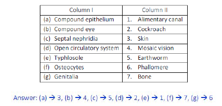
Question - 10. Match the terms in column I with those in column II:
Question 11. Mention breifly about the circulatory system of earthworm
Answer: Closed type blood vascular system is present in earthworm. The blood vascular system is composed of a heart, blood vessels and capillaries. Smaller blood vessels supply the gut, nerve cord and body wall. Blood glands are present on the 4th, 5th and 6th segments. The blood glands produce blood cells and haemoglobin. Blood cells are phagocytic in nature. Exchange of gases occurs through moist body surface into the blood stream.
Question 12. Mention the function of the following
(a) Ureters in frog
Answer: The ureters act as urinogenital duct male frogs. In females, the ureter is separate from the oviduct and only carries urine.
(b) Malpighian tubules
Answer: Excretion is performed by Malpighian tubules in cockroaches.
(c) Body wall in earthworm
Answer: The body wall of the earthworm facilitates exchange of gases.
Free study material for Biology
CBSE Class 11 Biology Chapter 7 Structural Organisation in Animals Study Material
Students can find all the important study material for Chapter 7 Structural Organisation in Animals on this page. This collection includes detailed notes, Mind Maps for quick revision, and Sure Shot Questions that will come in your CBSE exams. This material has been strictly prepared on the latest 2026 syllabus for Class 11 Biology. Our expert teachers always suggest you to use these tools daily to make your learning easier and faster.
Chapter 7 Structural Organisation in Animals Expert Notes & Solved Exam Questions
Our teachers have used the latest official NCERT book for Class 11 Biology to prepare these study material. We have included previous year examination questions and also step-by-step solutions to help you understand the marking scheme too. After reading the above chapter notes and solved questions also solve the practice problems and then compare your work with our NCERT solutions for Class 11 Biology.
Complete Revision for Biology
To get the best marks in your Class 11 exams you should use Biology Sample Papers along with these chapter notes. Daily practicing with our online MCQ Tests for Chapter 7 Structural Organisation in Animals will also help you improve your speed and accuracy. All the study material provided on studiestoday.com is free and updated regularly to help Class 11 students stay ahead in their studies and feel confident during their school tests.
FAQs
Our advanced study package for Chapter 7 Structural Organisation in Animals includes detailed concepts, diagrams, Mind Maps, and explanation of complex topics to ensure Class 11 students learn as per syllabus for 2026 exams.
The Mind Maps provided for Chapter 7 Structural Organisation in Animals act as visual anchors which will help faster recall during high-pressure exams.
Yes, teachers use our Class 11 Biology resources for lesson planning as they are in simple language and have lot of solved examples.
Yes, You can download the complete, mobile-friendly PDF of the Biology Chapter 7 Structural Organisation in Animals advanced resources for free.
Yes, our subject matter experts have updated the Chapter 7 Structural Organisation in Animals material to align with the rationalized NCERT textbooks and have removed deleted topics and added new competency-based questions.