
Read and download the NCERT Class 11 Biology Cell Cycle and Cell Division Important Notes. Designed for 2026-27, this advanced study material provides Class 11 Biology students with detailed revision notes, sure-shot questions, and detailed answers. Prepared by expert teachers and they follow the latest CBSE, NCERT, and KVS guidelines to ensure you get best scores.
Advanced Study Material for Class 11 Biology Chapter 10 Cell Cycle and Cell Division
To achieve a high score in Biology, students must go beyond standard textbooks. This Class 11 Chapter 10 Cell Cycle and Cell Division study material includes conceptual summaries and solved practice questions to improve you understanding.
Class 11 Biology Chapter 10 Cell Cycle and Cell Division Notes and Questions
Phases of Cell Cycle
The cell cycle is divided into two basic phases:
a. Interphase: The phase between subsequent cell divisions is called the interphase. The interphase lasts for more than 95% of the cell cycle.
b. M Phase (Mitosis phase): The actual cell division takes place in the M phase. The M phase lasts for less than 5% of the cell cycle. The M phase is composed of two major steps, viz. karyokinesis and cytokinesis. Division of nucleus happens during karyokinesis. Division of cytoplasm happens during cytokinesis.
The interphase is further divided into three phases, which are as follows:
a. G1 phase (Gap 1): During this phase, the cell is metabolically active and continuously grows.
b. S phase (Synthesis): During this phase, DNA synthesis or replication takes place. The amount of DNA becomes double during this phase, but the number of chromosomes remains the same.
c. G2 phase (Gap 2): During this phase, protein synthesis takes place.
Quiescent Stage (G0): Cells which do not divide further, exit G1 phase to enter an inactive stage. This stage is called quiescent stage (G0) of the cell cycle. The cells in this stage remain metabolically active but do not undergo division. But these cells can resume division as and when required.
M PHASE
Mitosis is divided into four stages, viz. Prophase, Metaphase, Anaphase and Telophase
Prophase
• Condensation of chromosomal material takes place. A chromosome is seen to be composed of two chromatids. The chromatids are attached together at the centromere.
• Spindle fibres are formed.
• Various cell organelles; like golgi bodies and ER cannot be seen during this staged. Nucleolus and nuclear envelope also disappear.
Metaphase
• All the chromosomes come to lie at the equator.
• In each chromosome, one chromatid is connected to the spindle fibre from one pole and another chromatid is connected to the spindle fibre from another pole.
• The plane of alignment of chromosomes during this phase is called metaphase plate.
Anaphase
• Centromeres split which results in separation of chromatids.
• After that, chromatids move to opposite poles.
Telophase
• The chromosomes form clusters at opposite poles. They become inconspicuous.
Cytokinesis
Division of cytoplasm is achieved by cytokinesis. In animal cell, a furrow appears in the plasma membrane. The furrow gradually deepens and finally joins in the centre. Thus, the cytoplasm is divided into two parts. In plant cells, cell wall formation begins in the centre. This grows outwards to meet the existing lateral walls and thus, the cytoplasm is divided into two parts.
Significance of Mitosis
• Mitosis results in the formation of new cells which are required for growth and repair.
• Mitosis results in the formation of two daughter cells; which have identical genetic makeup, similar to the mother cell.
MEIOSIS
• Meiosis involves two sequential cycles of nuclear and cell division, but only a single cycle of DNA replication. Meiosis is divided into meiosis I and meiosis II.
• Meiosis I begins after the S phase, and meiosis II follows meiosis I.
• Pairing of homologous chromosomes happens during meiosis which results in recombination of genes.
• Four haploid daughter cells are formed at the end of meiosis.
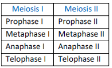
MEIOSIS I
Prophase I:
• Prophase in meiosis I is typically longer and more complex than the prophase in meiosis II. Prophase I is subdivided into five phases, viz. Leptotene, Zygotene, Pachytene, Diplotene and Diakinesis.
Leptotene:
• During this stage, the chromosomes become gradually visible under light microscope. Compaction of chromosomes continues throughout this phase.
Zygotene:
• Chromosomes start pairing together. This process is called synapsis. The paired chromosomes are called homologous chromosomes.
• Formation of synapsis is accompanied by the formation of synaptonemal complex.
•The synaptonemal complex by a pair of homologous chromosomes is called a bivalent or a tetrad.
Pachytene:
• Bivalent chromosomes clearly appear as tetrads, at this stage.
• Recombination nodules appear. These nodules are the sites at which crossing over takes place between non-sister chromatids of the homologous chromosomes.
• Exchange of genetic materials between two homologous chromosomes takes place during crossing over. This leads to recombination of genetic materials on the two chromosomes.
Diplotene:
• Synapotnemal complex is dissolved at this stage.
• The recombined homologous chromosomes of the bivalent separate from each other; except at the site of crossing over.
• The X-shaped structures; thus formed; are called chiasmata.
Diakinesis:
• Chiasmata is terminated at this stage.
• Meiotic spindles are formed to prepare the homologous chromosomes for separation.
• Nucleolus disappears and nuclear envelope breaks down by the end of diakinesis.
Metaphase I:
• The bivalent chromosomes are aligned on the equatorial plate.
• Spindle fibres from opposite poles attach to the pair of homologous chromosomes.
Anaphase I:
• Homologous chromosomes separate, but sister chromatids remain attached at their centromeres.
Telophase I:
• Nuclear membrane and nucleolus reappear.
• This is followed by cytokinesis and this stage is called the diad of cells.
• The stage between the two meiosis divisions is called interkinesis. Interkinesis is usually short lived.
MEIOSIS II
Prophase II: Meiosis II resembles the mitotic cell division. It begins immediately after cytokinesis. Nuclear membrane disappears. Chromosomes again become compact.
Metaphase II: The chromosomes align at the equator. Spindle fibres from the opposite poles get attached to the kinetochores of sister chromatids.
Anaphase II: Centromeres split and sister chromatids move towards the opposite poles.
Telophase II: The two groups of chromosomes get enclosed by nuclear envelope. This is followed by cytokinesis; resulting in the formation of four daughter cells.
Significance of Meiosis:
• Conservation of specific chromosome number of each species is achieved across successive generations in sexually reproducing organisms through meiosis.
• Meiosis helps in increasing the genetic variations in the population of organisms from one generation to the next.
NCERT Solution for Class 11 Biology Cell Cycle and Cell Division
Question. What is the average cell cycle span for a mammalian cell?
Answer: 24 hours
Question. Distinguish cytokinesis from karyokinesis.
Answer: Division of cytoplasm takes place during cytokinesis, while division of nucleus takes place during karyokinesis.
Question. Describe the events taking place during interphase.
Answer: The interphase is divided into three phases, which are as follows:
a. G1 phase (Gap 1): During this phase, the cell is metabolically active and continuously grows.
b. S phase (Synthesis): During this phase, DNA synthesis or replication takes place. The amount of DNA becomes double during this phase, but the number of chromosomes remains the same.
c. G2 phase (Gap 2): During this phase, protein synthesis takes place.
Question. What is G0 (quiescent phase) of cell cycle?
Answer: Quiescent Stage (G0): Cells which do not divide further, exit G1 phase to enter an inactive stage. This stage is called quiescent stage (G0) of the cell cycle. The cells in this stage remain metabolically active but do not undergo division. But these cells can resume division as and when required.
Question. Why is mitosis called equational division?
Answer: The number of chromosomes in daughter cells is same as in mother cell, so mitosis is called equational division.
Question. Name the stage of cell cycle at which one of the following events occur:
(a) Chromosomes are moved to spindle equator.
Answer: Metaphase
(b) Centromere splits and chromatids separate.
Answer: Anaphase
(c) Pairing between homologous chromosomes takes place.
Answer: Zygotene
(d) Crossing over between homologous chromosomes takes place.
Answer: Pachytene
Question. Describe the following: (a) synapsis (b) bivalent (c) chiasmata Draw a diagram to illustrate your answer.
Answer: The pairing of chromosomes during zygotene is called synapsis. The synaptonemal complex formed by a pair of homologous chromosomes is called bivalent. The X-shaped structure, formed during crossing over is called chiasmata.
Question. How does cytokinesis in plant cells differ from that in animal cells?
Answer: In animal cell, a furrow appears in the plasma membrane. The furrow gradually deepens and finally joins in the centre. Thus, the cytoplasm is divided into two parts. In plant cells, cell wall formation begins in the centre. This grows outwards to meet the existing lateral walls and thus, the cytoplasm is divided into two parts.
Question. Find examples where the four daughter cells from meiosis are equal in size and where they are found unequal in size.
Answer: The four daughter cells formed after microsporogenesis in flowering plants are equal in size. The four daughter cells formed after megasporogenesis in flowering plants are unequal in size.
Question. Distinguish anaphase of mitosis from anaphase I of meiosis.
Answer: The centromere splits during anphase of mitosis, while it does not split during anaphase I of meiosis.
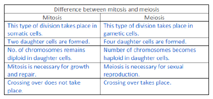
Question. List the main differences between mitosis and meiosis.
Answer:

Question. What is the significance of meiosis?
Answer: Significance of Meiosis:
• Conservation of specific chromosome number of each species is achieved across successive generations in sexually reproducing organisms through meiosis.
• Meiosis helps in increasing the genetic variations in the population of organisms from one generation to the next.
Question. Discuss with your teacher about
(a) Haploid insects and lower plants where cell-division occurs, and
Answer: Male bees, wasps and ants are haploid as they are produced from unfertilized eggs.
(b) Some haploid cells in higher plants where cell-division does not occur.
Answer: Cell division does not happen in synergids and antipodal cells; in the ovule.
Question. Can there be mitosis without DNA replication in ‘S’ phase?
Answer: There cannot be mitosis without DNA replication, because additional DNAs are required for the formation of new cells.
Question. Can there be DNA replication without cell division?
Answer: DNA replication can take place without cells division; as in case of formation of new mitochondria and chloroplasts.
Question. Analyse the events during every stage of cell cycle and notice how the following two parameters change
(a) Number of chromosomes (N) per cell
Answer: Number of chromosomes becomes half after meiosis.
(b) Amount of DNA content (C) per cell
Answer: Amount of DNA becomes double after S phase.
Cell division → Very important process in all living organisms. @ Continuity of life
During the division of a cell → DNA replication and cell growth take place.
Cell division, DNA replication, and Cell growth → Take place in a coordinated way to ensure correct division and formation of progeny cells containing intact genomes.
Cell cycle → The sequence of events by which a cell duplicates its genome, synthesises the other constituents of the cell and eventually divides into two daughter cells.
Cell growth (in terms of cytoplasmic increase) is a continuous process
Cell division requirement → The replicated chromosomes (DNA) are then distributed to daughter nuclei by a complex series of events. These events are under genetic control. (Cyclins & CDK kinase)
A typical eukaryotic cell cycle (Human cells) in culture →
• Divide once in every 24 hours (23 Hours Interphase + 1 Hour M Phase)
Duration of cell cycle →
• Vary from organism to organism and also from cell type to cell type.
• Yeast cell cycle in only about 90 minutes.
The cell cycle is divided into two basic phases:
• Interphase
• M Phase (Mitosis phase)
The M Phase → Actual cell division or mitosis occurs
Interphase → Represents the phase between two successive M phases.
• 95% of the duration of cell cycle.
• It is the resting phase → during which the cell is preparing for division
• Cell growth and DNA replication takes place in orderly manner.
• Three phases → G1 Phase + S Phase + G2 Phase = Interphase
• G1 – Means Gap 1; S – Means Synthesis; G2 – Means Gap 2
The M Phase →
• Starts with the nuclear division (karyokinesis)
• Ends with division of cytoplasm (cytokinesis).
G1 phase →
• Interval between mitosis and initiation of DNA replication (S phase)
• Cell is metabolically active and continuously grows @ Protein & RNA Synthesis extensively
S or synthesis phase
• DNA synthesis or replication takes place.
• Amount of DNA per cell doubles.
• 2C DNA becomes → 4C. (But chromosome number remain same)
• Each chromosome have double amount of DNA
• 2n Cell of G1 phase is Still 2n is S phase
• DNA replication begins in the nucleus
• Centriole duplicates in the cytoplasm. (In animal cell)
G2 phase
• Proteins are synthesised
• Preparation for mitosis (cell growth) continues. @ ATP storage takes
place & Spindle fibre synthesized
G0 Phase →
• The cells that do not divide further exit G1 phase to enter an inactive stage called quiescent stage (G0 ) of the cell cycle.
• Cells in this stage remain metabolically active
• Not proliferate unless required
In animals, mitotic cell division is only seen in the diploid somatic cells.
In plants mitotic divisions seen in both haploid and diploid cells.
M PHASE
• Most dramatic period of the cell cycle,
• Involving a major reorganisation of all components of the cell.
Equational division → Number of chromosomes in the parent and progeny cells is the same @ It’s MITOSIS
Mitosis divided into four stages of nuclear division, it is very essential to understand that cell division is a progressive process and very clear-cut lines cannot be drawn between various stages.
Mitosis is divided into the following four stages:
• Prophase
• Metaphase
• Anaphase
• Telophase
Prophase → First stage of mitosis
• Initiation of condensation of chromosomal material.
• The centriole begins to move towards opposite poles of the cell.
• The completion of prophase marked by the following events:
✓ Chromosomal material → Compact mitotic chromosomes.
✓ Chromosomes → Two chromatids attached at the centromere.
✓ Initiation of the assembly of mitotic spindle @ the microtubules
✓ In Late prophase → Golgi body, ER, nucleolus and the nuclear envelope disappears
Metaphase
• Begins when nuclear membrane completely disappeared
• Chromosomes are spread through the cytoplasm of the cell.
• Condensation of chromosomes is completed
✓ @ Maximum condensation ; Visible under microscope
• Morphology of chromosomes is most easily studied.
• Metaphase chromosome → two sister chromatids & a centromere.
• Small disc-shaped structures at the surface of the centromeres are called kinetochores.
✓ Sites of attachment of spindle fibres @ Protein Plates
• Metaphase is characterised by all the chromosomes coming to lie at the equator with one chromatid of each chromosome connected by its kinetochore to spindle fibres from one pole and its sister chromatid connected by its kinetochore to spindle fibres from the opposite pole • Metaphase plate
✓ The plane of alignment of the chromosomes at metaphase
• The key features of metaphase are:
✓ Spindle fibres attach to kinetochores of chromosomes.
✓ Chromosomes are moved to spindle equator and get aligned along metaphase plate through spindle fibres to both poles.
Anaphase
• At the onset of anaphase
✓ Splitting of chromosome @ Splitting of Centromere
✓ Two daughter chromatids, now referred to as chromosomes of the future daughter nuclei,
✓ Daughter chromatid / New chromosome begin their migration towards the two opposite poles.
• As each chromosome moves away from the equatorial plate, the centromere of each chromosome is towards the pole and hence at the leading edge, with the arms of the chromosome trailing behind .
Telophase
• Final stage of mitosis
• At the beginning of telophase
✓ The chromosomes that have reached their respective poles
✓ They de-condense and lose their individuality.
• The individual chromosomes can no longer be seen and chromatin material tends to collect in a mass in the two poles
• Nuclear envelope assembles around the chromosome clusters.
• Nucleolus, golgi complex and ER reform.
Cytokinesis
• Division of Cytoplasm (Cell divides into two after nucleus division)
• In an animal cell → appearance of a furrow in the plasma membrane.
• Plant cells → Wall formation starts in the centre of the cell and grows outward to meet the existing lateral walls.
• The formation of the new cell wall begins with the formation of a simple precursor, called the cell-plate that represents the middle lamella between the walls of two adjacent cells.
When karyokinesis is not followed by cytokinesis →
• Multinucleate condition ( formation of syncytium)
• e.g., liquid endosperm in coconut
Significance of Mitosis
• Mitosis results in the production of genetically similar daughter cells
• 2n to 2n ; n to n (No change in chromosome number)
• The growth of multicellular organisms is due to mitosis.
• Cell growth → nucleo-cytoplasmic ratio disturbs → The cell to divide to restore the nucleo-cytoplasmic ratio.
• A very significant contribution of mitosis is cell repair.
• The cells of the upper layer of the epidermis, cells of the lining of the gut, and blood cells are being constantly replaced.
• Mitotic divisions in the meristematic tissues – the apical and the lateral cambium, result in a continuous growth of plants throughout their life.
MEIOSIS
• Specialised kind of cell division that reduces the chromosome number by half results in the production of haploid daughter cells.
• Meiosis ensures the production of haploid phase in the life cycle of sexually reproducing organisms
✓ fertilisation restores the diploid phase.
• Meiosis involves two sequential cycles of nuclear and cell division called meiosis I and meiosis II but only a single cycle of DNA replication.
Meiosis I
• Four phases
✓ Prophase I (Largest Phase)
✓ Metaphase I
✓ Anaphase I
✓ Telophase I
Prophase I:
• Typically longer and more complex
• Subdivided into the following five sub phases based on chromosomal behaviour, i.e., Leptotene, Zygotene, Pachytene, Diplotene and Diakinesis.
• During leptotene stage
✓ Chromosomes become gradually visible under the light microscope.
✓ The compaction of chromosomes continues
• During zygotene stage
✓ Chromosomes start pairing together (synapsis)
✓ Paired chromosomes are called homologous chromosomes.
✓ Synaptonemal complex → Formed by a pair of synapsed homologous chromosomes is called a bivalent or a tetrad.
• During Pachytene stage
✓ Bivalent chromosomes now clearly appears as tetrads.
✓ Characterised by the appearance of recombination nodules
✓ Recombination nodules are the sites at which crossing over occurs between non-sister chromatids of the homologous chromosomes.
✓ Crossing over is the exchange of genetic material between two homologous chromosomes.
✓ Crossing over → An enzyme-mediated process and the enzyme involved is called recombinase.
✓ Crossing over leads to recombination of genetic material on the two chromosomes.
✓ At the end of pachytene, leaving the chromosomes linked at the sites of crossing over @ Diplotene begins
• During Diplotene stage
✓ Dissolution of the synaptonemal complex
✓ Recombined homologous chromosomes of the bivalents to separate from each other except at the sites of crossovers.
These X-shaped structures, are called chiasmata.
✓ Longest phase @ Can last months to years
• During Diakinesis stage
✓ The final stage of meiotic prophase I
✓ Terminalisation of chiasmata.
✓ Chromosomes are fully condensed
✓ Meiotic spindle is assembled
✓ Nucleolus disappears and the nuclear envelope also breaks down.
✓ Diakinesis represents transition to metaphase.
Metaphase I:
• The bivalent chromosomes align on the equatorial plate
• The microtubules from the opposite poles of the spindle attach to the pair of homologous chromosomes.
Anaphase I:
• The homologous chromosomes separate, while sister chromatids remain associated at their centromeresü @ Segregation of Mendelian characters
Telophase I:
• The nuclear membrane and nucleolus reappear,
• Cytokinesis follows and this is called as dyad of cells
INTERKINESIS →
• The stage between the two meiotic divisions I & II
• Generally short lived.
• Followed by prophase II
• Only G1 + G2 Phase ; No S Phase (DNA not duplicate)
Meiosis II
• Needed to normalise Chromosome (still have Double DNA because centromere not divided in Meiosis II so Chromosomes are Dyad)
• Have four phases
✓ Prophase II
✓ Metaphase II
✓ Anaphase II
✓ Telophase II
Prophase II:
• Resembles a normal mitosis prophase.
• The nuclear membrane disappears by the end of prophase II
• The chromosomes again become compact.
Metaphase II:
• At this stage the chromosomes align at the equator
• Microtubules from opposite poles of the spindle get attached to the kinetochores of sister chromatids. (Like Mitosis metaphase)
Anaphase II:
• Splitting of the centromere of each chromosome (which was holding the sister chromatids together),
• Chromosome (separated sister chromatid) move toward opposite poles of the cell (Like Mitosis Anaphase)
Telophase II:
• Meiosis ends with telophase II
• Nuclear envelope reappears
• Cytokinesis follows resulting in the formation of tetrad of cells i.e., four haploid daughter cells
SIGNIFICANCE OF MEIOSIS
• Maintain chromosome number generation after generation
✓ @ Specific chromosome number of each species
• Increases the genetic variability
✓ Crossing over brings variations
• Variations are very important for the process of evolution.
Free study material for Biology
CBSE Class 11 Biology Chapter 10 Cell Cycle and Cell Division Study Material
Students can find all the important study material for Chapter 10 Cell Cycle and Cell Division on this page. This collection includes detailed notes, Mind Maps for quick revision, and Sure Shot Questions that will come in your CBSE exams. This material has been strictly prepared on the latest 2026 syllabus for Class 11 Biology. Our expert teachers always suggest you to use these tools daily to make your learning easier and faster.
Chapter 10 Cell Cycle and Cell Division Expert Notes & Solved Exam Questions
Our teachers have used the latest official NCERT book for Class 11 Biology to prepare these study material. We have included previous year examination questions and also step-by-step solutions to help you understand the marking scheme too. After reading the above chapter notes and solved questions also solve the practice problems and then compare your work with our NCERT solutions for Class 11 Biology.
Complete Revision for Biology
To get the best marks in your Class 11 exams you should use Biology Sample Papers along with these chapter notes. Daily practicing with our online MCQ Tests for Chapter 10 Cell Cycle and Cell Division will also help you improve your speed and accuracy. All the study material provided on studiestoday.com is free and updated regularly to help Class 11 students stay ahead in their studies and feel confident during their school tests.
FAQs
Our advanced study package for Chapter 10 Cell Cycle and Cell Division includes detailed concepts, diagrams, Mind Maps, and explanation of complex topics to ensure Class 11 students learn as per syllabus for 2026 exams.
The Mind Maps provided for Chapter 10 Cell Cycle and Cell Division act as visual anchors which will help faster recall during high-pressure exams.
Yes, teachers use our Class 11 Biology resources for lesson planning as they are in simple language and have lot of solved examples.
Yes, You can download the complete, mobile-friendly PDF of the Biology Chapter 10 Cell Cycle and Cell Division advanced resources for free.
Yes, our subject matter experts have updated the Chapter 10 Cell Cycle and Cell Division material to align with the rationalized NCERT textbooks and have removed deleted topics and added new competency-based questions.