

Official CBSE Study Materials for Class 12 Biology
Review targeted academic resources with the CBSE Class 12 Biology Molecular Basis of Inheritance diagrams based questions. Built according to official CBSE standards for the 2026-27 term, these downloadable Class 12 Biology study materials support effective daily practice and deeper conceptual understanding for Chapter 05 Molecular Basis of Inheritance.
Advanced Resources for Biology
View or download the dedicated CBSE Class 12 Biology Molecular Basis of Inheritance diagrams based questions resource below. Engaging with these advanced study guides under focused conditions ensures continuous academic progress and mastery of the 2026-27 curriculum for Chapter 05 Molecular Basis of Inheritance.
CBSE Class 12 Biology Molecular Basis of Inheritance diagrams based questions. Please refer to the examination notes which you can use for preparing and revising for exams. These notes will help you to revise the concepts quickly and get good marks.
BASICS OF INHERITANCE
Biology: Chapter 6 – Molecular Basis of Inheritance (Diagram Based Questions)
This is a diagram of a polynucleotide. What do A and B represent? How are nucleotides linked? (
)Answer: A is the phosphate group at the 5′ end of the chain, and B is the free hydroxyl (–OH) group at the 3′ end of the chain. The nucleotides are linked by 3′–5′ phosphodiester bonds: the phosphate group of one nucleotide joins the 5′ carbon of its sugar to the 3′ carbon of the sugar of the next nucleotide.
(1) Label A and B. (2) What is the composition of B? (3) What is the length of the DNA wrapped? (4) What is this structure called? (
)Answer:
- A is the DNA and B is the histone octamer.
- B consists of eight histone molecules (two each of H2A, H2B, H3 and H4), which are positively charged basic proteins.
- The DNA wrapped is about 200 base pairs long (about 1.75 turns around the octamer; the core itself holds about 147 bp).
- The nucleosome.
(a) Identify this picture. (b) When can such structures be observed? (c) What are the dark round spots seen in the picture? (Electron micrograph of a thread with dark beads along it.)
Answer:
- It is the 'beads-on-string' structure of chromatin, seen under the electron microscope.
- It is observed in the interphase, when the chromatin is in its extended, thread-like form (before it condenses into chromosomes).
- The dark spots are the nucleosomes (the histone octamers with the DNA wrapped around them). The thread between them is the linker DNA.
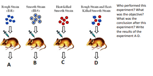
Who performed this experiment? What was the objective? What was the conclusion after this experiment? Write the results of the experiment A–D. (
)The experiment was performed by Frederick Griffith (1928), on Streptococcus pneumoniae and mice. His objective was to find out how the bacteria cause pneumonia and whether the bacteria can change their characters.
| Tube | Injected | Result |
|---|---|---|
| A | Rough (R) strain (non-virulent) | The mouse lives |
| B | Smooth (S) strain (virulent) | The mouse dies |
| C | Heat-killed S strain | The mouse lives |
| D | R strain + heat-killed S strain | The mouse dies; live S bacteria are found in its blood |
Conclusion: some 'transforming principle', transferred from the heat-killed S bacteria, had converted the R bacteria into the virulent S bacteria. (Later, Avery, MacLeod and McCarty showed that this principle is the DNA.)
(a) Who performed this experiment? (b) What was proved by this experiment? (Bacteriophages with sulphur-labelled protein coat or phosphorus-labelled DNA infect bacteria; infection, blending and centrifugation follow.)
Answer:
- Alfred Hershey and Martha Chase (1952).
- It proved that DNA, and not protein, is the genetic material. The phage labelled with 35S (protein) left its label outside, in the supernatant, and no sulphur was detected in the cells. The phage labelled with 32P (DNA) passed its label into the bacterial cells, and no phosphorus was detected in the supernatant.
(a) What was the objective of this experiment? (b) Who performed it? (c) How was the DNA separated into different layers? (d) Name any other scientist who performed an experiment to prove the same. (
)Answer:
- To prove that DNA replicates semi-conservatively.
- Matthew Meselson and Franklin Stahl (1958), using E. coli.
- By centrifugation in a caesium chloride (CsCl) density gradient. The DNA forms bands according to its density: 15N (heavy) DNA is lower, 14N (light) DNA is higher, and the hybrid DNA is in between. After one generation in 14N, all the DNA is hybrid; after two generations, half is hybrid and half is light.
- Taylor and his colleagues (1958), who used radioactive thymidine on the root tips of Vicia faba (broad bean).
What does this diagram represent? Label A–D. (
)Answer: It represents a transcription unit. A is the template strand (3′→5′), B is the coding strand (5′→3′), C is the promoter and D is the terminator. The structural gene lies between the promoter and the terminator; transcription starts at the start site near the promoter.
Study the diagram and elaborate what it explains. (
)Answer: It explains the cloning of a gene of interest using a vector. The plasmid is isolated from the bacterium (separate from the bacterial chromosome) and cut with a restriction enzyme. The gene of interest is cut from the DNA of the other cell with the same enzyme, and it is joined to the plasmid by DNA ligase, forming a recombinant plasmid. This is then introduced into the host bacterium (transformation), where it multiplies and makes many copies of the gene.
(a) What does the diagram represent? (b) Can you mention any other model proposed for the same? (A double helix of DNA with parental strands separating into two double helices, each with an old and a new strand.)
Answer:
- The semi-conservative replication of DNA: each daughter DNA has one old (parental) strand and one newly made strand.
- The conservative model (the parent DNA is retained and a completely new DNA is made) and the dispersive model (the daughter DNAs are mixtures of old and new segments).
(a) Mention the nonsense codons. (b) Which codon initiates translation? Does it have any other function? (c) The genetic code is degenerate. Take help of this table to explain it. (d) If an mRNA is written as below, how many amino acids will the polypeptide contain? 5′ AUGCAGGGUUCAAAAUAGGAUUCCGGACUA 3′ (
| First base | Second base: U | C | A | G | Third base |
|---|---|---|---|---|---|
| U | UUU – Phe | UCU – Ser | UAU – Tyr | UGU – Cys | U |
| UUC – Phe | UCC – Ser | UAC – Tyr | UGC – Cys | C | |
| UUA – Leu | UCA – Ser | UAA – Stop | UGA – Stop | A | |
| UUG – Leu | UCG – Ser | UAG – Stop | UGG – Trp | G | |
| C | CUU – Leu | CCU – Pro | CAU – His | CGU – Arg | U |
| CUC – Leu | CCC – Pro | CAC – His | CGC – Arg | C | |
| CUA – Leu | CCA – Pro | CAA – Gln | CGA – Arg | A | |
| CUG – Leu | CCG – Pro | CAG – Gln | CGG – Arg | G | |
| A | AUU – Ile | ACU – Thr | AAU – Asn | AGU – Ser | U |
| AUC – Ile | ACC – Thr | AAC – Asn | AGC – Ser | C | |
| AUA – Ile | ACA – Thr | AAA – Lys | AGA – Arg | A | |
| AUG – Met (start) | ACG – Thr | AAG – Lys | AGG – Arg | G | |
| G | GUU – Val | GCU – Ala | GAU – Asp | GGU – Gly | U |
| GUC – Val | GCC – Ala | GAC – Asp | GGC – Gly | C | |
| GUA – Val | GCA – Ala | GAA – Glu | GGA – Gly | A | |
| GUG – Val | GCG – Ala | GAG – Glu | GGG – Gly | G |
Answer:
- UAA, UAG and UGA. They do not code for any amino acid and act as the stop (termination) codons.
- AUG initiates the translation. It also codes for the amino acid methionine.
- Degeneracy means that more than one codon can code for the same amino acid. For example, leucine is coded by six codons (UUA, UUG, CUU, CUC, CUA and CUG), and serine and arginine are also coded by six; only methionine (AUG) and tryptophan (UGG) have a single codon.
- Read in triplets: AUG CAG GGU UCA AAA UAG GAU UCC GGA CUA. The codons are Met, Gln, Gly, Ser, Lys, and then UAG is a stop codon. So the polypeptide will contain 5 amino acids (Met–Gln–Gly–Ser–Lys).
What does the structure represent? What is its (model) called? What is the other one? Label A and B. (
)Answer: It represents a tRNA (transfer RNA) molecule. The structure shown is the clover-leaf model. The other model is the inverted L-shaped (three-dimensional) structure, which is the actual structure of the tRNA. A is the amino acid acceptor end (the 3′ end, with the CCA sequence, to which the amino acid binds), and B is the anticodon loop, which recognises the codon on the mRNA.
Name the three events A–C shown in this diagram. (
)Answer: A: transcription (formation of the primary transcript from the gene). B: splicing (removal of the introns and joining of the exons, along with capping and tailing) to form the mature mRNA. C: translation (formation of the protein from the mature mRNA).
(a) What is A doing? (b) Label B–D. (c) What would happen if A was not present? (
)Answer:
- A is the inducer (lactose or allolactose). It binds to the repressor protein and inactivates it, so that the repressor can no longer bind the operator.
- B, C and D are the three enzymes encoded by the genes z, y and a: B is β-galactosidase, C is permease and D is transacetylase.
- If A (the inducer) were not present, the repressor would remain active and bind to the operator. This would block the RNA polymerase, so the lac mRNA would not be transcribed and the enzymes would not be made.
The diagram shows steps in DNA fingerprinting. Some of the steps are unlabelled. Label steps 3, 5, 7 and 10. (
)Answer:
- (Step 3) The DNA is cut into fragments by restriction enzymes.
- (Step 5) The separated DNA fragments are transferred (blotted) from the gel on to a nylon or nitrocellulose membrane (Southern blotting).
- (Step 7) The radioactive probe is added to the membrane and hybridises with the complementary DNA fragments.
- (Step 10) The membrane is placed against X-ray film (autoradiography), and the film is then developed (step 11), which shows the DNA fingerprint.
(a) What is shown in this diagram? (b) Label A and B. (c) Place the primer(s) in the diagram. (d) How is the process different in prokaryotes and eukaryotes? (
)Answer:
- The replication fork of DNA, with the Okazaki fragments.
- A is the leading strand (synthesised continuously in the 5′→3′ direction), and B is the lagging strand (synthesised discontinuously, as the short Okazaki fragments).
- A short RNA primer is needed once at the start of the leading strand, and at the 5′ end of each Okazaki fragment of the lagging strand.
- In prokaryotes the circular DNA has a single origin of replication and the replication is done mainly by DNA polymerase III. In eukaryotes the linear chromosomes have many origins of replication, and several DNA polymerases (such as α, δ and ε) are involved; the replication is also more complex, with the DNA wrapped around histones.
Free study material for Biology
CBSE Class 12 Biology Study Material: Chapter 05 Molecular Basis of Inheritance
Core Study Kit: Class 12 Biology Chapter 05 Molecular Basis of Inheritance
Access comprehensive study material for Chapter 05 Molecular Basis of Inheritance, including revision notes, concept maps, and high-probability questions. These resources are designed in alignment with the latest 2026 CBSE syllabus for Class 12 Biology to support effective exam preparation.
NCERT-Aligned Solutions for Chapter 05 Molecular Basis of Inheritance
Designed around the official curriculum, these study guides guarantee standard compliance. Reviewing step-by-step solutions clarifies complex sub-topics within Chapter 05 Molecular Basis of Inheritance and demystifies standard marking schemes for Biology evaluations.
Next Steps in Your Exam Preparation
For peak performance in upcoming evaluations, integrate official Biology sample papers directly into your study schedule. Follow up your revision by attempting online MCQ tests for Chapter 05 Molecular Basis of Inheritance to refine calculation speed and precision.
FAQs
Our advanced study package for Chapter 05 Molecular Basis of Inheritance includes detailed concepts, diagrams, Mind Maps, and explanation of complex topics to ensure Class 12 students learn as per syllabus for 2026 exams.
The Mind Maps provided for Chapter 05 Molecular Basis of Inheritance act as visual anchors which will help faster recall during high-pressure exams.
Yes, teachers use our Class 12 Biology resources for lesson planning as they are in simple language and have lot of solved examples.
Yes, You can download the complete, mobile-friendly PDF of the Biology Chapter 05 Molecular Basis of Inheritance advanced resources for free.
Yes, our subject matter experts have updated the Chapter 05 Molecular Basis of Inheritance material to align with the rationalized NCERT textbooks and have removed deleted topics and added new competency-based questions.