

Class 12 Biology Study Guide: CBSE Class 12 Biology Human Reproduction Diagrams
Access comprehensive study materials and useful resources for Chapter 02 Human Reproduction using the CBSE Class 12 Biology Human Reproduction Diagrams. Designed to align with the 2026-27 CBSE academic guidelines, these advanced resources help Class 12 Biology students reinforce core concepts beyond standard textbooks.
Download Chapter 02 Human Reproduction Study Material PDF
Access the complete useful resource PDF for Class 12 Biology below. Regular practice with these targeted academic notes builds familiarity with complex topics and helps secure higher marks in final school evaluations.
This CBSE Class 12 Biology Chapter 3 Human Reproduction diagram notes cover the male and female reproductive systems, the structure of the seminiferous tubule and sperm, the sectional view of the ovary, oogenesis and spermatogenesis, the menstrual cycle, and fertilisation and development of the ovum to the blastocyst in the fallopian tube. Each labelled diagram is described with all its labels for quick revision.
CHAPTER 3: HUMAN REPRODUCTION
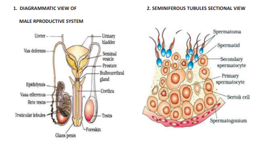
1. DIAGRAMMATIC VIEW OF MALE REPRODUCTIVE SYSTEM
[Figure: Labelled diagram of the male reproductive system with these parts: Ureter, Urinary bladder, Seminal vesicle, Prostate, Bulbourethral gland, Vas deferens, Epididymis, Vasa efferentia, Rete testis, Testicular lobules, Testis, Urethra, Glans penis, Foreskin.]
2. SEMINIFEROUS TUBULES SECTIONAL VIEW
[Figure: Sectional view of a seminiferous tubule showing the layers of developing sperm cells. The labels from the outside of the tubule towards the lumen are: Spermatogonium at the outer edge, Primary spermatocyte, Secondary spermatocyte, Spermatid, and Spermatozoa in the lumen. Large Sertoli cells are labelled among the developing cells.]
3. DIAGRAMMATIC VIEW OF FEMALE REPRODUCTIVE SYSTEM
[Figure: Labelled diagram of the female reproductive system with these parts: Uterine fundus, Uterine cavity, Endometrium, Myometrium, Perimetrium, Isthmus, Ampula, Infundibulum, Fimbriae, Fallopian tube, Ovary, Cervix, Cervical canal and Vagina.]
4. STRUCTURE OF SPERM
[Figure: Labelled diagram of a sperm with these parts from the top: Plasma membrane, Acrosome, Nucleus containing chromosomal material (in the head), Head, Neck, Middle piece with Mitochondria (energy source) spiralled around it, and a long Tail.]
5. SECTIONAL VIEW OF OVARY
[Figure: Sectional view of an ovary with these labelled structures: Blood vessels, Primary follicle (small follicles at various stages), Tertiary follicle showing antrum, Graafian follicle, Ovum released from the Graafian follicle, and Corpus luteum.]
6. OVUM SURROUNDED BY FEW SPERM (FERTILISATION)
[Figure: Ovum in the centre surrounded by cells of the corona radiata, with a zona pellucida around the ovum and a perivitelline space between the zona pellucida and the ovum membrane. Several sperm are shown around the outer cells, with labels for Sperm, Cells of the corona radiata, Zona pellucida, Perivitelline space and Ovum.]
7. SCHEMATIC REPRESENTATION OF OOGENESIS AND SPERMATOGENESIS
Spermatogenesis (left side):
Spermatogonia (at puberty) → Mitosis (differentiation) → Primary spermatocytes → 1st meiotic division → Secondary spermatocytes → 2nd meiotic division → Spermatids → Differentiation → Spermatozoa
Oogenesis (right side):
Oogonia (fetal life) → Mitosis (differentiation) → Primary oocyte → 1st meiotic division (completed prior to ovulation) → Secondary oocyte → Ovum, with the First polar body and the Second polar body formed during division
The chromosome number per cell is 46 up to the primary spermatocyte and primary oocyte stages, and it becomes 23 after the first meiotic division.
[Figure: Schematic diagram in two coloured panels. The left panel (pink) shows spermatogenesis with Spermatogonia, Primary spermatocytes, Secondary spermatocytes, Spermatids and Spermatozoa, with arrows labelled Mitosis differentiation, 1st meiotic division, 2nd meiotic division and Differentiation. The right panel (blue) shows oogenesis with Oogonia, Primary oocyte, Secondary oocyte, First polar body, Second polar body and Ovum. A central green column shows the chromosome number per cell: 46, 23 and 23. Along the side of the panels are the stages: Fetal life, Birth, Childhood, Puberty, and Adult reproductive life.]
8. DIAGRAMMATIC REPRESENTATION OF VARIOUS EVENTS DURING MENSTRUAL CYCLE
[Figure: Graph of the menstrual cycle over 28 days, with days 1 to 29 and Next cycle begins marked on the horizontal axis. The top section shows Pituitary Hormone levels for FSH and LH, with a sharp peak of LH at about day 14. The second section shows Ovarian events: Developing follicle, Mature follicle, Ovulation, Developing corpus luteum and Regressing corpus luteum. The third section shows Ovarian Hormone levels for Estrogen and Progesterone, with estrogen peaking before day 14 and progesterone rising in the luteal phase. The bottom section shows Uterine events: Menses at the start, then the thickening of the endometrium.]
Phases of the menstrual cycle:
1. Menstruation: days 1 to 5, when the uterine lining is shed.
2. Follicular (proliferative) phase: about days 6 to 14, when the endometrium grows again under the influence of estrogen.
3. Ovulation: about day 14, when the LH surge causes the release of the ovum.
4. Luteal (secretory) phase: about days 15 to 28, when the corpus luteum secretes progesterone that prepares the uterus for implantation.
9. FERTILISATION, TRANSPORT OF OVUM AND PASSAGE OF GROWING EMBRYO IN FALLOPIAN TUBE
[Figure: Series of stages of development. Stage (a) shows the ovum with its outer layer. Stage (b) shows the ovum being fertilised by sperm, with the sperm around its outer layer. Stage (c) shows the 2-cell stage. Stage (d) shows the 8-cell stage. Stage (e) is the Morula, a solid ball of cells. Stage (f) shows a later stage with more cells. Stage (g) is the Blastocyst, a hollow ball of cells with an inner cell mass. Below these, the diagram of the female reproductive tract shows the development of the ovum to the blastocyst as it travels along the fallopian tube, with the marked positions A to H from the ovary to the uterus, and the label Blastocyst implantation on the uterine wall.]
Key events shown in this diagram:
1. The ovum is fertilised in the upper part of the fallopian tube.
2. The zygote divides by mitosis as it moves down the tube, forming the 2-cell, 8-cell and morula stages.
3. The morula forms a blastocyst, which reaches the uterus.
4. The blastocyst gets implanted in the endometrium of the uterus.
Free study material for Biology
CBSE Class 12 Biology Study Material: Chapter 02 Human Reproduction
Quick Revision Material for Chapter 02 Human Reproduction
Review targeted study resources for Chapter 02 Human Reproduction tailored for Class 12 learners. Utilizing these structured notes and quick-revision tools ensures complete alignment with current CBSE evaluation standards.
Expert Notes and Solved Exam Questions
Designed around the official curriculum, these study guides guarantee standard compliance. Reviewing step-by-step solutions clarifies complex sub-topics within Chapter 02 Human Reproduction and demystifies standard marking schemes for Biology evaluations.
Additional Study Resources for Class 12
Maximize your grade potential by utilizing Class 12 Biology practice papers in tandem with these structured notes. All printable assignments and interactive mock tests on our platform are updated for the 2026 session and available free of charge.
FAQs
Our advanced study package for Chapter 02 Human Reproduction includes detailed concepts, diagrams, Mind Maps, and explanation of complex topics to ensure Class 12 students learn as per syllabus for 2026 exams.
The Mind Maps provided for Chapter 02 Human Reproduction act as visual anchors which will help faster recall during high-pressure exams.
Yes, teachers use our Class 12 Biology resources for lesson planning as they are in simple language and have lot of solved examples.
Yes, You can download the complete, mobile-friendly PDF of the Biology Chapter 02 Human Reproduction advanced resources for free.
Yes, our subject matter experts have updated the Chapter 02 Human Reproduction material to align with the rationalized NCERT textbooks and have removed deleted topics and added new competency-based questions.