

Download CBSE Class 12 Biology Useful Resources
Explore structured advanced study materials through the CBSE Class 12 Biology Human Reproduction diagrams based questions. Tailored for Class 12 learners, utilizing these Biology resources ensures thorough preparation and strengthens foundational knowledge before final CBSE evaluations.
Access CBSE Study Guides and Summaries
View or download the dedicated CBSE Class 12 Biology Human Reproduction diagrams based questions resource below. Engaging with these advanced study guides under focused conditions ensures continuous academic progress and mastery of the 2026-27 curriculum for Chapter 02 Human Reproduction.
This Class 12 Biology page gives diagram based questions with answers on Chapter 3 Human Reproduction. It covers the structure of a human sperm, the longitudinal section of the human testis, the seminiferous tubule and spermatogenesis, the human ovary and follicles, the menstrual cycle graph, the primary oocyte and the blastocyst.
3. Human Reproduction
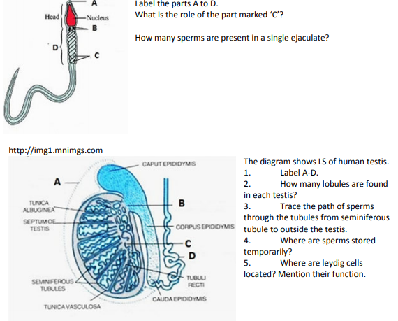
3.1 Label the parts A to D.
[Figure: Diagram of a human sperm. The top tip is marked A, the red Nucleus lies in the Head, B marks two small bodies just below the head, C marks the spirally coiled part with two arrows, and D is a bracket covering the part from below the head to the end of the coiled region. A long tail extends below]
Answer:
A - Acrosome
B - Centrioles (neck region)
C - Mitochondria
D - Middle piece
Teacher's Note:
a) The acrosome contains hydrolytic enzymes that help the sperm to penetrate the egg membranes.
b) Do not confuse the middle piece (D), which carries the mitochondria, with the head, which carries the nucleus and acrosome.
What is the role of the part marked 'C'?
Answer: The part C is the mitochondria. They produce energy (ATP) that is needed for the movement of the sperm tail, so that the sperm can swim towards the ovum.
Teacher's Note:
a) Mitochondria are the power houses of the cell and are packed in the middle piece to give energy for motility.
b) Write 'energy for sperm motility', not just 'respiration'.
c) This is why a sperm with damaged mitochondria cannot reach the egg.
How many sperms are present in a single ejaculate?
Answer: About 200 to 300 million sperms are present in a single ejaculate (about 60 percent of them must have normal shape and size and at least 40 percent must show vigorous motility for fertility).
Teacher's Note:
a) A normal ejaculate contains 200 to 300 million sperms; fertility needs about 60 percent normal and 40 percent motile sperms.
b) Students often write 200 to 300 thousand; the correct unit is million.
3.2 The diagram shows LS of human testis.
1. Label A-D.
[Figure: Longitudinal section of the human testis showing the Tunica albuginea, Septum of testis, Seminiferous tubules, Tunica vasculosa, Tubuli recti, Caput epididymis, Corpus epididymis and Cauda epididymis. The dotted outer covering is marked A. B points to the ducts between the testis and the caput epididymis. C points to the network of canals at the back of the testis. D points to the coiled duct that continues from the cauda epididymis]
Answer:
A - Scrotum
B - Vasa efferentia (efferent ductules)
C - Rete testis
D - Vas deferens
Teacher's Note:
a) The scrotum keeps the testis 2 to 2.5 degrees Celsius below body temperature, which is needed for sperm formation.
b) B and C are easily swapped; the rete testis lies inside the testis and the vasa efferentia lead out of it.
c) Always label from the figure, by following each line to the structure it touches.
2. How many lobules are found in each testis?
Answer: Each testis has about 250 testicular lobules (compartments).
Teacher's Note:
a) The testis is divided by septa into about 250 lobules, each containing one to three highly coiled seminiferous tubules.
3. Trace the path of sperms through the tubules from seminiferous tubule to outside the testis.
Answer: Seminiferous tubules → Tubuli recti → Rete testis → Vasa efferentia → Epididymis → Vas deferens → (ejaculatory duct) → Urethra → outside the body.
Teacher's Note:
a) Remember the sequence with the first letters: S T R V E V.
b) The vasa efferentia leave the testis and open into the epididymis.
c) Do not skip the rete testis, which is the commonly missed step.
4. Where are sperms stored temporarily?
Answer: Sperms are stored temporarily in the epididymis, where they also undergo maturation and gain motility.
Teacher's Note:
a) Sperms become motile and fertile only after they spend time in the epididymis.
b) The vas deferens is only a transport duct, not the main storage site.
5. Where are leydig cells located? Mention their function.
Answer: Leydig cells are located in the interstitial spaces (connective tissue) between the seminiferous tubules. They secrete androgens (mainly testosterone), which regulate spermatogenesis and develop and maintain the male secondary sexual characters.
Teacher's Note:
a) Leydig cells are also called interstitial cells; write the word 'interstitial' for full marks.
b) Do not confuse Leydig cells (secrete androgens) with Sertoli cells (nourish germ cells).
c) Their activity is stimulated by the luteinising hormone (LH) from the anterior pituitary.
3.3 1. This is a part of The Seminiferous tubule of human testis. Label A-D.
[Figure: Part of a seminiferous tubule. At the top, blue cells with tails are marked A (three cells with a line). Secondary spermatocyte is labelled with a pointer to a cell below. B points to a blue cell without a long tail near the secondary spermatocytes. Primary spermatocyte is labelled with a pointer to a large cell. C points to an elongated cell between the large cells. D points to a cell lying close to the basement membrane at the bottom, next to a blood vessel]
Answer:
A - Spermatozoa (sperms)
B - Spermatid
C - Sertoli cell
D - Spermatogonium
Teacher's Note:
a) Spermatogonia lie at the outer edge of the tubule and the later stages move towards the lumen.
b) The Sertoli cell is the tall supporting cell that gives nourishment to the germ cells.
c) Follow the order from the base to the lumen: spermatogonium, primary spermatocyte, secondary spermatocyte, spermatid, sperm.
2. What will be the number of chromosomes in Secondary spermatocyte, Spermatid?
Answer: The secondary spermatocyte has 23 chromosomes (haploid) and the spermatid has 23 chromosomes (haploid).
Teacher's Note:
a) Meiosis I reduces the chromosome number from 46 to 23, so the secondary spermatocyte is already haploid.
b) Do not write 46 for the secondary spermatocyte; that is the number in the primary spermatocyte.
3. What do the following terms mean: Spermatogenesis, Spermiogenesis, Spermiation?
Answer:
Spermatogenesis: the process of formation of sperms from the spermatogonia in the seminiferous tubules.
Spermiogenesis: the transformation of spermatids into spermatozoa (sperms).
Spermiation: the release of the sperms from the Sertoli cells into the lumen of the seminiferous tubule.
Teacher's Note:
a) Spermatogenesis includes the whole series, spermiogenesis is only the last change in shape, and spermiation is the release.
b) Students often write spermiation as the same as spermiogenesis; they are different steps.
c) Spermatogenesis starts at puberty under the action of GnRH, LH and FSH.
3.4 1. Label the parts A to D
[Figure: Section of an ovary with Blood vessels entering on the left, a Primary follicle and a Secondry follicle (printed label), a follicle marked A with a small cavity labelled Antrum, a large follicle marked B near the edge of the ovary, a small cell marked C leaving a ruptured follicle near the bottom, and yellowish structures marked D at the lower left]
Answer:
A - Tertiary follicle
B - Graafian follicle
C - Ovum (secondary oocyte released at ovulation)
D - Corpus luteum
Teacher's Note:
a) The Graafian follicle is the largest and most mature follicle and ruptures to release the ovum.
b) After ovulation the ruptured follicle turns into the corpus luteum, which secretes progesterone.
c) Write C as the released secondary oocyte, not as a primary oocyte.
2. How is the tertiary follicle identified?
Answer: The tertiary follicle is identified by the presence of a fluid-filled cavity called the antrum, and a layer of theca (theca interna and theca externa), with the secondary oocyte surrounded by granulosa cells.
Teacher's Note:
a) The antrum is the key identifying feature of a tertiary follicle.
b) The secondary follicle has no antrum yet, so do not mix the two.
3. When is Oogenesis initiated?
Answer: Oogenesis is initiated during the embryonic development stage, when the oogonia (about a couple of million in the foetus) divide to form primary oocytes that enter prophase I of meiosis and get temporarily arrested.
Teacher's Note:
a) Oogenesis begins before birth, whereas spermatogenesis begins only at puberty.
b) Primary oocytes remain arrested at prophase I until puberty.
4. How many primary follicles are present in each ovary during puberty? What happens to the remaining?
Answer: About 60,000 to 80,000 primary follicles are present in each ovary at puberty. The remaining follicles degenerate during the phase from birth to puberty (only about 400 follicles ovulate during the reproductive life of a woman).
Teacher's Note:
a) At birth each ovary has about 2 million primary follicles, which fall to 60,000 to 80,000 at puberty.
b) The loss of follicles is called atresia; it is not the same as ovulation.
3.5 Study the diagram and answer the following questions:
a) On which day the Graafian follicle ruptures to release the ovum? What is it called?
[Figure: Graph of the menstrual cycle over 28 days with four rows. Gonadotropic hormone levels: LH and FSH, with a sharp LH peak near day 14, and the follicular phase before it and the luteal phase after it. Ovarian cycle: the preovulatory phase with growing follicles, Ovulation at day 14, and the postovulatory phase with the corpus luteum. Ovarian hormone levels: Estrogens peak just before day 14 and show a second smaller rise, and Progesterone rises after day 14 to a peak near day 21 and then falls. Uterine cycle: the uterine wall breaks down during days 0 to 7 (MENSES), is thin and starts thickening in the PROLIFERATIVE PHASE (days 7 to 14), and is thickest in the SECRETORY PHASE (days 14 to 28). A dotted vertical line marks day 14]
Answer: The Graafian follicle ruptures on the 14th day of the cycle, which is mid-cycle. The release of the ovum is called ovulation.
Teacher's Note:
a) Ovulation occurs on about day 14, following a surge of LH (and FSH), called the LH surge.
b) Ovulation is the release of the ovum; do not call it fertilisation.
b) When does the level of Progesterone rise?
Answer: The level of progesterone rises after ovulation, during the luteal (secretory) phase, and reaches its peak at about the 21st day.
Teacher's Note:
a) Progesterone stays low in the follicular phase and rises only after ovulation.
b) The rise starts after the 14th day and the peak is near the 21st day.
c) From where is Progesterone secreted?
Answer: Progesterone is secreted by the corpus luteum, which is formed from the ruptured Graafian follicle after ovulation.
Teacher's Note:
a) The corpus luteum is a temporary endocrine gland formed from the remains of the follicle.
b) If pregnancy occurs, the placenta takes over progesterone secretion later.
d) What happens after 28th day of the cycle?
Answer: If fertilisation does not occur, the corpus luteum degenerates, the levels of progesterone and estrogen fall, the endometrium breaks down and menstruation begins again, so a new cycle starts.
Teacher's Note:
a) The fall in hormone levels is what causes the thick endometrium to shed.
b) The first day of bleeding is counted as day 1 of the next cycle.
e) In which phase of the menstrual cycle the thickness of the uterine wall is maximum?
Answer: The thickness of the uterine wall is maximum in the secretory phase (luteal phase), from day 14 to day 28.
Teacher's Note:
a) Under the action of progesterone the endometrium becomes thick, rich in blood vessels and glands to receive the embryo.
b) The proliferative phase is only the phase of growth, not the maximum thickness.
f) If pregnancy occurs, will menses continue?
Answer: No. If pregnancy occurs, menstruation stops, because the corpus luteum persists and the placenta keeps the endometrium intact through progesterone.
Teacher's Note:
a) Absence of menses is the first sign of pregnancy.
g) What will happen to the levels of Estrogen and progesterone in case of pregnancy?
Answer: In case of pregnancy the levels of estrogen and progesterone remain high (they rise further and are maintained by the corpus luteum and later by the placenta) and do not fall as in a normal cycle.
Teacher's Note:
a) High progesterone keeps the uterine lining intact and prevents new ovulation.
b) The placenta itself becomes a source of estrogen and progesterone during pregnancy.
c) Contrast this with the sudden fall of hormones in a cycle without pregnancy.
3.6 What does the diagram represent? Label A and B
[Figure: Large round cell with a nucleus and nucleolus in the centre, labelled Cytoplasm, Nucleus, Nucleolus and Plasma membrane. A clear ring surrounds the plasma membrane, marked B, and an outer layer of small follicle cells surrounds the whole cell, marked A]
Answer:
The diagram represents a primary oocyte enclosed in a primary follicle (an immature ovum).
A - Granulosa cells (follicle cells)
B - Zona pellucida
Teacher's Note:
a) The primary oocyte is surrounded by the zona pellucida and a layer of granulosa cells.
b) The zona pellucida is the glycoprotein layer that the sperm must penetrate during fertilisation.
c) Do not label B as the plasma membrane, which is already labelled in the diagram.
3.7 a) Name the structure.
[Figure: Hollow ball of cells. An outer single layer of cells is labelled Trophoblast. A cluster of cells attached to the inside at the bottom is labelled Inner cell mass. The central cavity is labelled Blastocoel]
Answer: The structure is a blastocyst.
Teacher's Note:
a) The blastocyst has an outer trophoblast, an inner cell mass and a cavity called the blastocoel.
b) The embryo with 8-32 blastomere is called ________
Answer: The embryo with 8 to 32 blastomeres is called a morula.
Teacher's Note:
a) The morula is a solid ball of cells formed by the cleavage divisions of the zygote.
c) What is the fate of the trophoblast and inner cell mass?
Answer:
The trophoblast attaches to the endometrium (implantation) and later forms the placenta and the extra-embryonic membranes (chorion).
The inner cell mass gets differentiated into the embryo, forming the three germ layers: ectoderm, mesoderm and endoderm.
Teacher's Note:
a) The trophoblast gives the structures that support the embryo, not the embryo itself.
b) The inner cell mass is the stem cell source of the embryo, which is why it is important in stem cell research.
c) Remember: implantation takes place at the trophoblast side of the blastocyst.
Free study material for Biology
Useful Resources and Notes for Class 12 Biology Chapter 02 Human Reproduction
Core Study Kit: Class 12 Biology Chapter 02 Human Reproduction
Review targeted study resources for Chapter 02 Human Reproduction tailored for Class 12 learners. Utilizing these structured notes and quick-revision tools ensures complete alignment with current CBSE evaluation standards.
NCERT-Aligned Solutions for Chapter 02 Human Reproduction
Designed around the official curriculum, these study guides guarantee standard compliance. Reviewing step-by-step solutions clarifies complex sub-topics within Chapter 02 Human Reproduction and demystifies standard marking schemes for Biology evaluations.
Next Steps in Your Exam Preparation
Maximize your grade potential by utilizing Class 12 Biology practice papers in tandem with these structured notes. All printable assignments and interactive mock tests on our platform are updated for the 2026 session and available free of charge.
FAQs
Our advanced study package for Chapter 02 Human Reproduction includes detailed concepts, diagrams, Mind Maps, and explanation of complex topics to ensure Class 12 students learn as per syllabus for 2026 exams.
The Mind Maps provided for Chapter 02 Human Reproduction act as visual anchors which will help faster recall during high-pressure exams.
Yes, teachers use our Class 12 Biology resources for lesson planning as they are in simple language and have lot of solved examples.
Yes, You can download the complete, mobile-friendly PDF of the Biology Chapter 02 Human Reproduction advanced resources for free.
Yes, our subject matter experts have updated the Chapter 02 Human Reproduction material to align with the rationalized NCERT textbooks and have removed deleted topics and added new competency-based questions.