

Official CBSE Study Materials for Class 12 Biology
Access comprehensive study materials and useful resources for important charts using the CBSE Class 12 Biology all chapters important charts. Designed to align with the 2026-27 CBSE academic guidelines, these advanced resources help Class 12 Biology students reinforce core concepts beyond standard textbooks.
Advanced Resources for Biology
View or download the dedicated CBSE Class 12 Biology all chapters important charts resource below. Engaging with these advanced study guides under focused conditions ensures continuous academic progress and mastery of the 2026-27 curriculum for important charts.
CBSE Class 12 Biology all chapters important charts. Please refer to the examination notes which you can use for preparing and revising for exams. These notes will help you to revise the concepts quickly and get good marks. For more study material for Biology please click here - eurythermal animals.
These CBSE Class 12 Biology all chapters important charts give concept maps, flow charts and labelled diagram notes for the whole syllabus. They cover reproduction in organisms and flowering plants, human reproduction and reproductive health, genetic disorders, molecular basis of inheritance, evolution, human health and disease, food production, biotechnology, organisms and populations, ecosystems, biodiversity and environmental issues. Use them for quick revision of key terms, sequences and examples before the board exam.
REPRODUCTION IN ORGANISMS
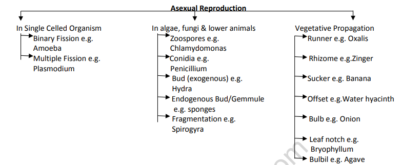
Asexual Reproduction
1. In single celled organisms: Binary fission (e.g. Amoeba) and Multiple fission (e.g. Plasmodium).
2. In algae, fungi and lower animals: Zoospores (e.g. Chlamydomonas), Conidia (e.g. Penicillium), Exogenous bud (e.g. Hydra), Endogenous bud or gemmule (e.g. sponges), and Fragmentation (e.g. Spirogyra).
3. Vegetative propagation: Runner (e.g. Oxalis), Rhizome (e.g. Zingiber), Sucker (e.g. Banana), Offset (e.g. Water hyacinth), Bulb (e.g. Onion), Leaf notch (e.g. Bryophyllum) and Bulbil (e.g. Agave).
Sexual Reproduction
1. Two parents each give a sex cell or gamete. Gametes may be similar (isogametes) or dissimilar (anisogametes). These are the pre-fertilisation events.
2. Offspring are NOT identical to their parents, so sexual reproduction gives variations in offspring.
3. Fertilisation is the fusion of sperm and egg to form a zygote. This is the fertilisation event.
4. The zygote develops into an embryo. This is the post-fertilisation event.
SEXUAL REPRODUCTION IN FLOWERING PLANTS
Microsporogenesis in the Anther
1. In the young anther, the corner cells become elongated and are called primary archesporial cells.
2. A periclinal division of the primary archesporial cell gives two cells: an outer parietal cell and an inner sporogenous cell.
3. The parietal cell divides mitotically to form four wall layers: the epidermis, the endothecium, the middle layer and the tapetum.
4. The sporogenous cell develops into the microspore mother cell (pollen mother cell).
5. Meiosis in the microspore mother cell gives a microspore tetrad, and each microspore develops into a pollen grain.
6. A microspore divides into a vegetative cell and a generative cell. The generative cell undergoes mitotic division to give two male gametes.
Wall Layers of the Anther
1. Epidermis: single layered.
2. Endothecium: single layered, and its cells are stony.
3. Middle layer: 2 to 3 layered.
4. Tapetum: single innermost layer, nutritive in nature, and polyploid.
HUMAN REPRODUCTION
Sexual Reproduction Events
1. Sexual reproduction events are divided into three groups: Pre-fertilisation (gametogenesis and gamete transfer), Syngamy and fertilisation, and Post-fertilisation.
Post Fertilisation Events
1. Zygote undergoes cleavage to form the 2, 4, 8 celled stage.
2. The 16 celled stage is the morula.
3. The morula develops into the blastocyst.
4. The blastocyst has an outer trophoblast and an inner cell mass.
5. The trophoblast forms the chorionic villi.
6. The inner cell mass forms three layers:
(i) Ectoderm: nervous system and skin.
(ii) Mesoderm: skeletal, urinary, circulatory and reproductive system.
(iii) Endoderm: digestive and respiratory system.
Menstrual Cycle
1. The menstrual cycle starts with menarche, the first period, at the age of 10 to 11 years. It continues through the reproductive years and stops at menopause, which occurs at the age of 45 to 55 years.
2. The endometrium has two parts: the stratum basale, which stays constant, and the stratum functionale, which changes cyclically.
3. The menstrual cycle has three phases: the proliferative phase, the secretory (luteal) phase and the menstrual phase.
Proliferative Phase
1. It is the first phase, starting just after menses.
2. Stratum basale remains as a thin layer.
3. Estrogens cause the endometrial glands to proliferate.
4. Epithelial cells from the glands cover the endometrium.
5. Stromal cells proliferate and secrete collagen and ground substance, forming new connective tissue.
6. Spiral arteries grow into the new endometrium, but not into the outer one-third.
7. The follicular phase (days 5 to 14) falls in this stage: 10 to 20 primary follicles develop. FSH stimulates granulosa cells and thecal cells to secrete estrogen. Days 1 to 8: FSH is the principal hormone; it stimulates granulosa and thecal cells to secrete estrogen. Later, LH rises, so progesterone is secreted. Then estrogen levels become high enough to be independent of FSH.
Ovulation and Luteal Phase
1. The surge of LH causes ovulation.
2. The luteal phase begins after ovulation. Granulosa and thecal cells transform into the corpus luteum.
3. The corpus luteum secretes estrogen and progesterone.
4. Progesterone controls the secretory phase, in which the stratum functionale expands. The glands enlarge, become corkscrew shaped and fill with secretory products. The mucoid secretion contains glycogen and nutrients for the development of the embryo.
5. If there is no fertilisation, the corpus luteum degenerates because hormone levels fall.
6. If fertilisation occurs, the corpus luteum is maintained, continues to secrete estrogen and progesterone, and hCG stimulates and maintains the corpus luteum through pregnancy.
Menstrual Phase
1. It is the last 5 days of the cycle.
2. Corpus luteum hormone levels fall, so the stratum functionale changes.
3. Spiral arteries contract, causing ischemia (lack of blood) to the stratum functionale.
4. The glands stop secreting, and the blood vessels rupture.
5. The surface epithelium, blood and tissue of the stratum functionale are shed.
6. Spiral arteries seal at the stratum basale.
Endometriosis
1. Pieces of endometrium present in other cavities of the body undergo cyclical changes, as they do in the uterus.
Hormonal control: the follicular phase (days 5 to 14, 10 to 20 primary follicles develop) is under hormonal control and leads into the proliferative phase. A surge of LH causes ovulation. The secretory (luteal) phase starts 1 to 2 days after ovulation. LH maintains the corpus luteum during the cycle. Menarche occurs at 10 to 11 years of life, and the cycle ends at menopause (perimenopause), at age 45 to 55, with gradual cessation of the menstrual cycle.
Oogenesis
1. Primary germ cells in the embryo differentiate into oogonia (2n), which divide mitotically.
2. The primary oocyte (2n) is arrested in prophase of meiosis I and is present at birth.
3. The primary oocyte completes meiosis I to form a secondary oocyte (n) and the first polar body. The secondary oocyte starts meiosis II and is arrested at metaphase of meiosis II.
4. Ovulation releases the secondary oocyte.
5. Entry of a sperm triggers the completion of meiosis II. This gives the ovum (n) and the second polar body.
Stages of Follicle Development
1. Oogonium in the ovary, then primary oocyte within the follicle, then growing follicle, then mature follicle.
2. The mature follicle ruptures and releases the ovulated secondary oocyte.
3. The ruptured follicle forms the corpus luteum, which later degenerates.
Figure: Oogenesis
[Figure: Flow diagram of oogenesis. An ovary with a primary germ cell in the embryo, which undergoes differentiation to an oogonium (2n). Mitotic division gives a primary oocyte (2n) arrested in prophase of meiosis I, present at birth. Completion of meiosis I and onset of meiosis II gives the secondary oocyte (n), arrested at metaphase of meiosis II, and the first polar body (n). Ovulation of the secondary oocyte follows. Entry of sperm triggers completion of meiosis II, giving the ovum (n) and the second polar body (n). Side panels show the oogonium in the ovary, the primary oocyte within the follicle, the growing follicle, the mature follicle, the ruptured follicle with the ovulated secondary oocyte, the corpus luteum and the degenerating corpus luteum.]
REPRODUCTIVE HEALTH
Path of Sperm Transfer
1. Seminiferous tubules, then rete testis, then vasa efferentia, then epididymis, then vas deferens, then urethra.
Hormones Involved in the Regulation of Spermatogenesis
1. The hypothalamus secretes GnRH, which stimulates the pituitary gland.
2. The pituitary secretes LH and FSH.
3. LH acts on Leydig cells, which secrete androgens.
4. FSH acts on Sertoli cells, which secrete factors that help spermiogenesis.
Population Explosion
Causes
1. Reduced infant mortality rate.
2. Decline in death rate.
3. Improved community health.
4. Improved agricultural practices.
Consequences
1. Scarcity of habitat.
2. Unemployment.
3. Shortage of food.
4. Ecological imbalance.
Control Measures
1. Birth control measures.
2. Creating awareness.
3. Legislation.
4. Family planning.
Methods of Birth Control
1. Natural methods: lactational amenorrhoea, coitus interruptus and periodic abstinence.
2. Barrier methods: condoms, diaphragms, cervical caps and vaults.
3. Intrauterine devices (IUDs): non medicated IUDs, copper releasing IUDs and hormone releasing IUDs.
4. Hormonal methods: oral contraceptives, injections and implants.
5. Surgical methods: vasectomy (male) and tubectomy (female).
PRINCIPLES OF INHERITANCE AND VARIATION
Genetic Disorders: Types
1. Genetic disorders are of two types: Chromosomal disorders and Mendelian disorders.
2. Chromosomal disorders are of two kinds: Aneuploidy and Polyploidy (mostly seen in plants).
3. Aneuploidy is divided into autosomal and sex chromosomal disorders.
4. Autosomal disorder: Down's syndrome, trisomy of the 21st chromosome.
5. Sex chromosomal disorders: Klinefelter's syndrome (XXY) and Turner's syndrome (XO).
6. Mendelian disorders: Phenylketonuria, Haemophilia, Thalassemia, Sickle cell anaemia, Colour blindness and Cystic fibrosis.
Mendelian Disorders
Haemophilia
1. Sex linked recessive trait (HbSHbS as written in the source) on the X chromosome.
2. A heterozygous (carrier) female may transmit the trait to her son.
3. A haemophilic daughter is rare, except from a cross between a carrier mother and a haemophilic father.
4. The mutant gene affects the clotting of blood. The defect leads to continuous bleeding, even from a minor injury, resulting in loss of blood.
Sickle Cell Anaemia
1. Autosomal recessive trait (HbSHbS).
2. Heterozygous parents (HbAHbS) may transmit the trait.
3. The mutant haemoglobin molecule changes the RBC into a sickle shape. Due to this shape, RBCs cannot pass through capillaries, which affects blood supply in low oxygen tension.
4. The defect is due to the substitution of glutamic acid by valine.
5. It is a point mutation, a single base substitution at the 6th codon of the beta globin chain of the haemoglobin gene, from GAG to GUG.
Phenylketonuria
1. Inborn error of metabolism, an autosomal recessive trait (HbSHbS as written in the source).
2. The mutant gene affects the enzyme phenylalanine hydroxylase, which converts phenylalanine to tyrosine in the liver.
3. Accumulated phenylalanine is converted into phenylpyruvic acid and other derivatives.
4. The defect causes black urine and mental retardation by affecting the brain.
Thalassemia
1. Autosomal recessive trait.
2. The disease is caused by mutation or deletion of one or more genes.
3. The mutant gene affects the rate of synthesis of one globin chain of haemoglobin, which results in anaemia.
4. Alpha thalassemia is caused by a defect in the HBA1 and HBA2 linked genes of chromosome 16.
5. Beta thalassemia is caused by a defect in the HBB linked genes of chromosome 11.
Colour Blindness
1. Sex linked recessive trait on the X chromosome.
2. A heterozygous female may transmit the trait to her son.
3. A colour blind daughter is rare, except from a cross between a carrier mother and a colour blind father.
4. The mutant gene affects the cone cells of the eye, so the person cannot discriminate between red and green colours.
5. The source also repeats the line "Defect leads to continuous bleeding even in minor injury resulting in loss of blood" here; this is copied from haemophilia and does not apply to colour blindness.
Chromosomal Disorders
Aneuploidy
1. Failure of segregation of chromatids during cell division.
2. Results in gain or loss of a chromosome.
Polyploidy
1. Failure of cytokinesis during cell division.
2. Results in an increase in a whole set of chromosomes.
3. Often found in plants.
Down's Syndrome (Autosomal, Trisomy 21)
1. Additional copy of chromosome 21 (trisomy).
2. Short statured, with a small round head.
3. Furrowed tongue and partially open mouth.
4. Broad palm with a characteristic palm crease.
5. Retarded mental development.
Klinefelter's Syndrome (Sex Chromosomal, XXY)
1. Additional copy of the X chromosome, giving a karyotype of 47 (XXY).
2. Overall masculine development, with feminine-like development of the breasts (gynaecomastia).
3. Sterile in nature.
[Figure: Ishihara colour vision test plates: two circular plates made of coloured dots, used to test colour blindness.]
Turner's Syndrome (Sex Chromosomal, XO)
1. Absence of one X chromosome, giving a karyotype of 45 (XO).
2. Sterile female with a rudimentary ovary.
3. Lack of secondary sex characters and reduced breast development.
4. Webbed neck and short stature with swollen fingers.
Figure: Inheritance of Turner's syndrome (non-disjunction)
- Female parent XX: meiosis with non-disjunction gives gametes XY... correct form: the XX female produces abnormal gametes XX (both X chromosomes together) and O (no sex chromosome). The male parent XY shows normal separation of chromosomes, giving gametes X and Y.
- Fertilisation of the female's XX gamete with X gives XXX zygote: sterile female but normal in all other respects.
- Fertilisation of the female's XX gamete with Y gives XXY zygote: Klinefelter's syndrome.
- Fertilisation of the female's O gamete with X gives XO zygote: Turner's syndrome.
- Fertilisation of the female's O gamete with Y gives YO zygote: not viable.
Mendel's and Morgan's Experiments and Sex Determination
1. Hybridisation experiments: Mendel's work in garden pea and Morgan's work in Drosophila.
2. Mendel's monohybrid cross (inheritance of one gene) gives the Law of Dominance and the Law of Segregation.
3. Mendel's dihybrid cross (inheritance of two genes) gives the Law of Independent Assortment.
4. Morgan's work in Drosophila gives the concepts of linkage and recombination.
5. Sex determination is of two kinds: autosomes and sex chromosomes.
6. Male heterogamety: in insects the male is XO and the female is XX. In human beings the male is XY and the female is XX.
7. Female heterogamety: in birds the male is ZZ and the female is ZW.
MOLECULAR BASIS OF INHERITANCE
DNA Replication: Enzymes and Their Functions
Figure: DNA replication in a eukaryotic cell
- Replication fork: the leading strand is made continuously (5' to 3'); the lagging strand is made as Okazaki fragments, each started by an RNA primer (RNA primers and replicating DNA are shown with different symbols).
- Removal of RNA primers by flap endonuclease (FEN1) and RNase H.
- Gaps between Okazaki fragments are filled by repair DNA polymerase.
- DNA fragments are sealed by DNA ligase. Action of DNA ligase: it joins the 3' OH end of one fragment to the 5' phosphate end of the next.
Figure: Replication fork with enzymes
- Leading strand: made by DNA polymerase (Pol delta). Lagging strand: made by DNA polymerase (Pol alpha) as Okazaki fragments, with RNA primers laid down by DNA primase and fragments joined by DNA ligase.
- Helicase unwinds the double helix, single strand binding proteins hold the strands apart, and topoisomerase relieves the strain ahead of the fork.
1. DNA helicase: also known as the helix destabilising enzyme. It unwinds the DNA double helix at the replication fork.
2. DNA polymerase: builds a new duplex DNA strand by adding nucleotides in the 5' to 3' direction. It also performs proof-reading and error correction.
3. DNA clamp: a protein which prevents DNA polymerase III from dissociating from the parent strand.
4. Single strand binding (SSB) proteins: bind to ssDNA and prevent the DNA double helix from re-annealing after DNA helicase unwinds it, thus maintaining strand separation.
5. Topoisomerase: relaxes the DNA from its super-coiled nature.
6. DNA gyrase: a specific type of topoisomerase which relieves the strain of unwinding by DNA helicase.
7. DNA ligase: re-anneals the semi-conservative strands and joins the Okazaki fragments of the lagging strand.
8. Primase: provides a starting point (a short RNA or DNA primer) for DNA polymerase to begin synthesis of the new DNA strand.
9. Telomerase: lengthens telomeric DNA by adding repetitive nucleotide sequences to the ends of eukaryotic chromosomes.
Eukaryotic RNA Polymerases
1. RNA polymerase I (Pol I, Pol A): transcribed in the nucleolus. Its product is the larger ribosomal RNA (rRNA) (28S, 18S and 5.8S).
2. RNA polymerase II (Pol II, Pol B): transcribed in the nucleus. Its product is messenger RNA (mRNA) and most small nuclear RNAs (snRNAs).
3. RNA polymerase III (Pol III, Pol C): transcribed in the nucleus, and possibly at the nucleolus-nucleoplasm interface. Its products are transfer RNA (tRNA) and other small RNAs, including the small 5S rRNA.
4. Other organisms utilise RNA polymerase I to transcribe certain protein-coding genes in addition to rRNAs.
Transcription in Prokaryotes
1. The promoter lies on the coding strand, upstream of the transcribed region.
2. RNA polymerase binds to the promoter with the help of sigma factor, and uses the template strand to make the RNA.
3. Ribonucleotides are added in the 5' to 3' direction.
4. Transcription stops at the terminator, with the help of rho factor.
Transcription and Processing in Eukaryotes
1. RNA polymerase transcribes the DNA template strand to form the primary transcript (hnRNA), which contains exons and introns.
2. Splicing: introns are removed and exons are joined.
3. Capping: a methylated guanine is added at the 5' end.
4. Tailing: 200 to 300 adenylate residues are added at the 3' end, forming the poly A tail.
5. The mature mRNA is released from the nucleus into the cytoplasm.
Central Dogma and Types of RNA
1. Central dogma: DNA undergoes replication, DNA is transcribed to RNA, and RNA is translated to protein.
2. Messenger RNA (mRNA) carries the message from DNA. It contains uracil.
3. Ribosomal RNA (rRNA) combines with proteins to form ribosomes.
4. Transfer RNA (tRNA) carries the amino acid to the ribosome, and has an anticodon.
[Figure: Central dogma diagram inside a cell with a nucleus and cytoplasm. Replication acts on DNA, transcription makes RNA from DNA, and translation makes protein in the cytoplasm. The three types of RNA are shown: messenger RNA with uracil bases, ribosomal RNA forming the ribosome with a large and small subunit, and transfer RNA with an amino acid attached at the top and an anticodon at the bottom.]
[Figure: Gene structure diagram. A DNA segment has a promoter with transcription factor binding sites (TATA box and CCAAT box) and a transcription start site (TSS), then Exon 1, Intron 1, Exon 2, Intron 2 and Exon 3, with splice junction sequences gt and ag at the boundaries. Transcription gives the primary transcript with a downstream element, a poly A signal and a cleavage site. Splicing removes the introns, giving mature mRNA with a 5' CAP, a 5' UTR, a start codon AUG, the coding sequence (CDS), a stop codon (UGA, UAA or UAG), a 3' UTR and a poly A tail. Translation then gives the protein.]
Figure: Translation
- A. Messenger RNA: mRNA is transcribed in the nucleus and moves out into the cytoplasm.
- B. Transfer RNA: the mRNA enters the cytoplasm and attaches to a ribosome. Translation begins at AUG, the start codon. Each tRNA has an anticodon whose bases are complementary to a codon on the mRNA strand. The ribosome positions the start codon to attract its anticodon, which is part of the tRNA that binds methionine. The ribosome also binds the next codon and its anticodon (tRNA carrying phenylalanine, lysine).
- C. The polypeptide assembly line: the ribosome joins the two amino acids, methionine and phenylalanine, and breaks the bond between methionine and its tRNA. The tRNA floats away, allowing the ribosome to bind to another tRNA. The ribosome moves along the mRNA (translation direction), binding new tRNA molecules and amino acids.
- D. Completing the polypeptide: the process continues until the ribosome reaches one of the three stop codons. The result is a growing polypeptide chain.
Translation
1. Translation is the process of joining amino acids by peptide bonds to form a polypeptide.
2. Activation of amino acids: AA + ATP + E (Mg2+) gives AA-AMP-E (aminoacyl adenylate enzyme complex) + PPi (pyrophosphate). AA-AMP-E + tRNA gives AA-tRNA (aminoacyl tRNA complex) + AMP + E.
3. Initiation: the small subunit (40S) of the ribosome binds to the mRNA. The charged tRNA for the initiation codon (methionine) reaches the P site. The large subunit (60S) then combines with the 40S-mRNA-tRNA complex in the presence of Mg2+.
4. Elongation: the second tRNA, charged with an amino acid, occupies the A site of the ribosome. A peptide bond forms between methionine and the second amino acid with the help of the enzyme peptidyl transferase. The ribosome moves over the mRNA in the 5' to 3' direction.
5. Termination: translation stops when a nonsense (stop) codon is reached. There is no tRNA for the stop codons UAA, UAG and UGA. The synthesised polypeptide is released with the help of a release factor.
[Figure: Cell diagram of gene expression. In the nucleus, the DNA double helix is transcribed into a newly formed messenger RNA (mRNA), which leaves through the nuclear membrane into the cytoplasm. Translation: the mRNA attaches to a ribosome, and amino acids bind to the growing protein chain. The role of the ribosome in translation: tRNAs bind to codons on the mRNA and bring amino acids (alanine, proline, arginine, leucine, etc.) to form the completed polypeptide.]
Genetic Code
1. Initiation codon: AUG.
2. Stop (nonsense) codons: UAA, UAG and UGA.
3. Features of the genetic code: triplet, commaless, universal, unambiguous (specific), degenerate, non overlapping and linear.
[Figure: Genetic code wheel. The circle is read from the centre outward: first base (G, U, A, C) in the centre, second base in the next ring, third base (U, C, A, G) in the outer ring, and the amino acid for each of the 64 codons around the edge (e.g. glycine, glutamic acid, aspartic acid, alanine, valine, arginine, serine, lysine, asparagine, threonine, methionine, isoleucine, glutamine, histidine, proline, leucine, cysteine, tryptophan, tyrosine, phenylalanine, with Stop codons). Question under the figure: for what amino acid does the codon UGC code? Answer: cysteine.]
Regulation of Gene Expression by Operon Model
Switch On Condition
1. The i-gene (regulator gene) makes the repressor protein.
2. The repressor protein combines with an inducer to form a repressor-inducer complex.
3. The structural gene is accessed by RNA polymerase, since there is no blockage at the operator.
4. Transcription of the structural gene takes place, and the enzyme or protein is formed.
Switch Off Condition
1. The repressor protein binds to the operator gene.
2. RNA polymerase cannot access the structural gene because of the repressor-operator complex.
3. There is no transcription of the structural gene, so no enzyme or protein is formed.
Figure: Lac operon
- Lactose absent: the regulator gene makes an active repressor, which binds to the operator (between the promoter and the structural genes lacZ, lacY, lacA). RNA polymerase cannot bind to the promoter, so the enzymes needed to take up and use lactose are not produced. If no inducer is bound to the repressor, the repressor can bind to the operator.
- Lactose present: lactose binds to the active repressor and makes it inactive. RNA polymerase can bind to the promoter and transcribes the structural genes into mRNA, which is translated into the enzymes. The enzymes to take up and use lactose are produced only when lactose is present.
Human Genome Project
Methodologies
1. Sequencing the whole genome, including coding and non-coding sequences.
2. Isolation of DNA from the cell.
3. Fragmentation by restriction endonucleases.
4. Fragments cloned in a suitable host using a suitable vector: BAC (bacteria) or YAC (yeast).
5. Fragments sequenced using automated DNA sequencers.
6. Sequences arranged on the basis of overlapping regions.
7. Alignment of the sequences by specialised computer based programmes.
8. Sequence annotation: identifying all the genes.
9. Expressed sequence tags (EST): identifying all the genes expressed as RNA.
Goals of HGP
1. Identification of all genes in human DNA.
2. Sequencing the 3 billion base pairs in human DNA.
3. Storing the information in data bases.
4. Improving tools for data analysis.
5. Using technologies in other sectors.
6. Addressing the ethical, legal and social issues (ELSI).
Salient Features of the Human Genome
1. The genome contains 3164.7 million nucleotide bases.
2. The average gene consists of 3000 bases.
3. The total number of genes is about 30,000, and these are 99.9 per cent the same in all humans.
4. Less than 2 per cent of the genes code for proteins.
5. About 50 per cent of the discovered genes have unknown functions.
6. Repetitive sequences of DNA contribute to evolution.
7. The largest gene is dystrophin, at 2.4 million bases.
8. Chromosome 1 has the maximum number of genes (2968). The Y chromosome has the minimum number of genes (231).
Applications of the Human Genome Project
1. Identification of defective genes.
2. Opportunity to offer early treatment.
3. Identification of genes that confer susceptibility to certain diseases.
4. Prediction of the proteins that the genes produce.
5. Drug designing to enhance or inhibit the activities of proteins.
DNA Fingerprinting
1. Technique developed by Dr. Alec Jeffreys. It is also known as DNA typing or DNA profiling.
2. The method uses short nucleotide repeats called Variable Number of Tandem Repeats (VNTR) as markers.
3. VNTRs are similar in closely related individuals, since they are inherited from ancestors.
Methodology
1. DNA isolation and extraction from the cell by a high speed refrigerated centrifuge.
2. Amplification of DNA by PCR (Polymerase Chain Reaction) to produce many copies.
3. DNA fragmentation (digestion) by restriction endonucleases.
4. Separation of DNA fragments by agarose gel electrophoresis.
5. Double stranded DNA is split into single stranded DNA.
6. Transfer of the separated DNA to a nylon or nitrocellulose sheet by Southern blotting.
7. The nylon sheet is immersed in a bath containing probes (markers) which bind to specific nucleotide sequences by hybridisation. Probes are radioactive synthetic DNA complementary to VNTR.
8. Hybridised DNA fragments are detected as dark bands on X-ray film by autoradiography.
9. The dark band develops at the probe site and is compared with the experimental segment.
Figure: Gel electrophoresis
- Restriction enzymes cleave the DNA sample into smaller segments of various sizes.
- DNA segments are loaded into wells in a porous gel. The gel floats in a buffer solution within a chamber between two electrodes.
- When an electric current is passed through the chamber, DNA fragments move toward the positively charged electrode (anode).
- Smaller DNA segments move faster and farther than larger DNA segments.
[Figure: Flow chart of DNA fingerprinting. DNA sample, then extraction of DNA, then cleavage of DNA by restriction enzyme, then gel electrophoresis to separate DNA fragments, then Southern blotting technique, then autoradiography to obtain DNA bands, then develop and visualise DNA fingerprints. Below it, a Southern blotting diagram for RFLP analysis: DNA extracted, restriction enzyme produces fragments, double stranded fragments separated by electrophoresis, denatured to single stranded fragments, transferred from gel to membrane, membrane covered with a radioactively labelled probe in single strand form and incubated, washed to remove excess probe, visualised by exposure to X-ray film, and the DNA typing pattern appears on the developed film.]
[Figure: Paternity test autoradiograms. Four sets of DNA band patterns are shown, each with three lanes labelled Mother, Child and Alleged Father. The bands are labelled A to O across the four sets. The child's bands are compared with the mother's bands and with the alleged father's bands to decide whether the alleged father is the biological father.]
EVOLUTION
Evidences of Evolution
1. Palaeontology: comparative study of fossils.
2. Embryology: study of the embryos of vertebrates.
3. Comparative anatomy: homologous organs, analogous organs and vestigial organs.
4. Connecting links: for example, Archaeopteryx.
[Figure: Adaptive radiation of marsupials of Australia. From a common marsupial ancestor (marsupial radiation), the diagram shows the Tasmanian wolf, tiger cat, sugar glider, banded anteater, marsupial mole, marsupial rat, koala, wombat, bandicoot and kangaroo.]
HUMAN HEALTH AND DISEASE
Types of Diseases
Infectious Diseases (e.g. Leprosy)
1. Transmitted by air, water, physical contact and animals.
2. Types of infectious diseases: viral, bacterial, fungal, worm (helminth) and protozoal.
3. Control measures: balanced diet, good personal hygiene, awareness about diseases, vaccination, and hygienic food and water.
Non-infectious Diseases (e.g. Cancer)
1. Causes: stress, no or less physical activity, genetic factors, unsuitable diet, wrong life style, and exposure to carcinogens.
2. Control measures: stress management, physical exercise, balanced diet, avoiding drugs, alcohol and tobacco.
Health Affecting Factors
1. Genetic factors.
2. Life style.
3. Infections.
Types of Acquired Immunity
Active Immunity
1. Antibodies are produced within one's own body.
2. It is long lasting and does not cause allergy.
3. It takes time to activate.
Passive Immunity
1. Antibodies are transferred from another individual.
2. It is not long lasting, and it sometimes causes allergy.
3. It provides immediate relief.
Cancer
Causes
1. Ionising radiation.
2. Chemicals such as tar from cigarettes.
3. Viruses, for example papilloma virus.
4. Predisposition.
Detection
1. Biopsy and histopathological study.
2. Imaging: CT, MRI and X-ray.
Treatment
1. Surgery.
2. Radiation.
3. Systemic treatment: (a) chemotherapy, (b) hormone therapy, (c) monoclonal antibodies, (d) alpha interferon.
4. Supportive care.
STRATEGIES FOR ENHANCEMENT IN FOOD PRODUCTION
Animal Breeding
1. Animal breeding is divided into out-breeding, controlled breeding and cross-breeding.
2. Out-breeding is divided into interspecific hybridisation and out-crossing.
[Figure: Fig 1: Concept map of animal breeding. Animal breeding branches into Out-breeding (with Interspecific hybridisation and Out-crossing), Controlled breeding and Cross-breeding.]
BIOTECHNOLOGY: PRINCIPLES AND PROCESSES
Features Required to Facilitate Cloning into a Vector
1. Origin of replication (ori).
2. Selectable markers.
3. Cloning sites.
4. Vectors for cloning genes in plants and animals.
Polymerase Chain Reaction (PCR)
1. PCR synthesises multiple copies of a gene of interest in vitro, using two sets of primers. Primers are small oligonucleotide strands complementary to the regions of DNA.
2. Steps: (i) the region to be amplified in ds DNA is separated by denaturation (heating), (ii) primers anneal to the single strands, (iii) DNA polymerase (Taq polymerase) extends the primers. These steps are repeated for 30 cycles, giving about 1 billion copies.
Palindromes in DNA
1. A palindrome is a sequence of base pairs that reads the same on the two strands when the orientation of reading is kept the same, in the 5' to 3' direction.
2. Example: 5'-GAATTC-3' on one strand pairs with 3'-CTTAAG-5' on the other strand, and both read the same in the 5' to 3' direction.
3. Another example in words: MALAYALAM reads the same in both directions.
[Figure: Pie charts of the global biodiversity slices are given without numerical values in the source.]
Genetically Modified Plants: Uses
1. Abiotic stress tolerant.
2. Enhance nutritional value.
3. Reduce post harvest loss.
4. Increase mineral usage.
5. Reduced reliance (on chemical inputs).
BIOTECHNOLOGY AND ITS APPLICATIONS
Genetically Engineered Insulin
1. Pro-insulin has an extra stretch of C-peptide in the middle, and is a single polypeptide chain.
2. Processing: the C-peptide is removed, and the A and B polypeptide chains are joined by disulphide bridges to give mature insulin.
RNA Interference (a Cellular Defence Mechanism)
1. DNA is transcribed to mRNA.
2. A double stranded RNA (dsRNA) is formed from the mRNA of the parasite.
3. The dsRNA prevents translation of the mRNA of the parasite.
Cry Protein of Bacillus thuringiensis (Bt)
1. Bacillus thuringiensis produces the Cry protein, which is an inactive toxin.
2. The alkaline pH of the gut of the insect (pest) activates the toxin.
3. The activated toxin causes swelling and lysis of the gut, and finally the death of the insect.
ORGANISMS AND POPULATIONS
How Do Organisms Live in Their Environment?
1. Four ways: Regulation, Conformation, Migration and Suspension.
Ecology Concept Map
1. Ecology is the study of the relationships between organisms and their environment.
2. The levels shown are organism, population, community, ecosystem and biosphere. Abiotic factors lead to the ecosystem and the biosphere.
3. Biotic factors are linked to community and population, and lead to biodiversity, population dynamics, conservation, trophic levels and food chain or food web.
4. Food chain and food web involve producers, consumers, decomposers, autotrophs and heterotrophs.
5. Symbiosis is shown with neutralism, mutualism, commensalism, parasitism and predator-prey relationships.
6. Other terms in the map: habitat, niche, adaptations, succession, biotic potential and carrying capacity.
ECOSYSTEM
Components of an Ecosystem
1. An ecosystem consists of abiotic components and biotic components.
2. Abiotic components are involved in cycling of matter, which sustains the biogeochemical cycles.
3. Biotic components comprise producers, macroconsumers and microconsumers. They form communities, which consist of populations, which consist of individuals of similar kind.
4. Biotic components depend on cycling of matter and flow of energy. The flow of energy is involved in the food chain. Food chains are interlocked with each other to make up the food web.
Energy Flow in an Ecosystem
1. Solar energy is captured by producers in photosynthesis, as chemical energy in organic food.
2. Herbivores obtain chemical energy in organic food from producers.
3. Carnivores obtain chemical energy in organic food from herbivores.
4. Excretion, death and decomposition return energy from the organisms.
5. Heat is released at every stage, from body activities and from decomposition.
[Figure: Energy flow in an ecosystem. Solar energy enters the producers box (chemical energy in photosynthesis), then herbivores (chemical energy in organic food), then carnivores (chemical energy in organic food), then excretion, death and decomposition. Heat is shown leaving at each step, and body activities produce heat at each trophic level.]
BIODIVERSITY AND ITS CONSERVATION
Conservation of Biodiversity
In-situ Strategies (conservation in the natural habitat)
1. Biosphere reserves.
2. National parks.
3. Wildlife sanctuaries.
4. Sacred forests.
Ex-situ Strategies (conservation outside the natural habitat)
1. Botanical gardens.
2. Zoological parks.
3. Gene banks.
4. Tissue culture.
Global Biodiversity
Vertebrates
1. Fishes, mammals, birds, reptiles and amphibians.
Plants
1. Fungi, mosses, ferns, angiosperms and lichens.
Invertebrates
1. Insects, molluscs, crustaceans and other animal groups.
Approximate proportions in the pie charts:
Vertebrates: fishes about one half; mammals, birds, reptiles and amphibians about one eighth each.
Plants: fungi about one third; angiosperms about one third; mosses, ferns and lichens each small (about 7%); one further small unlabelled segment.
Invertebrates: insects are the great majority (about 90%); molluscs, crustaceans and other animal groups are small slices (about 2%, 3% and 6%).
Reasons for More Biodiversity in the Tropical Region
1. Availability of more solar energy gives greater biomass production, and so greater species diversity.
2. Less seasonal variation gives a more constant and predictable environment. This allows niche specialisation and greater species diversity.
3. Less glaciation gives an undisturbed environment with more scope for evolution, and so more species diversity.
Number of Species in the Amazon Rain Forest in South America
1. Plant species: 40,000.
2. Fish species: 3,000.
3. Bird species: 1,300.
4. Mammal species: 427.
5. Amphibian species: 427.
6. Reptile species: 378.
7. Invertebrate species: 1,25,000.
MICROBES IN HUMAN WELFARE
Microbes in Sewage Treatment
Primary Treatment
1. Filtration.
2. Sedimentation.
3. Primary sludge (solids settle) and the supernatant form the effluent.
Secondary (Biological) Treatment
1. Waste water is passed to aeration tanks, where flocs form. Flocs are masses of bacteria associated with fungal filaments.
2. Flocs significantly reduce the biochemical oxygen demand (BOD).
3. In the settling tank, the sediments are called activated sludge.
4. The activated sludge is passed to anaerobic sludge digesters. During digestion, bacteria produce a mixture of gases such as methane, hydrogen sulphide and carbon dioxide.
Microbial Uses
Household
1. LAB (lactic acid bacteria) as starter: converts milk to curd, checks disease causing microbes, and synthesises vitamin B12.
2. Baker's yeast is used for making toddy from palm.
3. Swiss cheese: Propionibacterium sharmanii (bacteria).
4. Roquefort cheese: fungi.
Industrial
1. Fermented beverages: with distillation (whisky, brandy, rum) and without distillation (beer and wine).
2. Antibiotic production: penicillin (and its history), streptomycin and tetracycline.
3. Chemical production: Acetobacter gives acetic acid; Aspergillus niger (fungus) gives citric acid; Clostridium gives butyric acid; Lactobacillus gives lactic acid.
4. Enzymes: lipase is used as a detergent for removing oily stains; pectinase and protease are used as juice clarifiers; streptokinase (from Streptococcus) is a clot buster in myocardial infarction.
5. Bioactive compounds: Trichoderma polysporum (fungus) produces cyclosporin, an immunosuppressive agent used in organ transplants; Monascus purpureus (yeast) produces statin, which lowers blood cholesterol.
Bio-control
1. Bt (Bacillus thuringiensis) controls caterpillars and aphids.
2. Ladybird beetles control aphids and mosquitoes.
3. Trichoderma viride controls fungal diseases.
4. Nucleopolyhedrovirus controls insects and arthropods.
5. These are used in integrated pest management (IPM).
Biofertilisers
1. Symbiotic association: Rhizobium (in legume roots).
2. Free living: Azotobacter and Azospirillum.
3. Mycorrhiza: Glomus, which helps absorb phosphorus.
4. Cyanobacteria: Anabaena and Nostoc.
5. These are used in organic farming.
ENVIRONMENTAL ISSUES: POLLUTION
Air Pollution
1. Air Act (Prevention and Control) of 1981.
2. Causes: industries, vehicles, volcanic eruptions and household activities.
3. Pollutants: metallic and dust particles, aerosols and gases (CO, SO2, NO2).
4. Effects: harms the health of living organisms (asthma, bronchitis), causes global warming, and causes premature death of plants.
5. Control: electrostatic precipitator, scrubber, catalytic converter and fuel policy.
Water Pollution
1. Water Act (Prevention and Control) of 1974.
2. Causes: domestic sewage, agricultural waste (insecticides and pesticides), and industrial waste (PCBs, heavy metals such as lead, cadmium and mercury).
3. Effects: harms the health of living organisms (diarrhoea, naru), and retards the growth of plants.
4. Control: sewage treatment, awareness among people, and proper utilisation of media.
Miscellaneous Pollution
1. Solid wastes: domestic and industrial wastes.
2. E-wastes: electronic garbage such as worn out computers, mobiles and refrigerators.
3. Radioactive wastes: from utilised radioactive fuels.
Noise Pollution
1. The Air Act was amended in 1987 to include noise pollution.
2. Causes: vehicles, industries, loud speakers and jet planes.
3. Effects: sleeplessness, increased heart beat, stress and anxiety, and permanent hearing loss (at 150 dB).
4. Control: controlled utilisation of sound producing instruments.
Free study material for Biology
CBSE Class 12 Biology Study Material: important charts
Essential Notes for Class 12 Biology
Access comprehensive study material for important charts, including revision notes, concept maps, and high-probability questions. These resources are designed in alignment with the latest 2026 CBSE syllabus for Class 12 Biology to support effective exam preparation.
Verified Solutions for Class 12 Biology
Each resource draws directly from authorized textbooks to maintain academic accuracy. Evaluating solved examples allows Class 12 students to master the formal presentation and answer-writing standards expected in upcoming school exams.
Enhance Accuracy with Online Tests
Wrap up your chapter revision by testing your knowledge against standard objective question formats. Explore our full library of free, up-to-date sample papers and online assessments to secure top grades in your CBSE examinations.
FAQs
Our advanced study package for important charts includes detailed concepts, diagrams, Mind Maps, and explanation of complex topics to ensure Class 12 students learn as per syllabus for 2026 exams.
The Mind Maps provided for important charts act as visual anchors which will help faster recall during high-pressure exams.
Yes, teachers use our Class 12 Biology resources for lesson planning as they are in simple language and have lot of solved examples.
Yes, You can download the complete, mobile-friendly PDF of the Biology important charts advanced resources for free.
Yes, our subject matter experts have updated the important charts material to align with the rationalized NCERT textbooks and have removed deleted topics and added new competency-based questions.