

Official CBSE Study Materials for Class 12 Biology
Access comprehensive study materials and useful resources for Chapter 01 Sexual Reproduction In Flowering Plants using the CBSE Class 12 Biology Reproduction in Flowering Plants Diagrams. Designed to align with the 2026-27 CBSE academic guidelines, these advanced resources help Class 12 Biology students reinforce core concepts beyond standard textbooks.
Advanced Resources for Biology
Navigate directly to the advanced Biology study materials using the digital viewer below. Each resource pack includes detailed conceptual summaries and solved practice questions, allowing students to instantly reinforce their learning.
CBSE Class 12 Biology Reproduction in Flowering Plants Diagrams. Please refer to the examination notes which you can use for preparing and revising for exams. These notes will help you to revise the concepts quickly and get good marks.
BIOLOGY
CHAPTER - 2 REPRODUCTION IN FLOWERING PLANTS
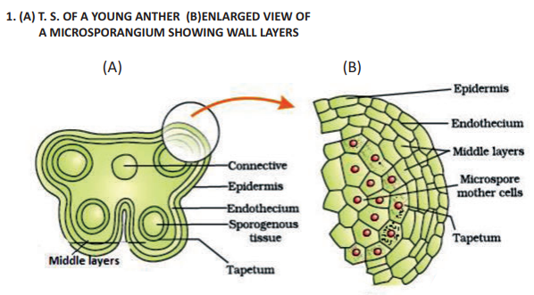
1. (A) T. S. OF A YOUNG ANTHER (B) ENLARGED VIEW OF A MICROSPORANGIUM SHOWING WALL LAYERS [2 Marks]
[Figure: Transverse section of a young anther showing four microsporangia, connective tissue, epidermis, endothecium, and middle layers, along with an enlarged view of a microsporangium showing Epidermis, Endothecium, Middle layers, Microspore mother cells, and Tapetum.]
Answer:
1. The anther is a four-sided (tetragonal) structure consisting of four microsporangia located in the corners, two in each lobe.
2. The microsporangium is generally surrounded by four wall layers namely epidermis, endothecium, middle layers, and tapetum.
Teacher's Note:
a) The outer three wall layers perform the function of protection and help in the dehiscence of anther to release the pollen.
b) The innermost wall layer is the tapetum which nourishes the developing pollen grains.
2. STAGES OF MICROSPORE MATURING INTO A POLLEN GRAIN [2 Marks]
[Figure: Stages showing a microspore developing a vacuole, followed by an asymmetric spindle division into a smaller generative cell and a larger vegetative cell inside the pollen grain.]
Answer:
1. As the microspore matures, its nucleus divides mitotically to form a vegetative cell and a generative cell.
2. The vegetative cell is bigger, has abundant food reserve and a large irregularly shaped nucleus, while the generative cell is small and floats in the cytoplasm of the vegetative cell.
Teacher's Note:
a) Asymmetric spindle division is crucial for the differentiation of vegetative and generative lineages.
b) In over 60 percent of angiosperms, pollen grains are shed at this 2-celled stage.
3. ANATROPUS OVULE [2 Marks]
[Figure: Structure of an anatropous ovule showing Hilum, Funicule, Micropyle, Micropylar pole, Outer integument, Inner integument, Nucellus, Embryo sac, and Chalazal pole.]
Answer:
1. The anatropous ovule is completely inverted such that the micropyle comes lying close to the funicle.
2. It consists of protective envelopes called integuments enclosing the nucellus, which houses the female gametophyte or embryo sac.
Teacher's Note:
a) Anatropous is the most common type of ovule found in flowering plants.
b) The point of attachment of the body of the ovule with the funicle is known as the hilum.
4. DIAGRAMMATIC REPRESENTATION OF DEVELOPMENT OF AN EMBRYO SAC [3 Marks]
[Figure: Diagrammatic representation showing successive stages of development of a female gametophyte or embryo sac from a single functional megaspore, depicting 1-nucleate, 2-nucleate, 4-nucleate and 8-nucleate stages leading to the mature 7-celled, 8-nucleate embryo sac with Micropylar end, Chalazal end, Synergids, Egg, Central cell, Polar nuclei, Antipodals, and Filiform apparatus.]
Answer:
1. The functional megaspore undergoes three successive mitotic divisions to form eight nucleated embryo sacs.
2. Out of the eight nuclei, six are surrounded by cell walls and organized into cells, while the remaining two polar nuclei reside in the central cell.
3. The mature embryo sac contains a 3-celled egg apparatus at the micropylar end, 3 antipodal cells at the chalazal end, and a single central cell with two polar nuclei.
Teacher's Note:
a) This illustrates monosporic development characteristic of flowering plants.
b) Students often confuse the number of cells with the number of nuclei in the central cell.
c) The filiform apparatus guides the pollen tube into the synergid.
5. FERTILISED EMBRYO SAC [3 Marks]
[Figure: Diagram of a fertilized embryo sac showing degenerating synergids, zygote (2n), primary endosperm cell (PEC), primary endosperm nucleus (3n) (PEN), and degenerating antipodal cells.]
Answer:
1. Syngamy results in the formation of a diploid zygote (2n) from the fusion of a male gamete and the egg cell.
2. Triple fusion involves the fusion of the second male gamete with the two polar nuclei to form the primary endosperm nucleus (PEN, 3n).
3. The central cell becomes the primary endosperm cell (PEC), and the synergids and antipodals begin to degenerate after fertilization.
Teacher's Note:
a) This represents double fertilization, a unique event to angiosperms.
b) Ensure correct ploidy levels are stated for zygote and PEN.
c) The note on degenerating cells highlights post-fertilization changes.
6. STAGES IN THE DEVELOPMENT OF DICOT EMBRYO [4 Marks]
[Figure: Stages in the development of a dicot embryo starting from Zygote, showing Proembryo stages, Globular Embryo, Heart-Shaped Embryo, and Mature embryo with Suspensor, Radicle, Plumule, and Cotyledons.]
Answer:
1. Embryogenesis starts with the division of the zygote into a proembryo.
2. The proembryo passes through globular and heart-shaped developmental stages.
3. The heart-shaped embryo eventually differentiates into a mature embryo containing plumule, radicle, and two cotyledons.
4. A suspensor is formed transiently to push the embryo into the nutritive endosperm.
Teacher's Note:
a) Dicot embryo development follows a predictable stereotypic sequence of stages.
b) Labeling both early and late structures like the suspensor and cotyledons is essential for full marks.
c) The heart-shaped stage clearly demarcates the two future cotyledons in dicots.
7. L.S. OF MONOCOT SEED [3 Marks]
[Figure: Longitudinal section of a monocot seed showing Pericarp, Endosperm, Scutellum, Coleoptile, Plumule, Radicle, and Coleorhiza]
Answer:
1. The monocot seed (such as maize) typically possesses a single cotyledon, which is large and shield-shaped, known as the scutellum.
2. The outer covering of the endosperm is called the aleurone layer, and the embryo consists of an embryonal axis with a plumule enclosed in a foliar sheath called coleoptile, and a radicle enclosed in an undifferentiated sheath called coleorhiza.
3. Endosperm is food-storing tissue that is bulky and nourishes the developing embryo during seed germination.
Teacher's Note:
a) Focus on the structural parts like scutellum and coleoptile.
b) Do not confuse coleoptile with coleorhiza; coleoptile covers the shoot apex/plumule while coleorhiza protects the root/radicle.
c) Monocot seeds like cereals are generally albuminous (endospermic).
8. L.S. OF GRASS EMBRYO [3 Marks]
[Figure: Longitudinal section of a grass embryo showing Scutellum, Coleoptile, Shoot apex, Epiblast, Radicle, Root cap, and Coleorhiza]
Answer:
1. The grass embryo is characterized by a single cotyledon called the scutellum situated towards one side of the embryonal axis.
2. At its lower end, the embryonal axis has the radical and root cap enclosed in an undifferentiated sheath called coleorhiza.
3. The portion of the embryonal axis above the level of attachment of scutellum is the epicotyl, which has a shoot apex enclosed in a hollow foliar structure called the coleoptile.
Teacher's Note:
a) Grass family (Poaceae) embryos have unique structures such as epiblast and coleoptile.
b) Ensure all labels including epiblast and root cap are clearly indicated.
c) Epiblast represents the rudimentary second cotyledon found in some grasses.
9. FALSE FRUIT OF APPLE [3 Marks]
[Figure: False fruit of apple showing Thalamus, Seed, Endocarp, and Mesocarp]
Answer:
1. In apple, the thalamus contributes significantly to fruit formation along with the ovary wall.
2. A false fruit (pseudocarp) is defined as a fruit in which some part of the plant other than the ovary, most commonly the thalamus, contributes to the edible and fleshy portion.
3. True fruits develop solely from the ovary, whereas in false fruits like apple and strawberry, the floral parts fuse to form the fruit structure.
Teacher's Note:
a) Differentiate clearly between true fruits and false fruits based on floral origin.
b) Students often miss labeling the thalamus, which is the key feature of an apple fruit.
c) Other examples of false fruits include strawberry and cashew nut.
10. A DICOT EMBRYO [3 Marks]
[Figure: A dicot embryo showing Plumule, Cotyledons, Hypocotyl, Radicle, and Root cap]
Answer:
1. A typical dicot embryo consists of an embryonal axis and two cotyledons.
2. The portion of the embryonal axis above the level of cotyledons is the epigeal epicotyl, which terminates with the plumule (stem tip), while the portion below the cotyledon level is the hypocotyl, which terminates at the lower end in the radicle (root tip).
3. The root tip is covered by a protective root cap at the terminal end of the radicle.
Teacher's Note:
a) Understand the polarity of the embryonal axis (epicotyl and hypocotyl).
b) Ensure correct distinction between plumule and radicle locations.
c) Dicot embryos store food reserves mostly in their fleshy cotyledons.
Free study material for Biology
CBSE Class 12 Biology Study Material: Chapter 01 Sexual Reproduction In Flowering Plants
Core Study Kit: Class 12 Biology Chapter 01 Sexual Reproduction In Flowering Plants
Review targeted study resources for Chapter 01 Sexual Reproduction In Flowering Plants tailored for Class 12 learners. Utilizing these structured notes and quick-revision tools ensures complete alignment with current CBSE evaluation standards.
NCERT-Aligned Solutions for Chapter 01 Sexual Reproduction In Flowering Plants
Each resource draws directly from authorized textbooks to maintain academic accuracy. Evaluating solved examples allows Class 12 students to master the formal presentation and answer-writing standards expected in upcoming school exams.
Next Steps in Your Exam Preparation
Maximize your grade potential by utilizing Class 12 Biology practice papers in tandem with these structured notes. All printable assignments and interactive mock tests on our platform are updated for the 2026 session and available free of charge.
FAQs
Our advanced study package for Chapter 01 Sexual Reproduction In Flowering Plants includes detailed concepts, diagrams, Mind Maps, and explanation of complex topics to ensure Class 12 students learn as per syllabus for 2026 exams.
The Mind Maps provided for Chapter 01 Sexual Reproduction In Flowering Plants act as visual anchors which will help faster recall during high-pressure exams.
Yes, teachers use our Class 12 Biology resources for lesson planning as they are in simple language and have lot of solved examples.
Yes, You can download the complete, mobile-friendly PDF of the Biology Chapter 01 Sexual Reproduction In Flowering Plants advanced resources for free.
Yes, our subject matter experts have updated the Chapter 01 Sexual Reproduction In Flowering Plants material to align with the rationalized NCERT textbooks and have removed deleted topics and added new competency-based questions.