

Advanced Study Material for Class 12 Biology: concept map
Review targeted academic resources with the CBSE Class 12 Biology all chapters concept map. Built according to official CBSE standards for the 2026-27 term, these downloadable Class 12 Biology study materials support effective daily practice and deeper conceptual understanding for concept map.
Practice Class 12 Biology Resources: concept map
Navigate directly to the advanced Biology study materials using the digital viewer below. Each resource pack includes detailed conceptual summaries and solved practice questions, allowing students to instantly reinforce their learning.

This CBSE Class 12 Biology concept map and diagram workbook covers the key diagrams, flow charts and crossword puzzles from all major chapters: vegetative propagation, sexual reproduction in flowering plants, human reproduction, reproductive health and birth control, principles of inheritance and variation, molecular basis of inheritance, evolution, and human health and disease, including tissue culture and plant breeding.
VEGETATIVE PROPAGATION
[Figure: Five labelled plant diagrams. (a) A potato tuber with eyes and germinating eye buds. (b) A rhizome with nodes, buds and adventitious roots. (c) A sprouting bulb. (d) A Bryophyllum leaf with adventitious buds along its edges. (e) A plant with an offset, labelled Offset.]
[Figure: Leaf bud of Bryophyllum. The main plant has a side stem (runner) that grows out from a bud at the base of the main stem. The labelled parts are Bud, Internode and Node. The runner grows along the soil, and new plants develop from the buds of the runner, with roots below them.]
[Figure: Leaf of Bryophyllum with the labels Notch, Shoot, Adventitious roots and Bud.]
CROSSWORD PUZZLE: SEXUAL REPRODUCTION
Across
1. The process of the fusion of the gametes.
6. The type of fertilization in hen.
7. The term used for bulges observed on the sides of the body of Hydra.
8. Eggs are produced here.
Down
2. Sperms are produced in these male reproductive organs.
3. Another term for the fertilized egg.
4. These animals lay eggs.
5. A type of fission in amoeba.
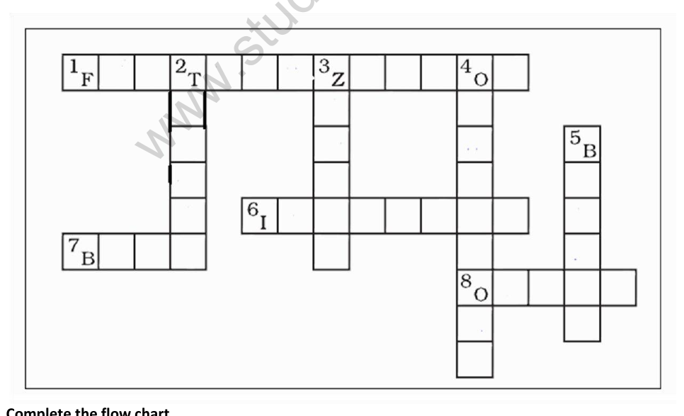
Crossword grid (with the given starting letters)
Answers
1 Across: FERTILIZATION
6 Across: INTERNAL
7 Across: BUDS
8 Across: OVARY
2 Down: TESTES
3 Down: ZYGOTE
4 Down: OVIPAROUS
5 Down: BINARY FISSION
FLOW CHART: POST-FERTILISATION EVENTS IN FLOWERING PLANTS
Complete the flow chart. The boxes are read in order:
Gametogenesis → A → Fertilization → Ovules transform into seeds → B → C
Hints printed in the PDF: A. Gamete transfer, B. Ovary transforms into fruit, C. Ovary wall transforms into pericarp.
A: Gamete transfer (pollination)
B: Ovary transforms into fruit
C: Ovary wall transforms into pericarp
CHAPTER 2: SEXUAL REPRODUCTION IN FLOWERING PLANTS
[Figure: Flower in longitudinal section with labels Stamen, Anther, Filament, Stigma, Style, Carpel, Ovary, Ovule, Petal, Sepal and Receptacle. A transverse section of an anther beside it shows pollen grains.]
Microsporogenesis
Pollen mother cell (diploid) → Meiosis I → Meiosis II → Tetrad of four haploid pollen cells → Secretion of walls → Separate pollen grains.
[Figure: One mature pollen grain with the labels Pit, Exine (thick, outer, sculptured wall), Intine (thin, inner wall), Generative nucleus (later forms two male gametes) and Pollen tube nucleus.]
[Figure: Lilium pollen grain germination in five stages. Stage 1 shows a pollen grain with a generative cell and a pollen tube nucleus. Stage 2 shows a germinating pollen grain with a pollen tube. Stages 3, 4 and 5 show the growing pollen tube. The labels are Pollen grain, Germinating pollen grain, Pollen tube, Generative cell, Male cells and Pollen tube nucleus.]
Ovule and megasporogenesis
[Figure: A typical anatropous ovule with the labels Hilum, Funicle, Micropyle, Micropylar pole, Outer integument, Inner integument, Nucellus, Embryo sac and Chalazal pole.]
[Figure: Megasporogenesis. Ovary → Ovule → Diploid cell (2n) → Meiosis → Four haploid cells (n), of which three die → Megaspore near the Micropyle → Surviving megaspore divides to form 2 haploid nuclei (n) → Each haploid nucleus divides twice more to form 8 haploid nuclei (n) → Egg (n). Labels on the right: Five nuclei die; three remain, Polar nuclei.]
[Figure: Stages of dicot embryo development. Zygote → Apical cell and Basal cell → Proembryo → Suspensor → Heart-shaped embryo with Cotyledons → Mature embryo with Shoot apical meristem, Root apical meristem, Vascular tissue, Ground tissue and Cotyledons.]
CHAPTER 3: HUMAN REPRODUCTION
Hormones at each stage of the gestation period
Ovulation → LH, FSH, Estrogen
Pregnancy → Estrogen, Progesterone by corpus luteum
Placenta → hCG, hPL, Estrogen, Progestogen
Foetal growth → Estrogen, Progestogen, Cortisol, Prolactin, Thyroxine
Parturition → Relaxin, Oxytocin
Lactation → Prolactin
[Figure: Menstrual cycle graph. The days run from 1 to 28, with the Follicular phase in the first half and the Luteal phase after Day 14. The rows show Basal body temperature, Hormone level (FSH, LH, Estrogen and Progesterone, with a peak at ovulation), the Ovarian cycle (follicles, Ovum, Ovulation) and the Uterine cycle (Menses, Proliferative and Secretory phases).]
CHAPTER 4: REPRODUCTIVE HEALTH
Population growth
[Figure: Line graph titled Population growth, some countries and regions. The y-axis shows population in millions from 0 to 2000 and the x-axis shows years from 1950 to 2050. The series are China, India, USA, Indonesia, S.S. Africa and Europe. India and S.S. Africa rise steeply, China rises and then levels off, Europe rises to about 700 million and then declines, and the USA and Indonesia rise gradually.]
Methods of birth control
Methods are arranged from most effective (top) to least effective (bottom). The percentages show the number of women out of every 100 who experienced an unintended pregnancy within the first year of typical use.
Reversible methods
Implant: 0.05%
Intrauterine device (IUD): LNG 0.2%, Copper T 0.8%
Permanent methods
Male sterilization (vasectomy): 0.15%
Female sterilization (abdominal, laparoscopic, hysteroscopic): 0.5%
Hormonal and device methods
Injectable: 6%
Pill: 9%
Patch: 9%
Ring: 9%
Diaphragm: 12%
Barrier, natural and other methods
Male condom: 18%
Female condom: 21%
Withdrawal: 22%
Sponge: 24% in parous women and 12% in nulliparous women
Fertility-awareness based methods: 24%
Spermicide: 28%
Notes from the chart: Vasectomy and hysteroscopic sterilization need another method for the first 3 months. Injectable: get repeat injections on time. Pills: take a pill each day. Patch and ring: keep in place and change on time. Diaphragm: use correctly every time you have sex. Condoms, sponge, withdrawal and spermicides: use correctly every time you have sex. Fertility-awareness based methods: abstain or use condoms on fertile days. The newest methods (Standard Days Method and TwoDay Method) may be the easiest to use and consequently more effective.
Condoms should always be used to reduce the risk of sexually transmitted infections.
Other methods of contraception: Lactational Amenorrhea Method (LAM) is a highly effective, temporary method of contraception. Emergency contraception: emergency contraceptive pills or a copper IUD after unprotected intercourse substantially reduces the risk of pregnancy.
In vitro fertilization (IVF)
1. Egg production stimulated by hormone therapy.
2. Eggs retrieved from ovary.
3. Sperm sample provided.
4. Eggs and sperm combined to allow fertilization.
5. Fertilized eggs introduced into uterus.
CHAPTER 5: PRINCIPLES OF INHERITANCE AND VARIATION
Amniocentesis
[Figure: Amniocentesis. An ultrasound probe is placed on the abdomen. A needle with a syringe is inserted through the uterus into the amniotic fluid around the foetus. Labels: Ultrasound probe, Amniotic fluid, Placenta, Uterus, Vagina, Cervix and Spine.]
1. A monohybrid cross
[Figure: Monohybrid cross. Pure tall (TT) and pure dwarf (tt) parents give F1 generation plants that are all tall (Tt). Self-pollination of the F1 gives an F2 generation with genotypes TT, Tt, Tt and tt, in the ratio 1 : 2 : 1, and phenotypes tall and dwarf in the ratio 3 : 1.]
2. Seven pairs of contrasting characters in Garden Pea (Pisum sativum)
Gregor Johann Mendel (photograph).
[Figure: Seven contrasting characters of pea plants: seed shape (round or wrinkled), seed colour (yellow or green), flower colour (violet or white), pod shape (inflated or constricted), pod colour (green or yellow), flower position (axial or terminal) and plant height (tall or dwarf).]
3. Turner syndrome
[Figure: Turner syndrome. Labels: Short stature, Low hairline, Shield-shaped thorax, Widely spaced nipples, Shortened metacarpal IV, Small finger nails, Brown spots (nevi), Characteristic facial features, Elbow deformity, Poor breast development, Rudimentary ovaries, Gonadal streak (underdeveloped gonadal structures) and No menstruation.]
4. Down syndrome
[Figure: Down syndrome. Labels on a labelled infant: Growth failure, Mental retardation, Flat back of head, Abnormal ears, Many "loops" on finger tips, Palm crease, Special skin ridge patterns, Unilateral or bilateral absence of one rib, Intestinal blockage, Umbilical hernia, Abnormal pelvis, Diminished muscle tone, Broad flat face, Staring eyes, Epicanthal eyefold, Short nose, Short and broad hands, Small and arched palate, Big wrinkled tongue, Dental anomalies, Congenital heart disease, Enlarged colon and Big toes widely spaced. Photographs of children and an adult with Down syndrome are also shown.]
5. Klinefelter syndrome
[Figure: Klinefelter syndrome. Labels: Frontal baldness absent, Poor beard growth, Tendency to grow fewer chest hairs, Breast development, Female-type pubic hair pattern, Small testicular size, Narrow shoulders, Wide hips and Long arms and legs. A karyotype beside it shows an extra X chromosome.]
6. Sickle cell anaemia
[Figure: Normal red blood cell, formation of sickled red blood cell and sickled red blood cell. A pedigree beside it uses the key male, female, unaffected carrier and affected. Outcomes for children of two carriers: one-in-four affected, one-in-two healthy carriers, one-in-four unaffected.]
7. Haemophilia
[Figure: Haemophilia pedigree. A carrier woman (X X-H) and a healthy man (X Y) have four children: a carrier girl (X X-H), a healthy girl (X X), a boy with haemophilia (X-H Y) and a healthy boy (X Y). Beside it, a father with haemophilia (X-H Y) and a mother who is a carrier (X-H X) have possible outcomes for each pregnancy, each with 25% chance: Son without haemophilia (X Y), Daughter with haemophilia (X-H X-H), Son with haemophilia (X-H Y) and Daughter carrier of haemophilia (X-H X).]
8. Phenylketonuria
[Figure: Phenylketonuria pathway. Dietary protein → Phenylalanine → Tyrosine → Melanin, Proteins and Dopamine. Phenylalanine is also converted to Phenylpyruvic acid and Phenylketones. Notes in the figure: Lack of phenylalanine hydroxylase blocks the transformation of phenylalanine into tyrosine; unmetabolized phenylalanine is shunted into the pathway that leads to the formation of phenylketones; excess phenylalanine also inhibits the formation of melanin from tyrosine.]
9. Colour blindness
[Figure: X-linked recessive inheritance in colour blindness. A pedigree with an unaffected father, a carrier mother, an unaffected son, an unaffected daughter, a carrier daughter and an affected son. Beside it, traffic lights are shown as seen by a normal person and by a colour-blind person.]
CHAPTER 6: MOLECULAR BASIS OF INHERITANCE
1. Structure of DNA
[Figure: Structure of DNA as a double helix. Base pairs shown are A-T and G-C, with the sugar-phosphate backbone, 3' and 5' ends, and labels Adenine, Thymine, Guanine, Cytosine and Phosphate-deoxyribose backbone.]
2. Structure of RNA
[Figure: Structure of RNA, with the nucleotide chain carrying bases A, G, U and C. A second diagram shows the nucleobases Cytosine, Guanine, Adenine and Uracil, with labels Nucleobase, Base pair and Helix of sugar-phosphates.]
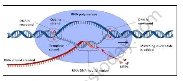
3. Transcription process
[Figure: Transcription. Labels: DNA is unwound, Coding strand, Template strand, RNA polymerase, RNA strand created, RNA-DNA hybrid region, Matching nucleotide is added and NTPs. A second diagram shows the three stages: Initiation (Promoter, Factor sigma, RNA polymerase), Elongation (RNA) and Termination (Stop sequence).]
4. Translation process
[Figure: Translation on a ribosome. Labels: mRNA, Ribosome, Start codon, Stop codon, Anticodon, tRNA, Amino acid, Polypeptide, Growing polypeptide, Peptidyl (P) site, Aminoacyl (A) site and the amino acids Tyr, Asp, Arg and Ser. A tRNA is shown with its amino acid attachment site, the anticodon UAC, and the sequence A C C at its end.]
5. DNA fingerprinting
Flow chart: DNA sample → Extraction of DNA → Cleavage of DNA by restriction enzymes → Gel electrophoresis to separate DNA fragments → Southern blotting technique → Autoradiography (label partly unclear in the PDF) → Develop and visualise DNA fingerprints.
[Figure: A DNA fingerprint pattern with several rows of coloured bands.]
6. Lac operon
[Figure: The lac operon. Labels: Regulatory gene (I), Promoter (P), Operator (O), Structural genes LacZ, LacY and LacA, mRNA, Ribosomes, Diffusible regulatory protein (produced by the regulatory gene) and RNA polymerase. The legend indicates Regions coding for proteins, Regulatory regions and Diffusible regulatory proteins. The beta-galactosidase (Z), permease (Y) and transacetylase (A) are shown as the proteins produced.]
7. DNA replication
Labels in the first diagram: Template strand, Replication fork, DNA polymerase, Okazaki fragments, DNA ligase, Leading strand and Lagging strand.
Labels in the second diagram: Leading strand, Lagging strand, Clamp, Polymerase III dimer, Helicase, Primase, Replication fork, RNA primer, Parent DNA, Leading strand template, Lagging strand template, Single-stranded DNA-binding proteins, DNA polymerase I, Ligase and Okazaki fragment.
CHAPTER 7: EVOLUTION
Evidence of evolution
Evidence of Evolution includes:
- The fossil record, which is composed of physical remains of organisms.
- Geographic distribution of living species, which indicates common ancestral species.
- Homologous body structures, which imply similar genes.
- Similarities in early development, which imply similar genes.
[Figure: Fossil formation in three steps. 1. Water carries small rock particles to lakes and seas. 2. Dead organisms are buried by layers of sediment, which forms new rock. 3. The preserved remains may later be discovered and studied.]
[Figure: Homologous forelimbs. The forelimbs of turtle, alligator, bird and mammal are shown, all arising from an Ancient lobe-finned fish. Note: example of homologous bones in forelimbs (forearms) in frogs, birds, bats, horses, whales and humans.]
Humans and chimpanzees: DNA is 98.5% the same.
Chromosomes of a human (left) and a bonobo chimpanzee are compared.
[Figure: Human karyotype showing chromosomes 1 to 22, X and Y, arranged in pairs.]
Comparing human and gorilla skeletons
| Feature | Modern Human | Modern Gorilla |
|---|---|---|
| Skull atop spine | S-shaped spine | C-shaped spine |
| Spinal cord exits | At bottom of skull | Near back of skull |
| Arms and legs | Arms shorter than legs; hands do not touch ground during walking | Arms longer than legs; hands touch ground during walking |
| Pelvis | Bowl-shaped | Long and narrow |
| Thigh bones | Angled inward, directly below body | Angled away from pelvis |
Human evolution
[Figure: Skulls of hominids. Australopithecus afarensis: large brow ridge, large canine teeth, face protrudes forward. Homo erectus: large brow ridge, large nose. Neanderthal: weak brow ridge, inflated cheeks, large nose. Cro-Magnon and Modern Homo sapiens: round high skull, large brain case, even teeth and strong chin.]
[Figure: World map showing the route of H.M.S. Beagle with its voyage around South America (Cape Horn, Galapagos Islands), Cape of Good Hope, Australia and New Zealand.]
CHAPTER 8: HUMAN HEALTH AND DISEASE
Malaria life cycle
Mosquito host: Female mosquito takes up gametocytes with blood meal → Fertilisation and development take place in the mosquito's intestine → Mature infective stages (sporozoites) escape from intestine and migrate to the mosquito's salivary glands → When the mosquito bites another human, sporozoites are injected with the bite.
Human host: Parasites (sporozoites) reach the liver through blood → The parasite reproduces asexually in liver cells, bursting the cell and releasing into the blood → Parasites reproduce asexually in red blood cells, bursting the red blood cells and causing cycles of fever and other symptoms; released parasites infect new red blood cells → Sexual stages (gametocytes), male and female, develop in red blood cells.
[Figure: Malaria life cycle drawn as a circle, with the Mosquito host and the Human host shown. Labels: Sporozoites, Salivary glands, Gametocytes, Female, Male, Parasites, Fertilisation and development.]
[Figure: Elephantiasis. A person with greatly swollen legs and lower body.]
Structure of an antibody molecule
[Figure: Antibody molecule with two heavy chains and two light chains. Labels: Antigen binding site (two places), Light chain, Heavy chain, and the N and C ends.]
HIV-1 life cycle
1. The free virus binds to a human CD4 receptor, commonly found on the cell surface. The virus fuses with the cell.
2. The virus penetrates the cell and its contents are emptied into the cell.
3. Single strands of viral RNA are converted to double-stranded DNA by HIV-1 Reverse Transcriptase (RT) enzyme.
4. Viral DNA is integrated into the human DNA by the integrase (IN) enzyme.
5. When the infected cell divides, HIV-1 protein chains are expressed (transcription). The transactivator of transcription (tat) protein binds to the viral RNA named TAR and triggers the replication of the entire viral genome.
6. The chains assemble together. The enzyme HIV-1 protease (PR) starts cleaving the HIV-1 protein chains at specific locations.
7. The assembly is pushed out of the cell. It takes with it part of the human membrane.
8. The virus breaks free from the cell.
9. HIV-1 PR finishes cleaving the protein chains. The viral particle is now mature and ready for attacking other human cells.
10. Free virus infects another cell.
PLANT TISSUE CULTURE AND SOMACLONES
[Figure: Chemical structure of morphine, and an opium poppy plant.]
Explant (any plant part) → Grown in a test tube under sterile condition in nutrient medium → Totipotent cells → Micropropagation → Somaclones (genetically identical plants).
[Figure: Fig 1: Tissue culture.]
STEPS IN PLANT BREEDING
1. Collection of variability.
2. Evaluation and selection of parents.
3. Cross hybridisation among the selected parents.
4. Selection and testing of superior recombinants.
5. Testing, release and commercialization of new cultivars.
Please click the link below to download pdf file of CBSE Class 12 Biology all chapters concept map.
Free study material for Biology
CBSE Class 12 Biology Study Material: concept map
Comprehensive Study Resources for concept map
Explore essential learning tools for Class 12 Biology concept map. This curated collection features in-depth notes and targeted practice questions built around the active 2026 curriculum to streamline your daily revision.
Understanding Marking Schemes
Built using official NCERT guidelines for Class 12 Biology, these materials provide reliable academic support. Integrating past examination questions and step-by-step solutions helps students understand official CBSE grading criteria.
Complete Revision for Biology
Maximize your grade potential by utilizing Class 12 Biology practice papers in tandem with these structured notes. All printable assignments and interactive mock tests on our platform are updated for the 2026 session and available free of charge.
FAQs
Our advanced study package for concept map includes detailed concepts, diagrams, Mind Maps, and explanation of complex topics to ensure Class 12 students learn as per syllabus for 2026 exams.
The Mind Maps provided for concept map act as visual anchors which will help faster recall during high-pressure exams.
Yes, teachers use our Class 12 Biology resources for lesson planning as they are in simple language and have lot of solved examples.
Yes, You can download the complete, mobile-friendly PDF of the Biology concept map advanced resources for free.
Yes, our subject matter experts have updated the concept map material to align with the rationalized NCERT textbooks and have removed deleted topics and added new competency-based questions.