
Read and download the CBSE Class 5 Science Sense Organs Worksheet in PDF format. We have provided exhaustive and printable Class 5 Science worksheets for Sense Organs, designed by expert teachers. These resources align with the 2026-27 syllabus and examination patterns issued by NCERT, CBSE, and KVS, helping students master all important chapter topics.
Chapter-wise Worksheet for Class 5 Science Sense Organs
Students of Class 5 should use this Science practice paper to check their understanding of Sense Organs as it includes essential problems and detailed solutions. Regular self-testing with these will help you achieve higher marks in your school tests and final examinations.
Class 5 Science Sense Organs Worksheet with Answers
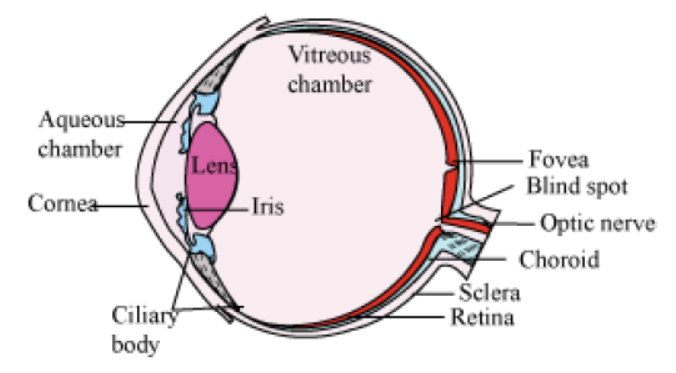
Structure of Human Eye
Sense Organs: Organs that helps us to be aware of our surroundings are known as sense organs. Some of the major sense organs of our body include eyes, ears, nose, tongue and skin.
Receptors: Any cell or tissue sensitive to a selective stimuli is known as receptor.
Some common receptors are:
• Mechanoreceptors: Receptors for touch or pressure; found in skin
• Thermoreceptors: Receptors fro temperature; found in skin
• Chemoreceptors: Receptors of taste (in tongue) and of smell (in nose)
• Photoreceptors: Receptors of light; found in eyes (rod and cone cells)
Eye
Eye is one of the most sensitive sense organs in the human body. Our eye enables us to see this beautiful world. It consists of a lens, which is made up of living tissues. How does our eye work? What are the nature, position and relative sizes of the images formed by the lens in the eye? In this section, we will learn about the structure and functioning of human eye. Structure of human eye
The human eye is roughly spherical in shape with diameter of about 2.3 cm. It is situated in the front side of the skull in bony sockets. It is covered by the eyelids that have eye lashes which prevent dust and other substances from entering into the eye. It consists of a convex lens made up of living tissues. Hence, human lenses are living organs contrary to the simple optical lenses. The inner region of the upper eye lid contains Lacrymal glands, that produces secretion known as tears, which keep eye surface moist and wash out dirt and other substances. Tears contain some salts and act as an antiseptic because of the presence of the enzyme lysozyme which kills the germs.
Structure of Human Eye: The wall of the eye consists of three layers namely sclera, choroid and retina.
The following table lists the main parts of the human eye and their respective functions.
| Human eye part | Function |
| Pupil | Opens and closes in order to regulate and control the amount of light |
| Iris | Controls light level similar to the aperture of a camera |
| Sclera | Protective outer coat |
| Cornea | Thin membrane which provides 67% of the eye’s focusing power |
| Crystalline lens | Helps to focus light into the retina |
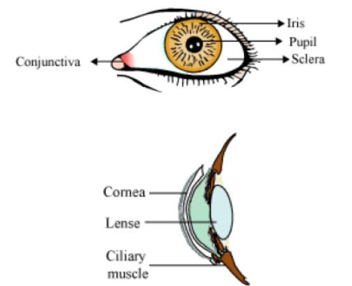
| Conjunctiva | Covers the outer surface (visible part) of the eye |
| Aqueous humour | Provides power to the cornea |
| Vitreous humour | Provides the eye its form and shape |
| Retina | Captures the light rays focussed by the lens and sends impulses to the brain via optic nerve |
| Optic nerve | Transmits electrical signals to the brain |
| Ciliary muscles | Contracts and extends in order to change the lens shape for focusing. |
The white of the eye is known as the sclera. It is the tough, opaque tissue that protects the outer layer of the eye. The bulged, transparent front portion of the sclera is called cornea. It is protected by thin, transparent tissue known as the conjunctiva.
The middle layer called choroid is supplied with nerves and blood vessels. It consists of the coloured layer of tissue called iris. It is responsible for the colour of the eye. Pupil is the black, circular hole that is located at the centre of the iris.
The lens consists of layers of tissues enclosed in a tough capsule. The focus of the lens is adjusted by the ciliary muscles that suspend and hold it. The lens focuses the light rays on retina where inverted image of the object is formed.
Retina contains two types of cells rods and cones. Rods are sensitive to dim light and cannot differentiate between various colours while as cones are sensitive to bright light and can distinguish various colours.
Functioning of the human eye
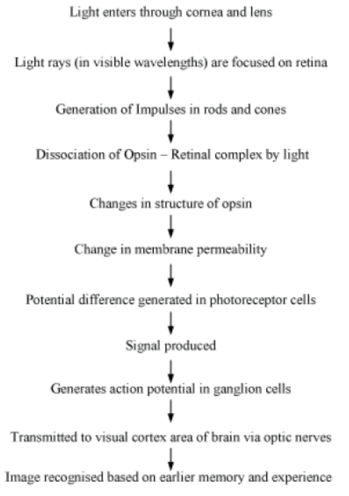
Light rays enter the eye through the cornea. The rays are bent, refracted, and focused by the cornea, lens, and the vitreous humour. The main function of the lens is to focus the light rays sharply on the retina. It is the outer surface of the cornea where most of the refraction of light occurs.
Iris controls the size of the pupil and the amount of light respectively. Since the eye lens is convex in nature, the resulting image is real, small, and inverted. This image is formed on the retina. The retina converts these light rays into electrical signals with the help of light sensitive cells. These signals are sent to the brain via translated and perceived objects in an erect or upright position.
The head of the optic nerve is devoid of photosensitive cells (rods and cones). Hence, no image is formed at that point called the blind spot of the eye.
Lateral to the blind spot, a yellow spot (fovea) is present that contains large number of cone cells. At this portion of retina a most clear and sharp image is formed.
Power of Accommodation
This is a special capacity of human eye to adjust its focus depending upon the object they are seeing. This happens because of the presence of flexible ciliary muscles around the eyes that helps in adjusting the focus of the eye lens. For distant vision the lens flattens whereas for near vision it becomes more convex.
Stereoscopic Vision
Humans and monkeys/apes have a special ability to perceive depth and relative distance as they can simultaneously focus on an object with both eyes. This results in the generation of a three dimensional image in our brain. This ability is known as stereoscopic vision.
On sunny days, when you enter a dimly lit room, you are unable to see clearly for a moment. Why does this happen?
In bright light, the iris expands, thereby contracting the pupil. This happens so that only a small quantity of light enters the eye. As a result, the retina is protected from exposure to excessive light.
On entering a dimly lit room after having been in the sun for some time, the iris contracts slowly to expand the pupil. Gradually, more light is able to enter the eye. Hence, it takes a few seconds before we are able to see the objects present in the dimly lit room.
Mechanism of Vision
• Photopigments in the eye consist of opsin (a protein) and retinal (an aldehyde of vitamin A).
Accommodation
Accommodation is the adjustment of the eye for a clear vision of objects at varying distances. Accommodation is brought about by changing the curvature of the elastic lens and making it more convex or concave with the help of suspensory ligaments and ciliary body.
When eye is at rest or is focused on distant objects, the suspensory ligaments are under great pressure. Therefore, the lens becomes flat and has maximum focal length. On the contrary, when we want to see the nearby objects, the ciliary muscles contract and pressure on the ligaments is released. The lens therefore bulges and becomes more round and convex.
Binocular Vision
Whenever we want to perceive depth or the relative distance of the objects, both our eyes focus simultaneously on the object.
Two separate images are formed, which the brain correlates and interprets as a single image. This results in a three-dimensional vision. It is also called stereoscopic vision.
Continuous Movement
The impression of an image remains on our brain for one-tenth of a second. If some other image is superimposed on the previous one, then the previous one is wiped off and we get an illusion of continuous movement.
Structure of Human Ear
Functions of the Ears
• Hearing
• Maintenance of body balance
Anatomy of the Ear
• Divided into three major sections:
• Outer ear
• Middle ear
• Inner ear
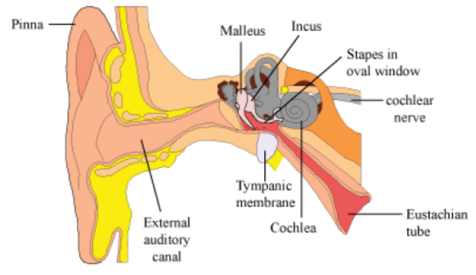
• Outer ear = Pinna + External auditory meatus (Canal)
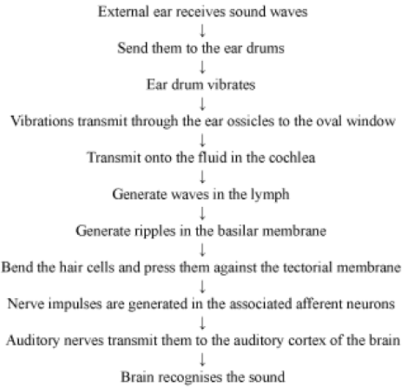
• Pinna − collects the vibrations that produce sound
• Canal − leads inwards and extends up to the tympanic membrane (Ear drum)
• Wax-secreting sebaceous glands are present in the skin of the pinna and the canal.
• Middle ear: Has 3 ossicles (Malleus, Incus, Stapes)
• Malleus is attached to the ear drum and stapes is attached to the oval window of the cochlea. Middle ear communicates to the inner ear through the oval window.
• Ossicles increase efficiency of transmission of sound waves to the inner ear.
• Eustachian tube − connects the middle ear cavity with the pharynx; equalises the pressure on either sides of the ear drum
• Inner ear (labyrinth): Has 2 parts (Bony Labyrinth and Membranous Labyrinth)
• Bony Labyrinth − series of channels in which the membranous labyrinth lies
• Membranous Labyrinth − surrounded by a fluid called perilymph and filled with a fluid called endolymph
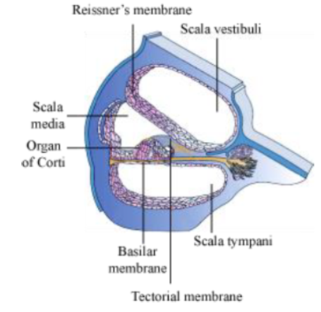
• Cochlea − coiled portion of the labyrinth
• 2 membranes surround cochlea, the reissner's membrane and the basilar membrane.
• These membranes divide the bony labyrinth into 3 parts -upper scala vestibuli, middle scala media and lower scala tympani.
• Scala media − filled with endolymph
• Scala vestibuli − ends at the oval window Scala tympani − ends at the round window that opens to the middle ear
• Organ of Corti − located on the basilar membrane and contains auditory receptors called hair cells (close contact with afferent nerve fibres)
• Tectorial membrane − elastic membrane present above the rows of hair cells
• Vestibular apparatus: Complex system located above the cochlea
• Composition − 3 semi-circular canals and otolith organ (has saccule and utricle)
• Base of the canals is swollen to form ampulla, which contains a projecting ridge called the crista ampullaris; it has hair cells
• Saccule and utricle − contain macula, which along with crista is responsible for maintaining the balance of the body and posture
Mechanism of Hearing
Role of Ear in Body Balancing
The fluid inside the semicircular canals moves when we turn our head. This moving fluid pushes against the sensory hair cells. This results in transmission of nerve impulse to brain through auditory nerve. The cells present in the semicircular canals are highly sensitive to dynamic equilibrium. Hence, they help us in maintaining balance of our bodies.
CBSE Science Class 5 Sense Organs Worksheet
Students can use the practice questions and answers provided above for Sense Organs to prepare for their upcoming school tests. This resource is designed by expert teachers as per the latest 2026 syllabus released by CBSE for Class 5. We suggest that Class 5 students solve these questions daily for a strong foundation in Science.
Sense Organs Solutions & NCERT Alignment
Our expert teachers have referred to the latest NCERT book for Class 5 Science to create these exercises. After solving the questions you should compare your answers with our detailed solutions as they have been designed by expert teachers. You will understand the correct way to write answers for the CBSE exams. You can also see above MCQ questions for Science to cover every important topic in the chapter.
Class 5 Exam Preparation Strategy
Regular practice of this Class 5 Science study material helps you to be familiar with the most regularly asked exam topics. If you find any topic in Sense Organs difficult then you can refer to our NCERT solutions for Class 5 Science. All revision sheets and printable assignments on studiestoday.com are free and updated to help students get better scores in their school examinations.
FAQs
You can download the latest chapter-wise printable worksheets for Class 5 Science Sense Organs for free from StudiesToday.com. These have been made as per the latest CBSE curriculum for this academic year.
Yes, Class 5 Science worksheets for Sense Organs focus on activity-based learning and also competency-style questions. This helps students to apply theoretical knowledge to practical scenarios.
Yes, we have provided solved worksheets for Class 5 Science Sense Organs to help students verify their answers instantly.
Yes, our Class 5 Science test sheets are mobile-friendly PDFs and can be printed by teachers for classroom.
For Sense Organs, regular practice with our worksheets will improve question-handling speed and help students understand all technical terms and diagrams.